Download presentation
Presentation is loading. Please wait.

1
Dr Wan Zaripah Wan Bakar School of Dental Sciences USM Health Campus BDS (Malaya), Grad. Dip. (Adelaide), D.Clin.Dent (Prosthodontics) (Adelaide), FRACDS
, D.Clin.Dent (Prosthodontics) (Adelaide), FRACDS.")
2
Objectives Knowledge of the basic root canal anatomy and its variations. Ability to gain good endodontic access. Ability to find the root canals. Use the right instruments and correct techniques.

3
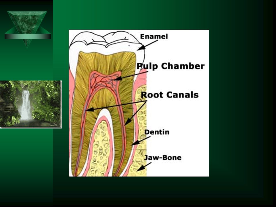
Access Cavity Preparation Access to pulp chamber and canals First step in root canal treatment

4
ROOT CANAL Anatomy Access

5
ANATOMY –A thorough knowledge of root canal anatomy and the possible variations for any given tooth is essential. –Variations:- in individuals between individuals between races unknown. –Each and every tooth should be assessed prior to treatment and expect the unexpected at times.

7
Methods of Determining Anatomic Configuration:- Diagnostic periapical radiographs Tube-shift radiographs Knowledge of root canal anatomy and typical variations Variations – ethnic Fibre optic light transillumination of the access cavity and pulp chamber Probing the floor of pulp chamber with an endodontic explorer Digital perception and tactile sensation with hand instruments.`

8
Diagnostic Radiographs Anatomy of pulp chamber and relationship to occlusal anatomy Estimated root canal lengths Root curvature (morphology) Root canal diameter Stage of root development – open apex Canal obstructions Root resorption Root fractures Periapical pathosis Previous root treatments or fillings Periodontal disease
 Root canal diameter Stage of root development – open apex Canal obstructions Root resorption Root fractures Periapical pathosis Previous root treatments or fillings Periodontal disease")
9
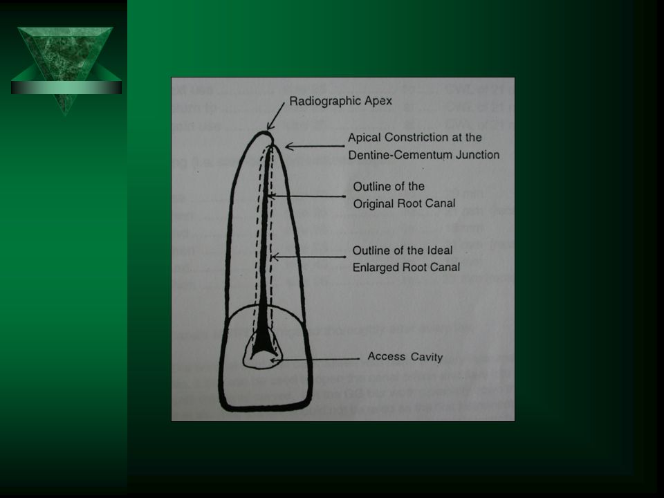
Anatomy of Root Apex

10
“Apical constriction “ – the point at which the root canal treatment is terminated apically. - the border between the pulp and the PDL. - narrowest part of the canal – should not be enlarged

11
ACCESS Visual and physical access to all canals. Good access for good endodontic treatment. Should not be compromised.

12
ACCESS

13
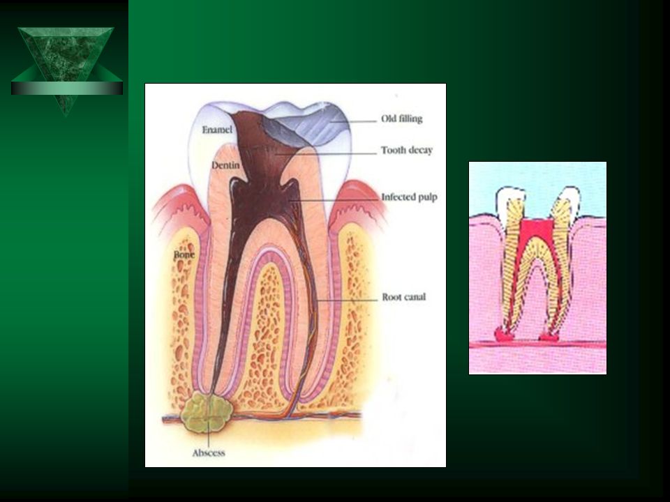
Principles Removing the entire existing restoration from the tooth. - recurrent caries. - enamel and dentine cracks - pathways for bacterial penetration and thus lead to failure. - assessed the feasibility of restoring the tooth again. - assessment of type of restoration required. - uninhibited access, allow more light. - for easy location of the canals. “ Dentin mapping ” - technique of finding other canals after find one canal - moving sideway following the path

15
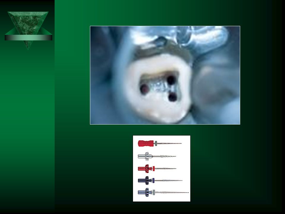
INSTRUMENTATION Rotary Equipment High speed: round diamond burs, TC - jet 330 to gain access. beaver burs (for metal) TC 169L Low speed: round burs Gates-Glidden burs spiral root filler Endo access bur Hand Instruments Mouth mirror Straight probe or endodontic explorer. Excavator Endodontic spreader Irrigating syringes with needles Metal ruler Blunted spreader or plugger or burnt plastic instrument. Files
 TC 169L Low speed: round burs Gates-Glidden burs spiral root filler Endo access bur Hand Instruments Mouth mirror Straight probe or endodontic explorer. Excavator Endodontic spreader Irrigating syringes with needles Metal ruler Blunted spreader or plugger or burnt plastic instrument. Files.")
16
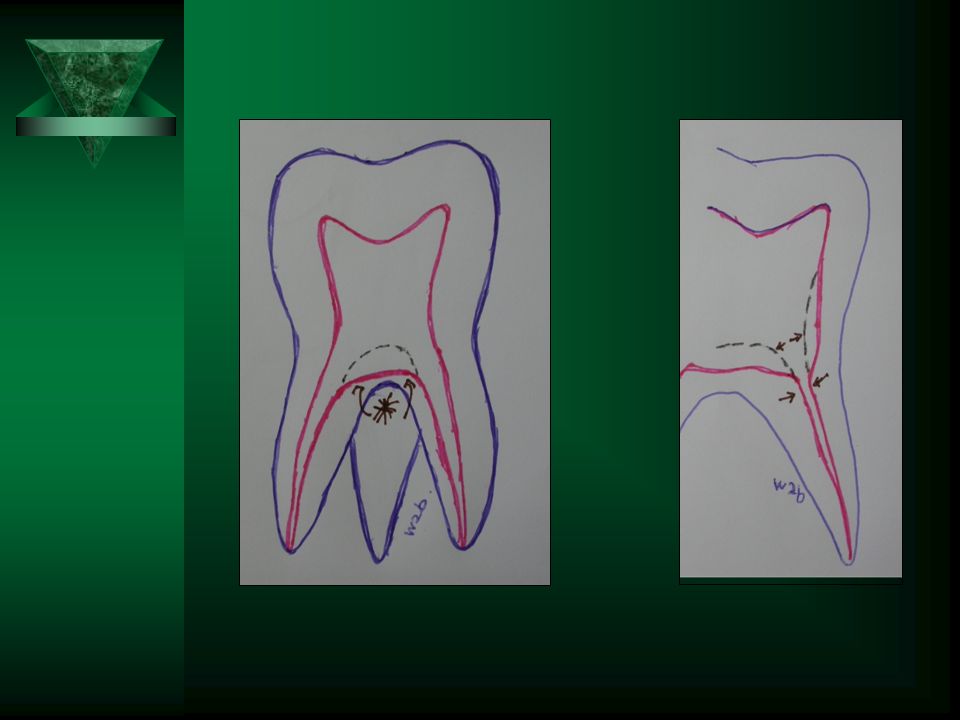
COMMON MISTAKES –Wrong alignment of the burs –Overremoved good dentin coronally –Overcut pulpal floor –Perforations – coronal - bifurcation - apical –Overextended preparation –Misdetection of canals –Accessory canals

17
CAUTION!!! Never ever removed sound pulpal floor. - increase difficulties in finding the canals (smaller). - weakness of the floor or bifurcation area may lead to fracture. - might perforate. - poor long term prognosis. - failure!! Do not give hard pressure to Gates-Glidden bur if feels retention. - might align the bur in wrong direction - can cause perforation.
. - weakness of the floor or bifurcation area may lead to fracture. - might perforate. - poor long term prognosis. - failure!. Do not give hard pressure to Gates-Glidden bur if feels retention. - might align the bur in wrong direction - can cause perforation..")
19
STEPS Radiographs Endodontic kit Local anaesthetics – optional Rubber dam –mandatory Removal of existing fillings or access gained Caries removal Canal access Canal preparation Medication – if needed Canal obturation Review Permanent filling. Monitoring

20
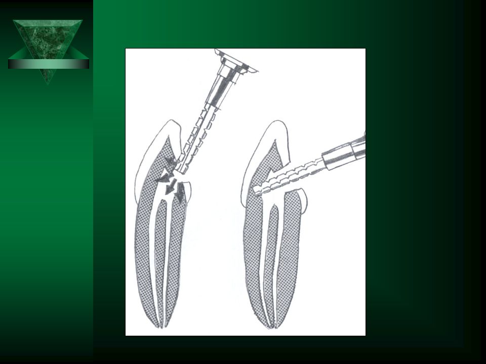
Gates Glidden burs

21
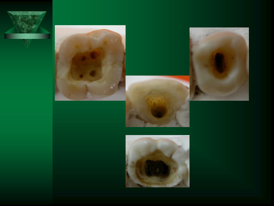
Access Remove coronal enamel with tungsten carbide or diamond bur at high speed. Use slow speed to enter pulp chamber with round bur by breaking the roof of pulp chamber. Widen the access cavity accordingly using TC169 bur. Dentin mapping to find other canals. Should lie in as near a straight line with apex as possible.

25
Radiographs

26
Endodontic pathfinder:- - to locate orifices - as an indicator of canal angulations - as a chipping tool to remove calcification.

29
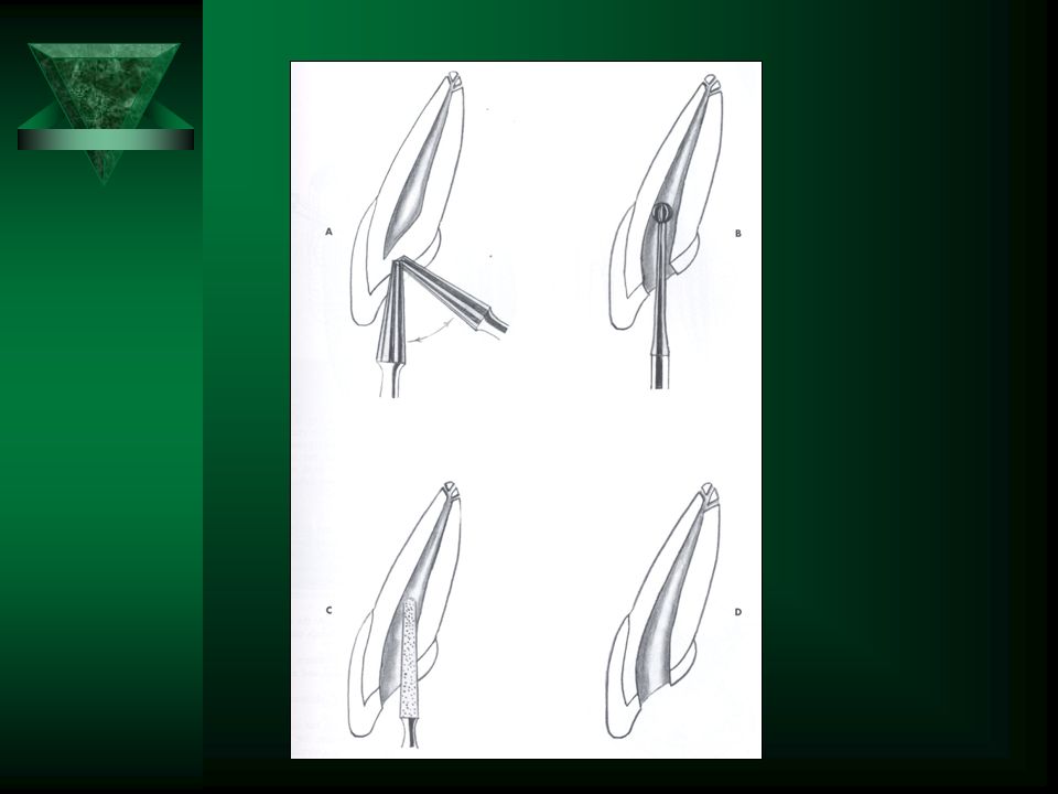
Pulp Extirpation Barbed broach inserted along canal wall short of apex, twisted and withdrawn in a smooth, gentle action. If tooth is non vital, necrotic tissue should be removed with a file and irrigated.

30
Upper Central Incisor One canal but the apical foramen may be towards the labial surface and short of the radiographic apex.

31
Upper Canine

32
Upper First Premolar May have three canals (MB< DB< Pal). Canals usually quite narrow.
. Canals usually quite narrow.")
33
Upper First Molar Four canals are very common (MB, MPal, DB, Pal) with MB root has two canals – MB & MPal.
 with MB root has two canals – MB & MPal.")
34
Lower Central Often two canals. Lingual can be difficult to find.

35
Lower Canine

36
Lower First Premolar Two or three canals can occur

37
Lower First Molar Usually three canals (MB, ML, D) or four canals.
 or four canals.")
38
Upper:- Canines: occasionally may have two canals. Second premolars: often just one canal but may have two or three canals. Second molars: usually three canals; occasionally only one or two canals. Third molars: variations from one to four canals. Lower:- Canines: commonly one canal but occasionally two canals. Premolars: commonly one canal but a second canal can occur. Second molars: usually three canals but four can occur. Occasionally only one or two canals and C-shaped variations. Third molars: variations from one to four canals. ***Always look for an extra canal.

39
Variations

40
References Paul V. Abbott. Endodontics and Dental Traumatology – An overview of modern endodontics; a teaching manual: 1998.

Similar presentations


. All rights reserved. Endodontics Chapter 54 Copyright 2003, Elsevier Science (USA). All rights reserved. No part.>")


