Download presentation
Presentation is loading. Please wait.

1
Cytotoxic T Lymphocyte Assays
Lab. 11

2
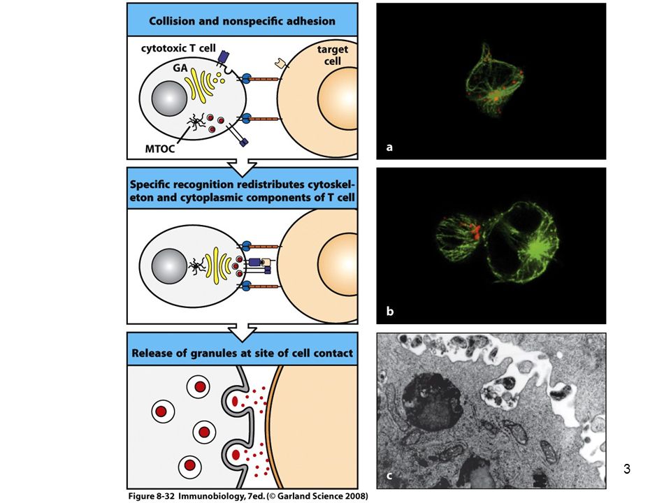
Cytotoxic T cells Cytotoxic T cells or lymphocytes (CTL) are one of the types of cells that develop from lymphoid stem cells. They are major component of the cellular mechanism by which an immune response leads to the destruction of foreign or infected tissue. T cells are antigen specific, and carry antigen specific receptors. T cells also carry receptors for major histocompatability complex (MHC) proteins, and will only function in the presence of the relevant antigen and the appropriate MHC protein. Cytotoxic T lymphocytes interact with a target cell carrying the relevant antigen e.g. a virus protein fragment on the surface of a cell infected with a particular virus and also carrying the appropriate MHC Class 1 protein (or human leukocyte antigen (HLA) Class 1 molecule), and the CTL release cytotoxic enzymes which act to kill the target cell.
 proteins, and will only function in the presence of the relevant antigen and the appropriate MHC protein. Cytotoxic T lymphocytes interact with a target cell carrying the relevant antigen. e.g. a virus protein fragment on the surface of a cell infected with a particular virus. and also carrying the appropriate MHC Class 1 protein (or human leukocyte antigen (HLA) Class 1 molecule), and the CTL release cytotoxic enzymes which act to kill the target cell.")
4
Cytotoxic T cells T lymphocytes play a central role in:
host defense directed against infectious agents and certain malignancies. play a role in transplant rejection and organ-specific autoimmune disease, uncontrolled T cell immune responses can be pathogenic. Despite significant progress, the cellular and molecular mechanisms that mediate these disease states are only partially understood. Thus, the development of methods to describe and quantify T cell immunity in humans may provide novel insights into our basic understanding of human disease. Such methodology will aid in developing strategies directed at improving clinical outcomes in a wide range of disorders.

5
T cell immune response T cell immune response determine whether and how the response is effective, ineffective or inappropriately pathogenic. Clearly, clonal size (the number of antigen-specific cells induced) is a core characteristic, as low frequency responses may be inadequate to control certain infections and therapy might be aimed at boosting the frequency. In contrast, high frequency pathogenic T cell immune responses may mediate more severe pathology and therapy should be directed at inhibiting the function of these cells.
 is a core characteristic, as low frequency responses may be inadequate to control certain infections and therapy might be aimed at boosting the frequency. In contrast, high frequency pathogenic T cell immune responses may mediate more severe pathology and therapy should be directed at inhibiting the function of these cells.")
6
Specificity of antigen-reactive T cells
By determining the fine specificity for antigen-reactive T cells, this could provide specific targets for immunotherapy aimed at appropriately manipulating the quality and quantity of the response, so as ultimately to improve clinical outcome. Independent of clonal size and specificity, the cytokine secretion pattern, as well as the ability to mediate alternate effector functions such as cytotoxicity, can have a large impact on the outcome of T cell immunity While a certain type of cytokine-secreting phenotype may protect against certain intracellular infections, a T cell immune response of similar specificity and frequency, but producing IL-4 or IL-5, may not be able to control the same pathogen.

7
Techniques for cytotoxicity measurment
Cell mediated cytotoxicity can be mediated through antibody or occur without a need for antibodies (CTL). Several different ways of target cell labeling for the assessment of cytotoxic activity have been used. The two most common techniques use 51Cr release from 51Cr-labeled targets or an enzymatic assay measuring release of intracellular substances. All these methods are based on measuring cell death judged from plasma membrane disintegration and the consequent release of cytoplasm. DNA fragmentation, a common event occurring during apoptosis, is also an early event in the cell death caused by cytotoxic cells. The JAM test is a sensitive and easy test for DNA fragmentation and cell death.
. Several different ways of target cell labeling for the assessment of cytotoxic activity have been used. The two most common techniques use 51Cr release from 51Cr-labeled targets or an enzymatic assay measuring release of intracellular substances. All these methods are based on measuring cell death judged from plasma membrane disintegration and the consequent release of cytoplasm. DNA fragmentation, a common event occurring during apoptosis, is also an early event in the cell death caused by cytotoxic cells. The JAM test is a sensitive and easy test for DNA fragmentation and cell death.")
8
Techniques for cytotoxicity measurment
Another technique for measuring cytotoxicity is ELISPOT assay. This identifies T cells that not only recognize their target, but react to it by producing cytokines, such as interferon. It offers a measure of functionality other than cytotoxicity.

9
51Chromium (Cr) Release Assays
The CTLs Assay is used to detect the cytolytic activity of Ag-specific lymphocytes. The classical assay for CTL activity is the chromium release assay. CTL’s function by destroying cells those express foreign antigens (Ag) such as virally infected cells: typically mediated by CD8+ T-lymphocytes.
 such as virally infected cells: typically mediated by CD8+ T-lymphocytes.")
10
51Chromium (Cr) Release Assays
Cytotoxicity is relatively easy to measure, there are straightforward ways to measure cell death. If lymphocytes are taken from a mouse (or a person) that was previously infected with a virus, and mixed with other lymphocyte cells infected with the same virus, the infected cells will be killed.
 that was previously infected with a virus, and mixed with other lymphocyte cells infected with the same virus, the infected cells will be killed.")
11
51Chromium (Cr) Release Assays
Target cells are stimulated with an appropriate peptide to activate the cells Target cells expressing the epitope of interest are labeled with 51Cr Cells are incubated with CTL effector cells As the CTL effector cells bind to the antigen specific target, the cells are lyzed and the 51Cr is released into the culture supernatant The cells are centrifuged and the level of 51Cr in the supernatant is measured by liquid scintillation.

12
Assay for cytotoxic T cell function: Chromium release
Release of perforin & granzymes

13
Applications The method has been shown to work well in detecting human immunodeficiency virus (HIV) specific CTL, using peptide fragments of HIV. It is also applicable to detection of CTL specific for other viruses. There is evidence that HIV specific CTL activity in patients infected with HIV varies with development of acquired immunodeficiency syndrome (AIDS) or related conditions: many healthy HIV seropositive patients have a vigorous anti-HIV CTL response, but there is evidence that HIV specific CTL activity declines as disease progresses. Measurement of HIV specific CTL in samples from patients infected with HIV may thus provide useful information in following disease progression.
 or related conditions: many healthy HIV seropositive patients have a vigorous anti-HIV CTL response, but there is evidence that HIV specific CTL activity declines as disease progresses. Measurement of HIV specific CTL in samples from patients infected with HIV may thus provide useful information in following disease progression.")
14
Disadvantages The main drawback to using 51Cr release assays is the involvement of radioactive reagents which: requires specialist laboratory certification and specially trained users. In addition the protocol can only be carried out on fresh cells

15
The ELISPOT Assay Versions of the enzyme-linked immunospot assay (ELISPOT) have been used for ~20 years to detect antibody-secreting B cells and more recently, cytokine-secreting lymphocytes. Several technical advances, including development of synthetic membranes and computer-assisted image analysis hardware and software, have improved the reliability and reproducibility of the assay and have facilitated the data analysis.
 have been used for ~20 years to detect antibody-secreting B cells and more recently, cytokine-secreting lymphocytes. Several technical advances, including. development of synthetic membranes. and computer-assisted image analysis hardware and software, have improved the reliability and reproducibility of the assay and have facilitated the data analysis.")
16
The ELISPOT Assay To detect cytokine-secreting lymphocytes, commercially available 96-well ELISPOT plates that have a white synthetic membrane as a floor are coated with a primary antibody specific for the cytokine to be detected. Responding lymphocytes, in most clinical situations peripheral blood lymphocytes (PBLs) or purified peripheral blood T cells (or T cell subsets), are added to the wells at varying dilutions
 or purified peripheral blood T cells (or T cell subsets), are added to the wells at varying dilutions.")
17
Principle These assays take advantage of the relatively high concentration of a given protein (such as a cytokine) in the environment immediately surrounding the protein-secreting cell. These cell products are captured and detected using high-affinity antibodies. The ELISPOT assay utilizes two high-affinity cytokine-specific antibodies directed against different epitopes on the same cytokine molecule: either two monoclonal antibodies or a combination of one monoclonal antibody and one polyvalent antiserum.
 in the environment immediately surrounding the protein-secreting cell. These cell products are captured and detected using high-affinity antibodies. The ELISPOT assay utilizes two high-affinity cytokine-specific antibodies directed against different epitopes on the same cytokine molecule: either two monoclonal antibodies. or a combination of one monoclonal antibody and one polyvalent antiserum.")
18
Results ELISPOT generates spots based on a colorimetric reaction that detects the cytokine secreted by a single cell. The spot represents a “footprint” of the original cytokine-producing cell. Spots are permanent and can be quantitated visually, microscopically, or electronically.

19
Steps of ELISPOT assay The ELISPOT assay involves five specific steps:
(1) coating a purified cytokine-specific antibody to a nitrocellulose-backed microtiter plate; (2) blocking the plate to prevent nonspecific absorption of any other proteins; (3) incubating the cytokine-secreting cells at several different dilutions; (4) adding a labeled second anti-cytokine antibody; (5) and detecting the antibody-cytokine complex.
 coating a purified cytokine-specific antibody to a nitrocellulose-backed microtiter plate; (2) blocking the plate to prevent nonspecific absorption of any other proteins; (3) incubating the cytokine-secreting cells at several different dilutions; (4) adding a labeled second anti-cytokine antibody; (5) and detecting the antibody-cytokine complex.")
20
In vitro assays for production of cytokines: ELISPOT assays

21
ELISPOT Assay Principle
Prepare PBMC and count Add PBMC Wash out cells, add detector Ab wash 24 h 1 h Coat plate with anti-cytokine Ab Add Ag Schematic protocol for the ELISPOT assay. 15 min Count on microscope or Analyze on automated reader Wash, add substrate 21

22
Applications Universities, research centers, cancer centers, pharmaceutical companies, biotechnology companies, clinical research laboratories and government related research centers all have the potential to use ELISPOT. ELISPOT assays can be used for the quantitative analysis of T cell responses to infections, identification of T cell epitopes, and for monitoring immunogenicity in vaccine trials. Uses might include monitoring of clinical trials involving vaccinations against HIV, cancer, hepatitis B and C, and other auto-immune disorders; monitoring of disease status and specific immune status; and other basic scientific research, such as inflammation, cell biology, transplantation, vaccine development,

23
MLR as a measure of homograft reactivity
MLR has multiple applications. One area is still in assessing histocompatibility between the donor and recipient of a graft in order to gauge the likely immunological stimulation on grafting. The main histocompatibility differences can be assessed from tissue typing but the responses observed in MLR will include additional effects. The relevance of disparities in major histocompatibility antigens and others induced by minor antigens, not generally assessed by tissue typing, can be more directly assessed by using an MLR

24
Mixed Lymphocyte Reaction
Measure of histocompatibility at the HLA locus. Peripheral blood lymphocytes from two individuals are mixed together in tissue culture for several days. Lymphocytes from incompatible individuals will stimulate each other to proliferate significantly (measured by tritiated thymidine uptake) whereas those from compatible individuals will not. In the one-way MLC test, the lymphocytes from one of the individuals are inactivated (usually by treatment with MITOMYCIN or radiation) thereby allowing only the untreated remaining population of cells to proliferate in response to foreign histocompatibility antigens.
 whereas those from compatible individuals will not. In the one-way MLC test, the lymphocytes from one of the individuals are inactivated (usually by treatment with MITOMYCIN or radiation) thereby allowing only the untreated remaining population of cells to proliferate in response to foreign histocompatibility antigens.")
Similar presentations


 CBA: cytometric bead array DC: dendritic.>")

 Features of immune.>")


 The adaptive immune system:>")
