Download presentation
Presentation is loading. Please wait.

1
Jaw relationship part II
صناعة نظري (ج 2) ثاني اسنان كركوك 24 / 2 / 2016 Jaw relationship part II
 ثاني اسنان كركوك. 24 / 2 / Jaw relationship part II.")
2
Horizontal relations Centric relation Eccentric relation
Protrusive relation Lateral relation Left lateral Right lateral

3
Horizontal relations:- are relations of mandible to maxilla in a horizontal plane or in anteroposterior direction. Centric relation:- the most retruded relation of the mandible to the maxilla when the condyles are in the most posterior unstrained position in the glenoid fossa from which lateral movement can be made, at any given degree of jaw separation It’s a bone relation

4
This position is independent of tooth contact
It is restricted to a purely rotary movement about the transverse horizontal axis. CR is anatomically determined; it is repeatable and reproducible. It is the most reliable reference point for accurately recording the relationship of the mandible to the maxilla.

5
In CR the mandible has Purely rotary movement (hing motion) about transverse Horizontal axis.
 about transverse Horizontal axis.")
6
Eccentric relation :- any relation of the mandible to the maxilla other than centric relation
Protrusive relation:- is the relation of the mandible to maxilla when the mandible is thrust forward

7
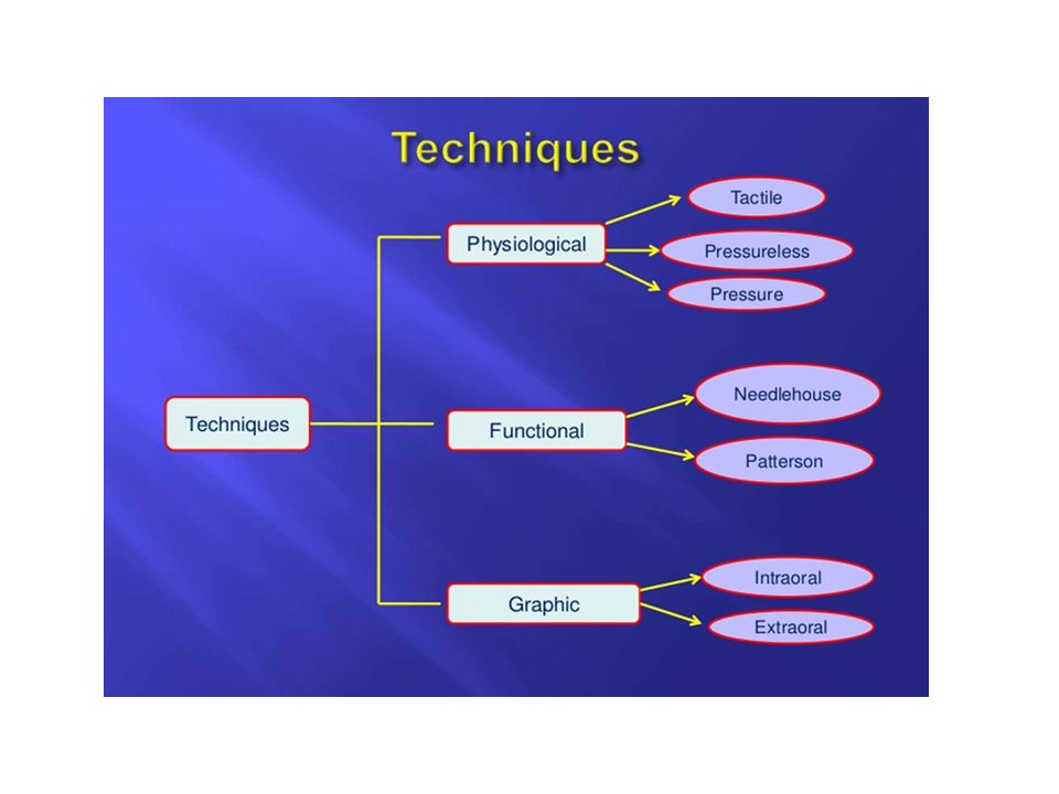
PHYSIOLOGICAL FUNCTIONAL GRAPHIC RADIOGRAPHIC METHOD
METHODS TO RECORD CENTRIC RELATION: PHYSIOLOGICAL tactile or inter occlusal check record method Pressureless method Pressure method FUNCTIONAL Needle house method Patterson method GRAPHIC Intra oral Extra oral RADIOGRAPHIC METHOD

9
Physiologic method Based on: Proprioceptive impulse of patient
Kinesthetic sense of mandibular movement Visual perception and sense of touch of patient Tactile sense or inter occlusal check record method. Tentative jaw relation is recorded. Ask the patient to retrude the mandible. Casts are articulated based on this tentative record

10
INDICATIONS Abnormally related jaws. Displaceable flabby tissue. Large tongue Uncontrolled mandibular movements. In patients already using a complete denture Material used Waxes: low fusing Impression compound Dental plaster Zinc Oxide eugenol paste

11
PROCEDURE: Recording tentative jaw relation: Upper and lower trial dentures are inserted into the mouth. keep a piece of cotton to prevent contact of opposing members. Aluwax is added on the occlusal surface of mandibular occlusal rim Patient asked to retrude mandible and close on the wax till occlusal rim contact occurs. Or ask the patient to roll the tongue backwards towards the posterior border of upper denture and close the rims until they meet Trial dentures removed and allowed to cool.

13
Guide lines (midline, high lip line and canine lines).
.")
14
2)Static or pressureless method
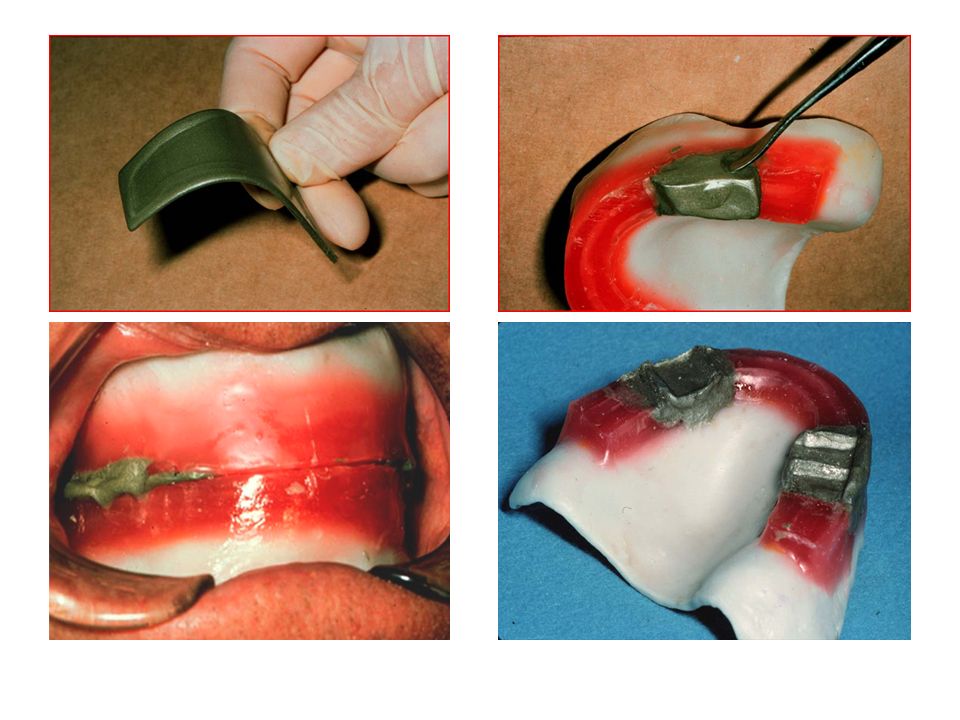
Nick notch method: Patient asked to retrude mandible in position. Up to 3mm of wax removed from mandibular occlusal rim from the premolar region till the distal end 1 or 2 notches are cut on the corresponding area of maxillary occlusal rim. One nick is cut anterior to the notch, a V shaped valley Nick : prevent lateral movement Notch: anteroposterior movement

15
Nick and notch are lubricated with petroleum.
Prepared occlusal rim are inserted into patient’s mouth and taught to close his mandible in maximum retruded position. Aluwax is placed on the trough created in mandibular rim. Mandibular occlusal rim is cooled and inserted into patients mouth and closed in centric relation.

16
3)Pressure method The lower occlusal rim is fabricated to be of excess height Upper occlusal rim inserted. lower occlusal rim is softening in water bath. Insert it into patients mouth. Patient asked to close mouth in centric relation on soft wax in predetermined vertical dimension and then articulated.

17
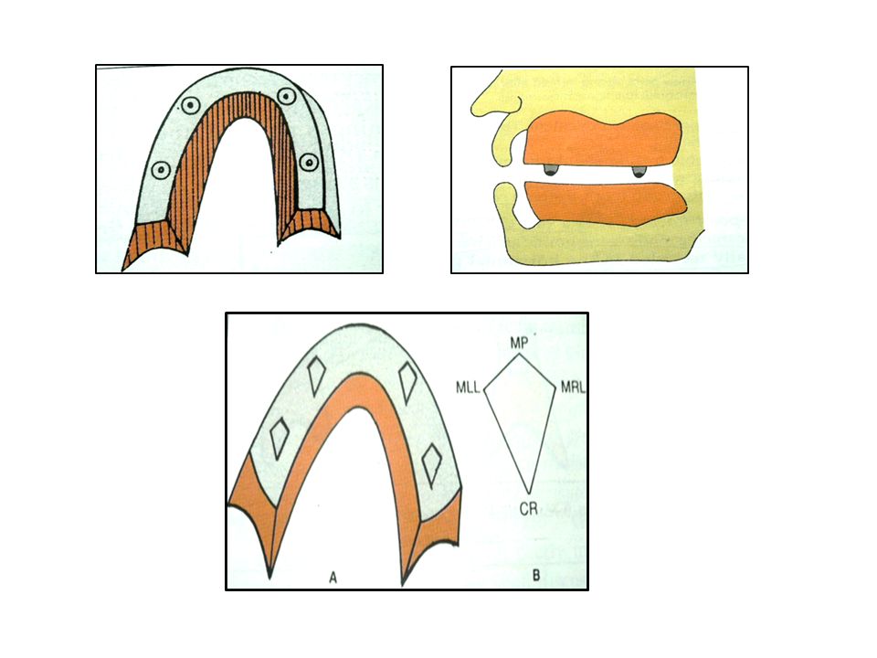
Functional method Method utilize the functional movements of jaws to record the centric relation. Patient asked to perform border movements such as protrusive and lateral excersion movement. Needle house method Fabrication of occlusal rim made from impression compound Four metal beads are embedded into premolar and molar areas of maxillary occlusal rim. Occlusal rim inserted into patients mouth and asked to close on occlusal rim and make protrussive,retrussive ,right and left movement of mandible. When movements are made “diamond shaped marking pattern rather than a line is formed on the mandibular occlusal rim.

19
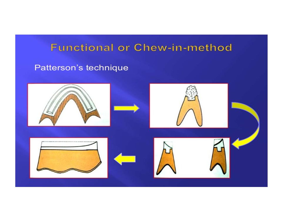
b)Patterson method Occlusal rim made of modeling wax. trench or trough is made along the length of mandibular occlusal rim. A mixture of carborun dum and dental plaster is loaded into the trench. Perform mandibular movement till predetermined vertical dimension. Movement generates compensative curves in plaster

21
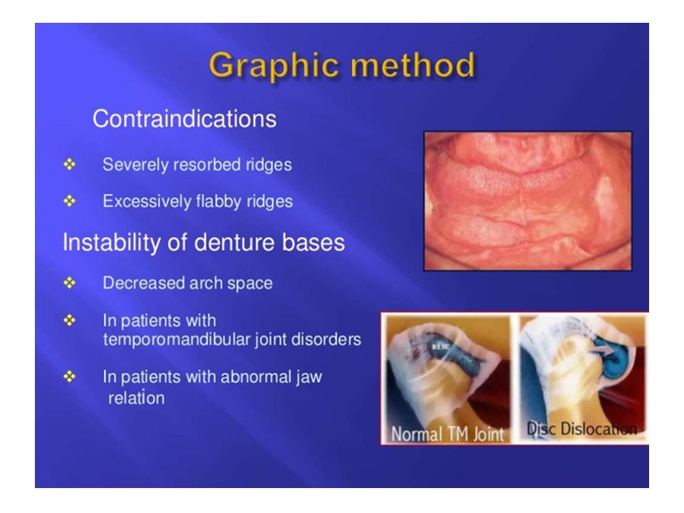
Graphic method The graphic method record a tracing of mandibular movements in one plane 2 types: Arrow point tracing Pantograph Arrow point tracing: is a graphic record measured across single plane Pantograph: is measured three dimensionally

22
Factors to be considered while carrying out tracing
Stability of denture base Resistance of rims Difficulty in placing central bearing device Height of residual alveolar ridge Tongue interference Efficiency of recording device Lack of coordinated movements

23
Arrow point tracing or gothic arch tracer
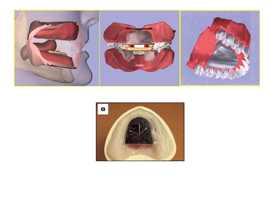
Recorded in horizontal plane. Consists of central bearing device: a device that provide central point of bearing or support between the maxillary & mandibular dental arches. consists of contacting point attached to one dental arch and plate attached to opposing dental arch Plate provide surface on which the tracing of mandibular movements is recorded.

24
TYPES OF ARROW POINT TRACERS:
INTRA ORAL TRACING POINT: Central bearing device is located intra orally. Tracer is placed within the mouth. Central bearing point & plate is inserted into patients mouth. Central bearing point is adjusted such that it contact the central bearing plate at predetermined vertical dimension. Ask to make anteroposterior and lateral movements. Central bearing point will draw the tracing pattern on central bearing plate Tracing should resemble an arrow point with a sharp apex

26
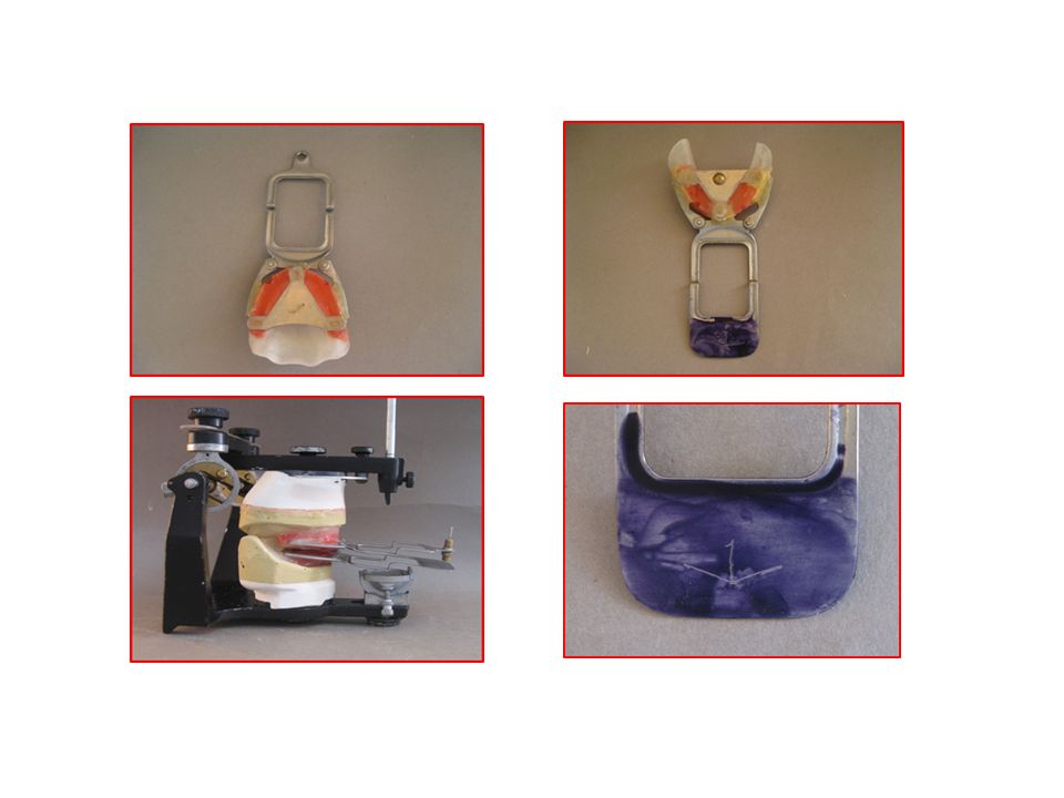
2)EXTRA ORAL POINT TRACER:
Concept similar to intra oral tracer. Additionally have an attachment that project outside mouth. Record bases attached to recording devices inserted in patients mouth. Central bearing point is retracted to conduct training exercises. Recording plate which projects extra orally is coated with precipitated chalk and denatured alcohol. Patient asked to perform all movements. Examine for sharp apex.

29
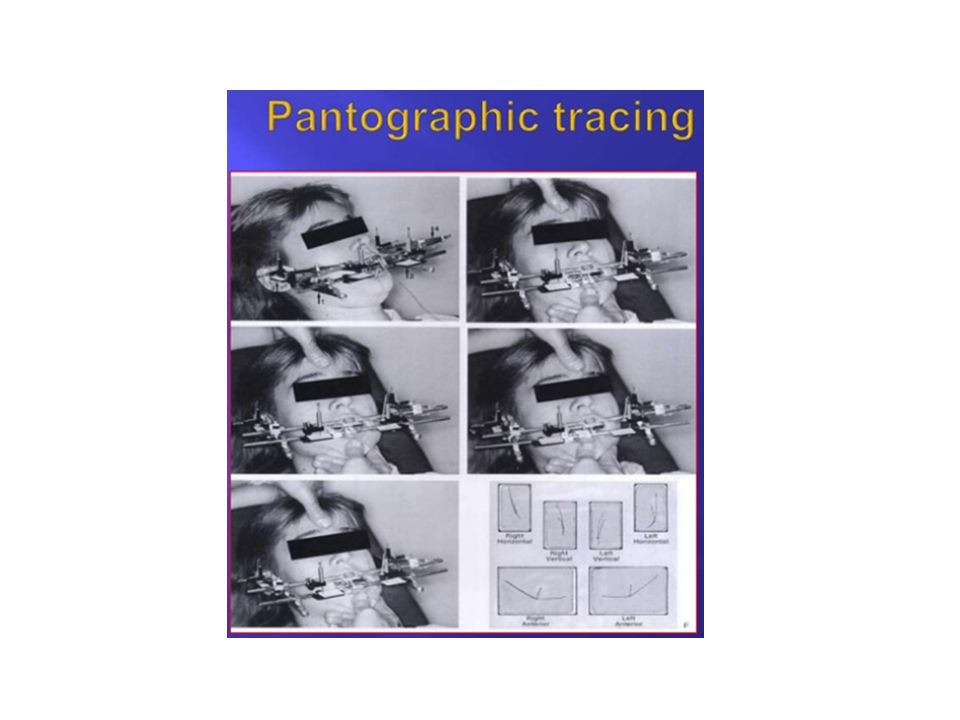
Pantographic tracing A graphic record of mandibular movements in three planes as registered by styli on recordable tables of a pantograph, tracing of mandibular movement recorded on plates in horizontal and sagittal planes. Make the rim contact at desired vertical relationship. Strips of celluloid paper are placed between the rim and pulled out. patient is asked to close and restrain the celluloid from slipping a way, mandible goes to centric relation. Softened wax is placed on mandibular occlusal rim and patient is asked to bite in centric relation

Similar presentations