Download presentation
Presentation is loading. Please wait.

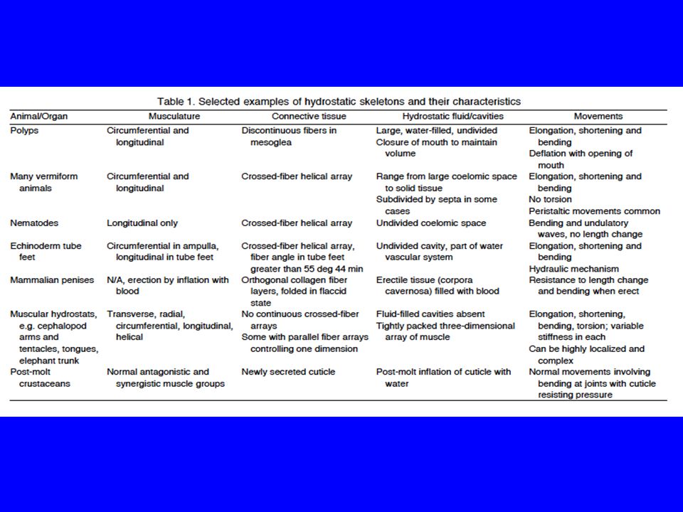
1
Fig. 5 Kier The body of a sea anemone is “a hollow column...closed at the base...at the top with an oral disc that includes a ring of tentacles surrounding the mouth and pharynx”. “By closing the mouth, the water in the internal cavity –the coelenteron/GV – cannot escape, and thus the internal volume remains essentially constant. The walls of an anemone include a layer of circular muscle fibres. Longitudinal muscle fibres are found on the vertical partitions called septa that project radially inward into the coelenteron, including robust longitudinal retractor muscles along with sheets of parietal longitudinal muscle fibres adjacent to the body wall.”

2
Phylum Cnidaria sea anemones, corals, jellyfish etc. “With the mouth closed, contraction of the circular muscle layer decreases the diameter and thereby increases the height of the anemone. Contraction of the longitudinal (or R =retractor) muscles shortens the anemone and re-extends the circular muscle fibres.” “...with this simple muscular arrangement a diverse array of bending movements and height change can be produced.”
 muscles shortens the anemone and re-extends the circular muscle fibres. ...with this simple muscular arrangement a diverse array of bending movements and height change can be produced. .")
3
Connective tissue fibre reinforcement “The walls of many hydrostatic skeletons are reinforced with layers of connective tissue fibres that control and limit shape change. The fibres are typically arranged as a ‘crossed-fibre helical connective tissue array’ in which sheets of connective tissue fibres (often collagenous) wrap the body or structure in right- and left-handed helices. Even though the connective tissue fibres are typically stiff in tension and are thus relatively inextensible, such an arrangement actually allows length change. Elongation and shortening is possible because the pitch of the helix changes during elongation...”
 wrap the body or structure in right- and left-handed helices. Even though the connective tissue fibres are typically stiff in tension and are thus relatively inextensible, such an arrangement actually allows length change. Elongation and shortening is possible because the pitch of the helix changes during elongation... .")
4
Phylum Echinodermata radially rather than bilaterally symmetrical, with oral and aboral surfaces. Ambulacral groove on underside of each arm lined with tube feet also called podia. Water vascular system is unique to these animals among phyla; it is a vessel system filled with coelomic fluid. ‘Arm vessels’ arise from a tubular ring canal. In an asteroid (starfish) five radial canals branch, one into each of the arms.
 five radial canals branch, one into each of the arms..")
5
Asteroid echinoderms have an exoskeleton. In their dermis are embedded calcareous plates called ossicles (inorganic salt Calcium Carbonate is the material). The skin thus consists of stiff relatively shape-stable elements, in a matrix of flexible collageous fibres (i.e., connective tissue): tough, solid, thick yet flexible protective armour. Above each tube foot, within the arm, is a vesicle called an ampulla, encircled by ampullar muscles; contraction of ampullar muscles will displace fluid out of the ampulla into the tube foot because fluid is incompressible. There is a valve in the side branch to the radial canal – a one-way valve -- that closes to prevent backflow of the fluid into the water vascular system. So for each ampulla +tube foot as it operates the volume of fluid in the ampulla and tube foot lumen is fixed because the one-way valve closed. But this fixed volume of fluid moves back and forth from ampulla to tube-foot lumen; the system in the starfish is HYDRAULIC rather than hydrostatic as in earthworm segments.
. The skin thus consists of stiff relatively shape-stable elements, in a matrix of flexible collageous fibres (i.e., connective tissue): tough, solid, thick yet flexible protective armour. Above each tube foot, within the arm, is a vesicle called an ampulla, encircled by ampullar muscles; contraction of ampullar muscles will displace fluid out of the ampulla into the tube foot because fluid is incompressible. There is a valve in the side branch to the radial canal – a one-way valve -- that closes to prevent backflow of the fluid into the water vascular system. So for each ampulla +tube foot as it operates the volume of fluid in the ampulla and tube foot lumen is fixed because the one-way valve closed. But this fixed volume of fluid moves back and forth from ampulla to tube-foot lumen; the system in the starfish is HYDRAULIC rather than hydrostatic as in earthworm segments..")
6
From Brown, Selected Invertebrate Types

7
Circumferential stress in a pressurized cylindrical vessel (e.g., worm, tube foot) is exactly double the longitudinal stress ‘Kier’s Law’. Imagine it as it isn’t: no helical array in the tube foot wall. When the ampulla pushes fluid into the podium lumen there will be an increase in diameter rather than a lengthening Rosette of ossicles with intrinsic musculature that pulls up the disc middle creating suction to substratum. Stress distribution in a fluid-filled cylinder is not uniform (as per annelid metameres): hoop stress [force acting to increase diameter] is twice as large as longitudinal stress.
: hoop stress [force acting to increase diameter] is twice as large as longitudinal stress..")
8
Santos, R. et al. 2005. Adhesion of echinoderm tube feet to rough surfaces. J. exp. Biol. 208: 2555- 2567. Fig. 6 External morphology of unattached pedal discs of Paracentrotus lividus (left) [sea urchin] and Asterias rubens [starfish] (right). End of extensible cylinder is the disc, larger in diameter than the stem. There is a central depression. Temporary adhesion: the epidermis of the disc contains glands which produce two secretions: glue/bonder and de-bonder, i.e., adhesive secretions and de-adhesive secretions. The glue is delivered through the disc cuticle to the substratum where it forms a thin film bonding the foot. The debonding secretions act as enzymes, detaching the upper coat of the glue and leaving the rest of the adhesive material behind attached to the substratum as a footprint.
 [sea urchin] and Asterias rubens [starfish] (right). End of extensible cylinder is the disc, larger in diameter than the stem. There is a central depression. Temporary adhesion: the epidermis of the disc contains glands which produce two secretions: glue/bonder and de-bonder, i.e., adhesive secretions and de-adhesive secretions. The glue is delivered through the disc cuticle to the substratum where it forms a thin film bonding the foot. The debonding secretions act as enzymes, detaching the upper coat of the glue and leaving the rest of the adhesive material behind attached to the substratum as a footprint..")
9
Pulling with tube feet adhering and starfish arm muscles to open the protective valves of shellfish Mollusca Virginia Living Museum ‘off the beaten path’ Importance of tube foot in predation

10
An interesting picture of razor clams packaged for sale in a chinatown market in Philadelphia Phylum Mollusca

11
Razor clam burrowing Winter A.G. et al. 2012. Localized fluidization burrowing mechanics of Ensis directus. Journal of experimental Biology 215: 2072-2080. (See also Inside JEB, Kathryn Knight. 2012. Razor clams turn soil into quicksand to burrow.) Blood and foot sinuses serve as hydraulic burrowing mechanism.
 Blood and foot sinuses serve as hydraulic burrowing mechanism..")
12
Cycle of burrowing movements Fig. from Winter: In stage A, adductor relaxed so shells braced on surrounding sand by ligaments; protractors start to contract (B) pushing blood into foot and the foot probes down, gaining ground into the mud; pushing force of foot makes body move up a little (C) (relative to dashed line). Stage D, adductors contract, pulling valves together, (red indicates the space they DID occupy), pushing blood into foot to make an anchor, simultaneously squirting seawater out around valves from mantle; this water* ‘puddles’ sand; foot swells with blood into this quicksand region– the localized fluidation; displaced blood is swelling the foot maximally into the bottom anchor of the TWO ANCHOR SYSTEM. Cycle renews (F). *Its not clear whether this involves ocean water drawn in by siphons; perhaps it does if the clam is burrowing near the surface and perhaps if lower down it oscillates (?) its valves to draw in pore water (Winter)
 pushing blood into foot and the foot probes down, gaining ground into the mud; pushing force of foot makes body move up a little (C) (relative to dashed line). Stage D, adductors contract, pulling valves together, (red indicates the space they DID occupy), pushing blood into foot to make an anchor, simultaneously squirting seawater out around valves from mantle; this water* ‘puddles’ sand; foot swells with blood into this quicksand region– the localized fluidation; displaced blood is swelling the foot maximally into the bottom anchor of the TWO ANCHOR SYSTEM. Cycle renews (F). *Its not clear whether this involves ocean water drawn in by siphons; perhaps it does if the clam is burrowing near the surface and perhaps if lower down it oscillates ( ) its valves to draw in pore water (Winter).")
13
Kier p.1252 Tongues, tentacles, trunks: “lack the fluid-filled cavities and fibre-reinforced containers that characterize... hydrostatic skeletal support systems” rather they are: “a densely packed, three-dimensional array of muscle and connective tissue fibres” Muscular hydrostats

14
Transverse sections showing the muscular arrangement of three examples of muscular hydrostats. Kier W M J Exp Biol 2012;215:1247-1257 ©2012 by The Company of Biologists Ltd A. Squid tentacle: T, transverse muscle fibres; L, longitudinal; transverse in the tentacle core, “and extend to interdigitate with bundles of longitudinal muscle fibres, notice the suckers.

15
Transverse sections showing the muscular arrangement of three examples of muscular hydrostats. Kier W M J Exp Biol 2012;215:1247-1257 ©2012 by The Company of Biologists Ltd B. Elephant Trunk: R, radials ‘extend from centre of the trunk between bundles of longitudinal muscle that are more superficial, notice nasal passages.

16
C. Monitor lizard tongue. Circular muscle fibres surround two large bundles of longitudinal fibres.

17
“The muscle fibers are typically arranged so that all three dimensions of the structure can be actively controlled, but in several cases such as the mantle of the squid [of which more later] and some frog tongues, one of the dimensions is constrained by connective tissue fibers.” “Because muscle tissue, like most animal tissues lacking gas spaces, has a high bulk modulus, selective muscle contraction that decreases one dimension of the structure must result in an increase in another dimension. This simple principle serves as the basis upon which diverse deformations and movement of the structure can be achieved” (Kier 2012). Read carefully all the section on muscular hydrostats by Kier: complex bending achieved by interplay of contracting muscles --more subtle than a passive uniform fluid in a chamber – i.e., some muscles by contracting can affect the bulk modulus presented to other muscles that are acting upon its incompressibility. *bulk modulus of a substance measures its resistance to uniform compression Wikki
![The muscle fibers are typically arranged so that all three dimensions of the structure can be actively controlled, but in several cases such as the mantle of the squid [of which more later] and some frog tongues, one of the dimensions is constrained by connective tissue fibers. Because muscle tissue, like most animal tissues lacking gas spaces, has a high bulk modulus, selective muscle contraction that decreases one dimension of the structure must result in an increase in another dimension.](http://images.slideplayer.com/35/10470496/slides/slide_17.jpg "This simple principle serves as the basis upon which diverse deformations and movement of the structure can be achieved (Kier 2012). Read carefully all the section on muscular hydrostats by Kier: complex bending achieved by interplay of contracting muscles --more subtle than a passive uniform fluid in a chamber – i.e., some muscles by contracting can affect the bulk modulus presented to other muscles that are acting upon its incompressibility. *bulk modulus of a substance measures its resistance to uniform compression Wikki.")
18
Muscular hydrostats (Kier contin.) Selective contraction: “This simultaneous contractile activity is necessary to prevent the compressional forces generated by the longitudinal muscle from simply shortening the structure, rather than bending it, and can actually augment the bending by elongating the structure along the outside radius of the bend.” “The longitudinal muscle bundles are frequently located near the surface of the structure, as this placement away from the neutral plane increases the bending moment.” Helically arranged muscle fibres can be present and generate torsion.
 Selective contraction: This simultaneous contractile activity is necessary to prevent the compressional forces generated by the longitudinal muscle from simply shortening the structure, rather than bending it, and can actually augment the bending by elongating the structure along the outside radius of the bend. The longitudinal muscle bundles are frequently located near the surface of the structure, as this placement away from the neutral plane increases the bending moment. Helically arranged muscle fibres can be present and generate torsion.")
20
https://youtu.be/K2G7L5hcEt8 See this URL for an animation of Echinodermata body plan, especially water vascular system

21
Hydrostatic skeletons of Nematoda Pseudocoelom locomotion CFHCTA and adaptive volume of a fluid-filled cavity “…if all the matter in the universe except the nematodes were swept away, our world would still be dimly recognizable, and if, as disembodied spirits, we could then investigate it, we should find its mountains, hills, vales, rivers, lakes and oceans represented by a film of nematodes. The location of towns would be decipherable, since for every massing of human beings there would be a corresponding massing of certain nematodes. Trees would still stand in ghostly rows representing our streets and highways. The location of the various plants and animals would still be decipherable, and had we sufficient knowledge, in many cases even their species could be determined by an examination of their erstwhile nematode parasites." from "Nematodes and Their Relationships", 1915 Nathan Augustus Cobb, father of American nematology. What is the most famous nematode and what is it most famous for? Nematodes Speciose: 20,000 described More than a million to go. See next slide: C. elegans: for being the first Animalia whose genome was entirely sequenced.

22
Pseudocoelom as fluid skeleton Caenorhabditis elegans* Phylum Nematoda Roundworms pinworms: most too small (<2mm) to leave any impression: ‘never seen a nematode’. Most free-living in soil; some parasitic. Nematodes in the ocean? They are internal parasites of whales. Unsegmented, i.e., not metameric Circular/ellipitical body in transverse section, pointed ends (burrowing?) HAVE NO CIRCULAR MUSCLES, ONLY LONGITUDINAL. Very high internal pressure of pseudocoel fluid. * Jon Eisenback Locomotes lying on Its side and arching Its body about.
 HAVE NO CIRCULAR MUSCLES, ONLY LONGITUDINAL. Very high internal pressure of pseudocoel fluid. * Jon Eisenback Locomotes lying on Its side and arching Its body about..")
23
Though the number of nematode species is so great, they are unusually (in comparison to other phyla) uniform in body design, i.e., they seem to conform to some important physical constraint (?). “Characteristic features …are largely independent of size, of diet and of stage of development [of free-living or parasitic existence].” “…the elementary student may be forgiven …for thinking …there is only one nematode, but that the model comes in different sizes and with a great variety of life histories. (Harris & Crofton 1957)”. Recall D’Arcy Thompson is associated with the idea that along with evolution mechanical principles constrain form. “A bridge, a ship, an aeroplane are recognizable at once because their design is based necessarily and largely on the mechanical forces which play such a predominant part in their economy” (Harris & Crofton 1957). It is suggested that the conservative nature of nematode shape is dictated by mechanical considerations. Being an elongated unsegmented, high internal pressure pointy ended cylinder with no circular muscles is the result of physical constraints related to its locomotion. Uniformity of body design
 . Recall D’Arcy Thompson is associated with the idea that along with evolution mechanical principles constrain form. A bridge, a ship, an aeroplane are recognizable at once because their design is based necessarily and largely on the mechanical forces which play such a predominant part in their economy (Harris & Crofton 1957). It is suggested that the conservative nature of nematode shape is dictated by mechanical considerations. Being an elongated unsegmented, high internal pressure pointy ended cylinder with no circular muscles is the result of physical constraints related to its locomotion. Uniformity of body design.")
24
“a structure composed of inextensible fibres [can] accommodate large extensibility” (Shadwick 2008) After Kier, start with this paper which explains the essence of nematode movement and from a historical perspective. Shadwick R.E. 2008. Foundations of animal hydraulics: geodesic fibres control the shape of soft bodied animals. J. exp. Biol. 211: 289-291. Shadwick is discussing the importance of classic work by Clark & Cowey. Clark R.B., Cowey J.B. 1958. Factors controlling the change of shape of certain nemertean and turbellarian worms. J. exp. Biol. 731-748. Harris J.E., Crofton H.D. 1957. Structure and Function in the nematodes: internal pressure and cuticular structure in Ascaris. J. exp. Biol. 34: 116-130.
![a structure composed of inextensible fibres [can] accommodate large extensibility (Shadwick 2008) After Kier, start with this paper which explains the essence of nematode movement and from a historical perspective.](http://images.slideplayer.com/35/10470496/slides/slide_24.jpg "Shadwick R.E Foundations of animal hydraulics: geodesic fibres control the shape of soft bodied animals. J. exp. Biol. 211: Shadwick is discussing the importance of classic work by Clark & Cowey. Clark R.B., Cowey J.B Factors controlling the change of shape of certain nemertean and turbellarian worms. J. exp. Biol Harris J.E., Crofton H.D Structure and Function in the nematodes: internal pressure and cuticular structure in Ascaris. J. exp. Biol. 34:")
25
Worm as a fibre-reinforced cylinder Clark-Cowey model “one of the key design principles of structural systems in biology” (Shadwick 2008) From the 1950s the concept has existed of a hydrostatic skeleton: “a system in which muscles shorten to act against a contained volume of fluid, rather than rigid skeletal elements [sclerites, bones] to maintain shape and effect movement” (op. cit.) The important role of collagen helices (‘springs’) is more recent and begins with Clark & Cowey. “…a structure composed of inextensible fibres [can] accommodate large extensibility” is the basic idea of the classic Clark & Cowey paper (Shadwick 2008). “…how [did a pseudocoelomate]… move if it had only one set of muscles as in the case of nematodes” (op. cit.) “…the geometry of the reinforcing fibres in the body wall was the key to the solution…”. An antagonist of the longitudinal muscles of nematodes is a collagen fibre helix in the cuticle. [ memonic device ‘CFHCTA’ Canadians For Happy Carefree TAs : crossed fibre helical connective tissue arrays (Kier).] Screen demo
![Worm as a fibre-reinforced cylinder Clark-Cowey model one of the key design principles of structural systems in biology (Shadwick 2008) From the 1950s the concept has existed of a hydrostatic skeleton: a system in which muscles shorten to act against a contained volume of fluid, rather than rigid skeletal elements [sclerites, bones] to maintain shape and effect movement (op.](http://images.slideplayer.com/35/10470496/slides/slide_25.jpg "cit.) The important role of collagen helices (‘springs’) is more recent and begins with Clark & Cowey. …a structure composed of inextensible fibres [can] accommodate large extensibility is the basic idea of the classic Clark & Cowey paper (Shadwick 2008). …how [did a pseudocoelomate]… move if it had only one set of muscles as in the case of nematodes (op. cit.) …the geometry of the reinforcing fibres in the body wall was the key to the solution… . An antagonist of the longitudinal muscles of nematodes is a collagen fibre helix in the cuticle. [ memonic device ‘CFHCTA’ Canadians For Happy Carefree TAs : crossed fibre helical connective tissue arrays (Kier).] Screen demo.")
26
The Clark-Cowey model as explained by Kier, but see also Shadwick A: imagine the worm as a fluid-filled cylinder one unit long, its wall stiffened by one turn of a collagen fibre helix. There is a (helical) fibre angle ø: as the segment lengthens, cylinder diameter and ø both decrease [at 0° cylinder is a thread]; as the segment shortens, its diameter and ø both increase [at 90° it is a flat disc]. If one plots the volume of the cylinder (y axis) vs fibre angle ø (x axis), V will vary following this curve, the imaginary worm’s cylindrical body remaining circular in transverse section. B represents the unit length of the worm cut along the top and laid open, D being the fibre length. Continued next lecture
 fibre angle ø: as the segment lengthens, cylinder diameter and ø both decrease [at 0° cylinder is a thread]; as the segment shortens, its diameter and ø both increase [at 90° it is a flat disc]. If one plots the volume of the cylinder (y axis) vs fibre angle ø (x axis), V will vary following this curve, the imaginary worm’s cylindrical body remaining circular in transverse section. B represents the unit length of the worm cut along the top and laid open, D being the fibre length. Continued next lecture.")
Similar presentations


 Finishing echinoderm tube feet Moving on to squids and jetting Sources: Kier W.M. 2012.>")


