Download presentation
Presentation is loading. Please wait.

1
SFA 2073 NUCLEOTIDES: STRUCTURE & METABOLISM
Nik Norma Nik Mahmood (Ph.D) U.N.S.W Sydney
 U.N.S.W Sydney.")
2
Objectives At the end of the lecture, you should be able to: 1- List the precursors of purine synthesis. 2- Describe purine de novo synthesis & its regulation. 3-Describe salvage synthesis of purine. 4-Describe purine catabolism & urate formation. 5-Explain biochemical basis of diseases of purine metabolism defects such as gout. 6-Explain the action of Allopurinol in treatment of gout.

3
Discussion flow Nucleotide? - structure components - types Nucleotide pool: - incomming by (i) de novo pathways (ii) salvage pathways - outgoing : conversion to other form or breakdown to smaller end products
 salvage pathways. - outgoing : conversion to other form or breakdown to smaller end products.")
4
NUCLEOTIDES - introduction
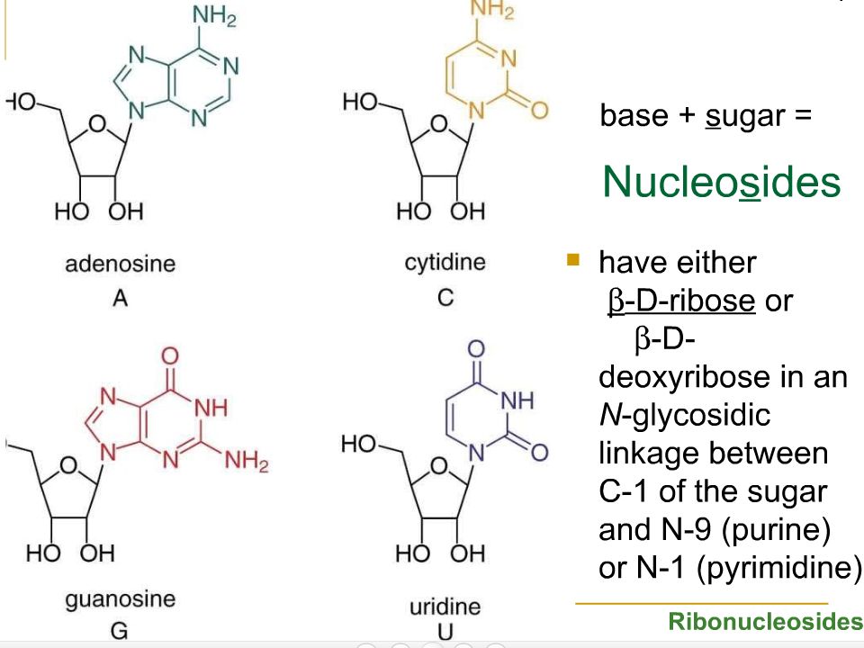
A nucleotide a molecule comprises of phosphoric acid, sugar ribose (in RNA) or deoxyribose (in DNA), and an organic base (derivative of purines or pyrimidines). There are 2 types ; deoxyribose nucleotides (in DNA ) and ribose nucleotides ( in RNA). Nucleotides are phosphorylated nucleosides. In the nucleoside, the base is bonded through a β-N-glycosidic bond to the anomeric carbon (C1)of the ribose/deoxyribose and N9 of purine or N1 of pyrimidine. The base is in the anti-orientation*)
 or deoxyribose (in DNA), and an organic base (derivative of purines or pyrimidines). There are 2 types ; deoxyribose nucleotides (in DNA ) and ribose nucleotides ( in RNA). Nucleotides are phosphorylated nucleosides. In the nucleoside, the base is bonded through a β-N-glycosidic bond to the anomeric carbon (C1)of the ribose/deoxyribose and N9 of purine or N1 of pyrimidine. The base is in the anti-orientation*)")
5
Skeleton derivatives purine: adenine guanine pyrimidine uracil thymine cytocine

6
deoxyribonucleosides

8
Adenine : (RNA) AMP, ADP, ATP; (DNA ) dAMP, dADP, dATP
Beside the sugar unit, the base component in DNA and RNA differ slightly: DNA: adenine, guanine, cytosine and thymine RNA: adenine, guanine, cytosine and uracil Nucleotide ( XMP; XDP; XTP):- X= nucleoside ;M= mono, D= di, T= tri, P=phosphate Adenine : (RNA) AMP, ADP, ATP; (DNA ) dAMP, dADP, dATP Guanine : (RNA) GMP, GDP, GTP; (DNA ) dGMP, dGDP, dGTP Cytosine : (RNA) CMP, CDP, CTP; (DNA ) dCMP, dCDP, dCTP Thymine : (DNA ) dTMP, dTDP, dTTP Uracil : (RNA) UMP, UDP, UTP Naming : CMP: cytosine mono phosphate , GDP: guanosine di phosphate; ATP: adenosine tri phosphate
:- X= nucleoside ;M= mono, D= di, T= tri, P=phosphate. Adenine : (RNA) AMP, ADP, ATP; (DNA ) dAMP, dADP, dATP. Guanine : (RNA) GMP, GDP, GTP; (DNA ) dGMP, dGDP, dGTP. Cytosine : (RNA) CMP, CDP, CTP; (DNA ) dCMP, dCDP, dCTP. Thymine : (DNA ) dTMP, dTDP, dTTP. Uracil : (RNA) UMP, UDP, UTP. Naming : CMP: cytosine mono phosphate , GDP: guanosine di phosphate; ATP: adenosine tri phosphate.")
10
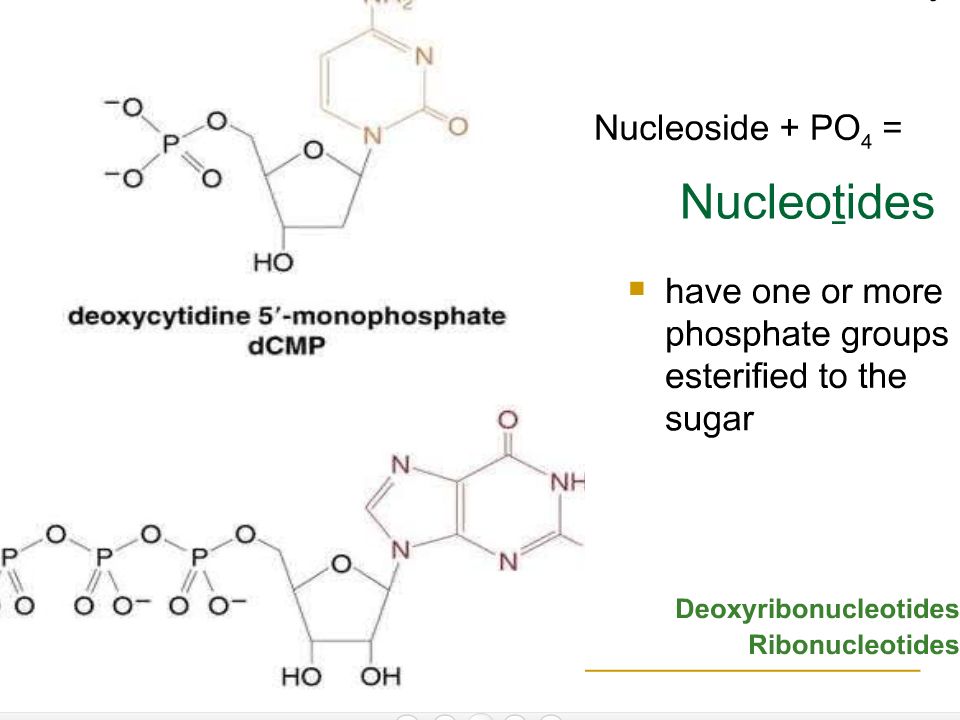
A nucleotide can be mono, di or tri phosphorylated.
The first phosphate group is bonded to the 5'-carbon of the sugar unit. 3rd 1st 2nd

11
Adenosine Triphosphate (ATP)
deoxyguanosine Triphosphate (GTP) Adenosine Triphosphate (ATP) Guanosine Triphosphate (GTP) Cytosine Triphosphate (CTP) Uridine Triphosphate (UTP)
 Adenosine Triphosphate (ATP) Guanosine Triphosphate (GTP) Cytosine Triphosphate (CTP) Uridine Triphosphate (UTP)")
12
syn-Adenosine anti-Adenosine Orientation of sugar/base in adenosine

13
Two nucleotides are condensed by the reaction between the alcohol of a 5'-phosphate of one nucleotide and the 3'-hydroxyl of a second, with the elimination of H2O, forming a phosphodiester bond. NUCLEOTIDE also are required for numerous other important functions within the cell. These functions include 1. serving as energy stores for future use in phosphate transfer reactions. These reactions are predominantly carried out by ATP. 2. forming a portion of several important coenzymes such as NAD+, NADP+, FAD and coenzyme A. 3. serving as mediators of numerous important cellular processes such as second messengers in signal transduction events. The predominant second messenger is cyclic-AMP (cAMP), a cyclic derivative of AMP formed from ATP.
, a cyclic derivative of AMP formed from ATP.")
14
4. controlling numerous enzymatic reactions through allosteric effects on enzyme activity.
5. serving as activated intermediates in numerous biosynthetic reactions. Eg of activated intermediates (i) S-adenosylmethionine (S-AdoMet) involved in methyl transfer reactions (ii) sugar coupled nucleotides involved in glycogen and glycoprotein synthesis 6. precursors to DNA & RNA synthesis Purine bases and purine nucleosides are toxic to humans so must be readily eliminated. Purine and pyrimidine nucleotides, is metabolized via its specific pathway
 S-adenosylmethionine (S-AdoMet) involved in methyl transfer reactions (ii) sugar coupled nucleotides involved in glycogen and glycoprotein synthesis. 6. precursors to DNA & RNA synthesis. Purine bases and purine nucleosides are toxic to humans so must be readily eliminated. Purine and pyrimidine nucleotides, is metabolized via its specific pathway.")
15
NUCLEOTIDE METABOLISM
Discusses pathways that lead to: i - breakdown of nucleotides ii - [ ] of nucleotides ▪ de novo pathway i.e synthesis from precursor ▪ salvage pathway ( recycling) Salvage (recycling) DNA/RNA synthesis Nucleotides Pool Biosynthesis from precursor degradation
 Salvage. (recycling) DNA/RNA synthesis. Nucleotides Pool. Biosynthesis from precursor. degradation.")
16
After dissociation, the protein is metabolized like any other protein.
Most, but not all, nucleic acids in cell (animal or plant) are associated with protein═> nucleoprotein eg chromatin, ribosomes, viruses. Dietary nucleoprotein is split by pancreatic enzymes (in stomach) and tissue nucleoprotein by lysosomal enzymes. After dissociation, the protein is metabolized like any other protein. The nucleic acids are hydrolyzed randomly by nucleases to yield a mixture of shorter polynucleotides. Cellular nucleoprotein Lysosomal enz Protein + N.A Poly nucleotides Endo & exo Nucleases, and nucleases phosphodiesterases Pacreatic enz Triphospho, monophosphon’tide, Diet nucleoprotein nucleotidase free bases deaminase Salvage (recycling)
 are associated with protein═> nucleoprotein eg chromatin, ribosomes, viruses. Dietary nucleoprotein is split by pancreatic enzymes (in stomach) and tissue nucleoprotein by lysosomal enzymes. After dissociation, the protein is metabolized like any other protein. The nucleic acids are hydrolyzed randomly by nucleases to yield a mixture of shorter polynucleotides. Cellular nucleoprotein. Lysosomal enz. Protein + N.A. Poly. nucleotides. Endo & exo. Nucleases, and. nucleases. phosphodiesterases. Pacreatic enz. Triphospho, monophosphon’tide, Diet nucleoprotein. nucleotidase. free bases. deaminase. Salvage. (recycling)")
17
shorter polynucleotides are hydrolyzed by endo & exonucleases(endo & exonucleotidase) to yield tri and mononucleotides (nucleotidase = diesterase ) The triP cleaved by phosphodiesterases to the mononucleotides: AMP, GMP, CMP, UMP and TMP The pathway of purine and pyrimidine n’tide differ after this point The mononucleotides are hydrolyzed by nucleotidases and purine/pyrimidine nucleoside phosphorylase to free base. The free base undergoes either catabolic pathway or salvage pathway. release of free base from N.A occurs in 2 stages: i- hydrolysis of phosphoester bonds in N.A nucleotides ii- hydrolysis of phosphoester and glycosidic bonds in nucleotide free base

18
Catabolism/Degradation of purine and pyrimudine (fate of free base) - IN LIVER.
free bases end products breakdown of base structure conversion to 5’ mononucleotide – Salvage pathway

19
I. Catabolism(Degradation) of the Bases.
Purine and paramidine are metabolized differently: End product of purine base catabolism/degradation is uric acid. End product of pyrimidine base catabolism (degradation) is β- alanine and β- amino Isobutyrate II. Characteristic of catabolism of purine base : - Pathway for adenine differs from that for guanine. - Pathway lead to similar final product that is Uric Acid. - No base ring-cleavage
 is β- alanine and β- amino Isobutyrate. II. Characteristic of catabolism of purine base : - Pathway for adenine differs from that for guanine. - Pathway lead to similar final product that is Uric Acid. - No base ring-cleavage.")
20
Catabolism of Purine Nucleotides
Groups attached to the purine ring are sequentially removed from AMP & GMP by parallel pathways: 1- Phosphate groups are removed by nucleotidase. 2- Amino groups are released by:adenosine deaminase & guanine deaminase (or guanese). 3-The pentoses are removed by purine nucleoside phosphorylase . The sum of these reaction converts AMP & GMP to hypoxanthine & xanthine respectively. 20
. 3-The pentoses are removed by purine nucleoside phosphorylase . The sum of these reaction converts AMP & GMP to hypoxanthine & xanthine respectively. 20.")
21
Guanine Nucleotide (GMP)
GMP is acted upon by nucleotidase producing guanosine and Pi. ii) Guanosine is further acted upon by purine nucleoside phosphorylase liberating free guanine + dribose iii)Guanese acts upon guanine to create Xanthine. iv) Xanthine oxidase acts upon xanthine to create Uric acid. This enzyme is clinically important
 Guanosine is further acted upon by purine nucleoside phosphorylase liberating free guanine + dribose. iii)Guanese acts upon guanine to create Xanthine. iv) Xanthine oxidase acts upon xanthine to create Uric acid. This enzyme is clinically important.")
22
Adenine Nucleotide (AMP) Degradation occurs by either
(i) AMP is acted upon by nucleotidase liberating adenosine which is further acted by adenosine deaminase producing inosine OR (ii) AMP is acted upon by AMP deaminase producing inosine monophosphate (IMP) - IMP is then acted upon by nucleotidase liberating inosine INOSINE is further acted to form final product xanthine: AMP deaminase nucleotidase AMP inosine IMP phosphorylase inosine Xanthine oxidase hypoxanthine xanthine
 AMP is acted upon by nucleotidase liberating adenosine which is further acted by adenosine deaminase producing inosine. OR. (ii) AMP is acted upon by AMP deaminase producing inosine monophosphate (IMP) - IMP is then acted upon by nucleotidase liberating inosine. INOSINE is further acted to form final product xanthine: AMP deaminase. nucleotidase. AMP. inosine. IMP. phosphorylase. inosine. Xanthine oxidase. hypoxanthine. xanthine.")
23
Catabolism of Guanine Nucleotide (GMP) & Adenosine Nucleotide AMP
nucleotidase Pi ribose guanese excrete Catabolism of Guanine Nucleotide (GMP) & Adenosine Nucleotide AMP
 & Adenosine Nucleotide AMP.")
24
Xanthine is acted upon by Xanthine oxidase to form urate.
Urate is transported to and excreted by the kidney into the urine. Urate is not very soluble but is not a problem to kidney for excretion. When the urine is very acid or has high [Ca2+]; [Na+], urate salts co precipitate with calcium or sodium salts and can form stones in kidney or bladder. A very high concentration of urate in the blood leads to a fairly common group of diseases referred to as gout (intense pain with swelling). To reduce [plasma urate], is to reduce urate synthesis which is catalysed by Xanthine oxidase. This is key enzyme. Its activity is inhibited by drug ‘allopurinol’ which is structurally similar to xanthine In birds, uric acid is further degraded to a high water soluble end product, allantoin
. To reduce [plasma urate], is to reduce urate synthesis which is catalysed by Xanthine oxidase. This is key enzyme. Its activity is inhibited by drug ‘allopurinol’ which is structurally similar to xanthine. In birds, uric acid is further degraded to a high water soluble end product, allantoin.")
25
Adenine Guanine A nuclease frees the nucleotide
Summary Guanine A nuclease frees the nucleotide A nucleotidase creates guanosine Purine nucleoside phosphorylase converts guanosine to guanine Guanase converts guanine to xanthine Xanthine oxidase converts xanthine to uric acid (urate) Adenine A nucleotidase creates adenosine, then adenosine deaminase creates inosine Alternatively, AMP deaminase creates inosinic acid, then a nucleotidase creates inosine Purine nucleoside phosphorylase acts upon inosine to create hypoxanthine Xanthine oxidoreductase acts upon hypoxanthine to create xanthine Xanthine oxidoreductase acts upon xanthine to create uric acid
 Adenine. A nucleotidase creates adenosine, then adenosine deaminase creates inosine. Alternatively, AMP deaminase creates inosinic acid, then a nucleotidase creates inosine. Purine nucleoside phosphorylase acts upon inosine to create hypoxanthine. Xanthine oxidoreductase acts upon hypoxanthine to create xanthine. Xanthine oxidoreductase acts upon xanthine to create uric acid.")
26
Gout (hyperuricemia) Clinical Significances of Purine Metabolism
Clinical problems result of abnormal catabolism of purine is due to insolubility of uric acid. Gout (hyperuricemia) Excess accumulation of uric acid . GOUT results from the precipitation of sodium urate crystals (tophi) in the synovial fluid of joints, leading to severe inflammation,arthritis & severe degeneration of joints. Often attacks first metatarsophalangeal joint of big toe. Gout is treated by allopurinol. Allopurinol is a structural analog of hypoxanthine that strongly inhibits xanthine oxidase. 26
 Excess accumulation of uric acid . GOUT results from the precipitation of sodium urate crystals (tophi) in the synovial fluid of joints, leading to severe inflammation,arthritis & severe degeneration of joints. Often attacks first metatarsophalangeal joint of big toe. Gout is treated by allopurinol. Allopurinol is a structural analog. of hypoxanthine that strongly inhibits xanthine oxidase. 26.")
27
Uric acid normal limits are 4-7mg/dl for males &
3 - 6mg/dl for females. Severe gout in the fingers resulting in large, hard deposits of crystals of uric acid. These deposits are called tophi. 27

28
Causes of hyperuricemia
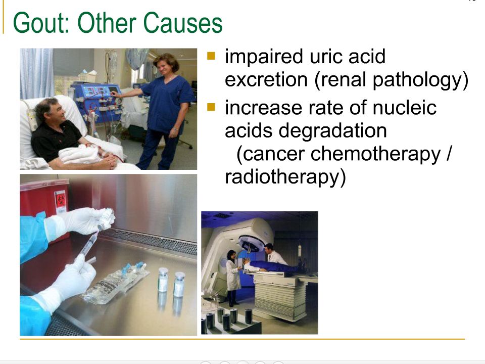
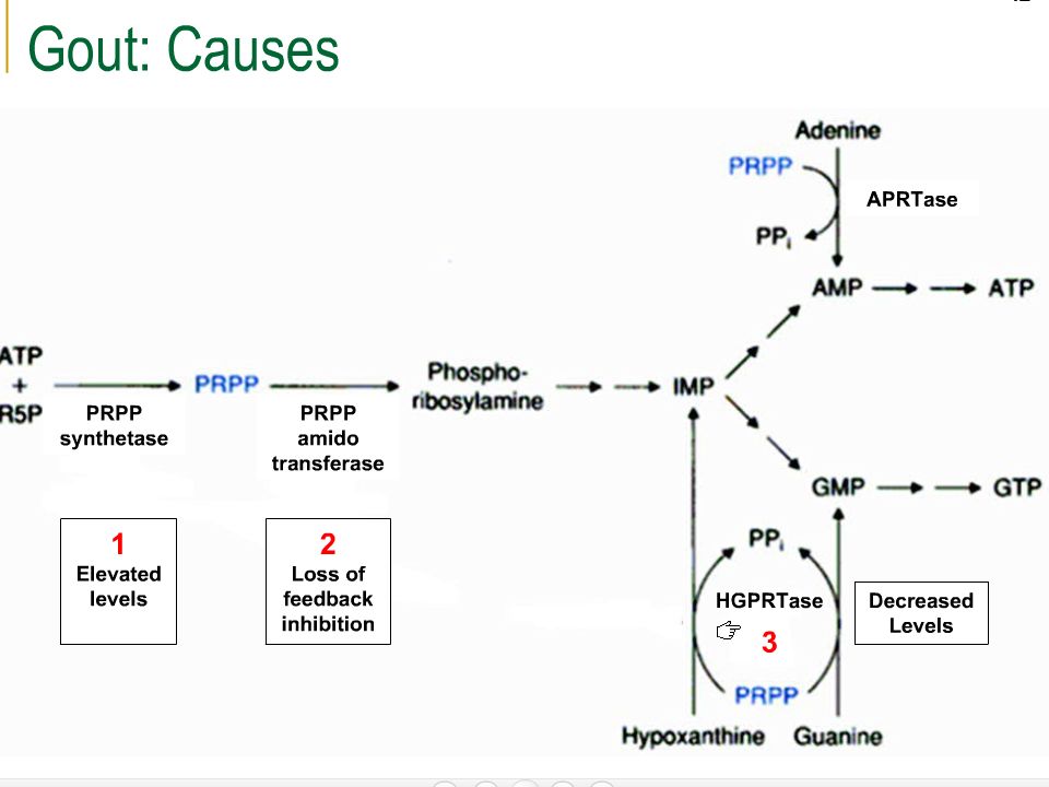
I- Overproduction of purine due to: 1-Specific enzyme defects: A- Increased activity of PRPP synthase. B- PRPP amidotransferase is less sensitive to the feedback inhibition by purine nucleotides. C-Deficiency of salvage enzymes ( HGPRT), so consumption of PRPP is decreased leading to its accumulation as in Lesch Nyhan syndrome. D-Deficiency of glucose-6-phosphatase & enhanced conversion of g-6-p to ribose 5- phosphate and PRPP as in Von Gierke’s disease 2- Secondary to other diseases e.g. cancer that enhance tissue turnover and overload of purines. II- Defective elimination of urate ( renal disorder) 28
, so consumption of PRPP is decreased leading to its accumulation as in Lesch Nyhan syndrome. D-Deficiency of glucose-6-phosphatase & enhanced conversion of g-6-p to ribose 5- phosphate and PRPP as in Von Gierke’s disease. 2- Secondary to other diseases e.g. cancer that enhance tissue turnover and overload of purines. II- Defective elimination of urate ( renal disorder) 28.")
31
III- Genetic defect 1. Lesch-Nyhan syndrome
Due to loss of a functional HGPRT gene, so consumption of PRPP is decreased leading to uric acid accumulation. Patients exhibit severe gout & severe malfunction of the nervous system, mental retardation, spasticity & self harm (self-mutilation). Death usually occurs before the age of 20 year. 2. Hypouricemia Severe combined immunodeficiency disease (SCID) SCID is a group of inherited disorders characterized by the lack of immune response to infectious diseases. This is due to the inability of B &T lymphocytes to proliferate & produce antibodies. SCID patients suffer from a deficiency in the enzyme adenosine deaminase (ADA) (~30%). In the absence of ADA, deoxyadenosine is not degraded and converted into dAMP and then into dATP.
. Death usually occurs before the age of 20 year. 2. Hypouricemia. Severe combined immunodeficiency disease (SCID) SCID is a group of inherited disorders characterized by the lack of immune response to infectious diseases. This is due to the inability of B &T lymphocytes to proliferate & produce antibodies. SCID patients suffer from a deficiency in the enzyme adenosine deaminase (ADA) (~30%). In the absence of ADA, deoxyadenosine is not degraded and converted into dAMP and then into dATP.")
32
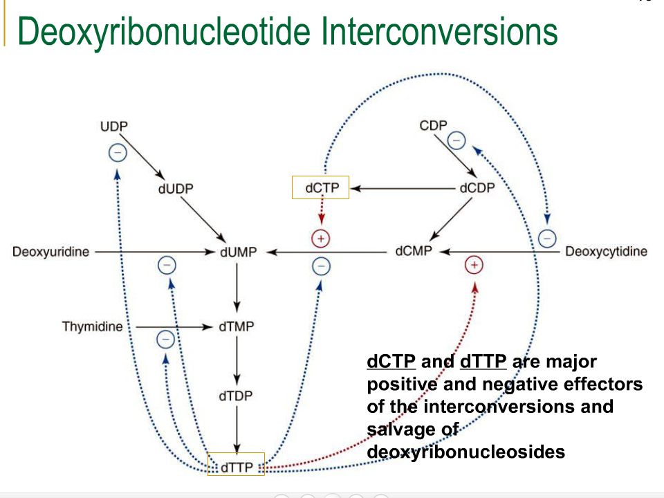
The disease is usually fatal in infancy .
continuous dATP is a potent feedback inhibitor of deoxynucleotide biosynthesis. So, DNA synthesis is impaired. Rapidly proliferating lymphocytes are particularly susceptible if DNA synthesis is impaired, & seriously impairs the immune responses. The disease is usually fatal in infancy . A less severe immunodeficiency results when there is a lack of purine nucleoside phosphorylase (PNP) (ribonucleotide reductase & DNA synthesis are inhibited due to accumulation of dGTP). 32
 (ribonucleotide reductase & DNA synthesis are inhibited due to accumulation of dGTP). 32.")
33
Catabolism/Degradation of pyrimidine nucleotides
nucleotidases pyrimidine nucleoside pyrimidine nucleoside phosphorylase pyrimidine

34
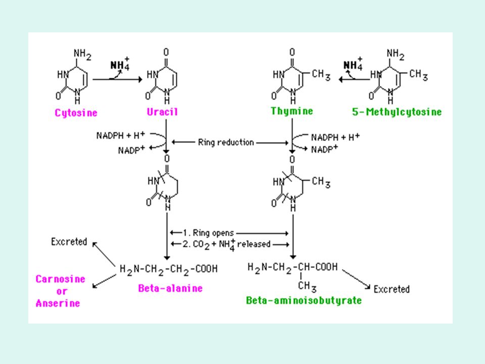
The pyrimidine are then degraded further into β- alanine and β- amino isobutyrate involving ring cleavage: - Atoms 2 and 3 of both rings are released as ammonia and carbon dioxide. -The rest of the ring is left as a beta-amino acid. Beta-amino isobutyrate from thymine or 5-methyl cytosine is largely excreted. Beta-alanine from cytosine or uracil may either be excreted or incorporated into the brain and muscle dipeptides, carnosine (his-beta-ala) or anserine (methyl his-beta-ala).
 or anserine (methyl his-beta-ala).")
36
Salvage pathway: Is a metabolic pathway that uses substrates other than the usual biosynthetic intermediates for a product, eg. free purines from the hydrolysis of nucleotides (from diet & intracellular N.A) is salvaged for the generation of new nucleotides PRPP is the donor of phosphorylribose to the base. The reaction is catalyzed by phosphoribosyl transferase enzyme. It requires far less energy than de novo synthesis. Mammalian liver provides purine bases & nucleosides for salvage to tissues incapable for their biosynthesis e.g- brain cells, RBCs, & WBCs
 is salvaged for the generation of new nucleotides. PRPP is the donor of phosphorylribose to the base. The reaction is catalyzed by phosphoribosyl transferase enzyme. It requires far less energy than de novo synthesis. Mammalian liver provides purine bases & nucleosides for salvage to tissues incapable for their biosynthesis e.g- brain cells, RBCs, & WBCs.")
39
Mechanisms of salvage pathway
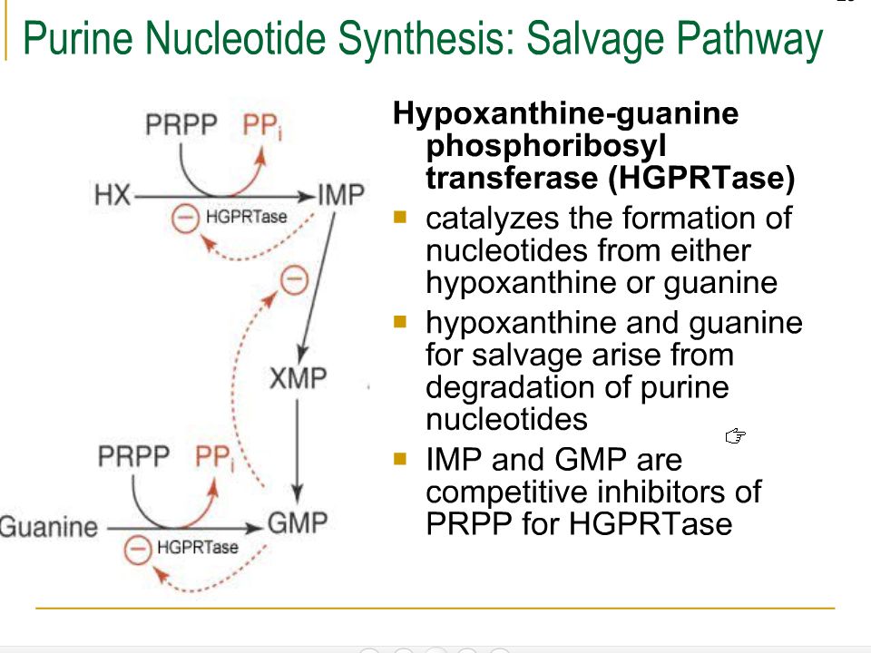
1- Phosphoribosylation of purines: A- Hypoxanthine-Guanine Phosphoribosyl transferase [HGPRT]. This enzyme transfers ribose 5- phosphate from PRPP to the purine ring (hypoxanthine & guanine) resulting in IMP & GMP respectively. Hypoxanthine PRPP PPi IMP HGPRT 39
 resulting in IMP & GMP respectively. Hypoxanthine. PRPP. PPi. IMP. HGPRT. 39.")
40
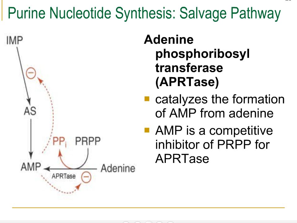
Guanine GMP AMP B- Adenine Phosphoribosyl transferase [APRT]. Adenine
continue Guanine B- Adenine Phosphoribosyl transferase [APRT]. Adenine PPi PRPP GMP HGPRT PRPP PPi AMP APRT 2- Direct phosphorylation of purine nucleosides: AMP ATP ADP ADP Adenosine kinase 40
![Guanine GMP AMP B- Adenine Phosphoribosyl transferase [APRT]. Adenine](http://slideplayer.com/slide/10456466/35/images/40/Guanine+GMP+AMP+B-+Adenine+Phosphoribosyl+transferase+%5BAPRT%5D.+Adenine.jpg "continue. Guanine. B- Adenine Phosphoribosyl transferase [APRT]. Adenine. PPi. PRPP. GMP. HGPRT. PRPP. PPi. AMP. APRT. 2- Direct phosphorylation of purine nucleosides: AMP. ATP. ADP. ADP. Adenosine kinase. 40.")
41
Anabolism of base Synthesis of purine bases from precursors
- The starting step is formation of PRPP from ribose 5’-phosphate & ATP - the final form of purines in its synthesis process is as the ribonucleotides. The synthesis starts of with Phosphoribosyl-1-pyrophosphate (PRPP) which is the activated form of ribose 5-phosphate. - occurs in the cytosol of the liver cells.
 which is the activated form of ribose 5-phosphate. - occurs in the cytosol of the liver cells.")
42
Origin of atoms in the purine

43
─ This rxn occurs in many tissue types
─ This rxn occurs in many tissue types – sensitive to di- and tri-phosphates, and 2,3-DPG

44
replacement of the pyrophosphate of PRPP by the amide group of glutamine, 5-phosphoribosylamine is formed. This rxn is catalysed by glutamine PRPP amidotranferase (a dimer ) and is the rate determining step (control/regulated point) RDS Regulation of

45
A series of additions take place to make first the 5- and then the 6-membered ring.
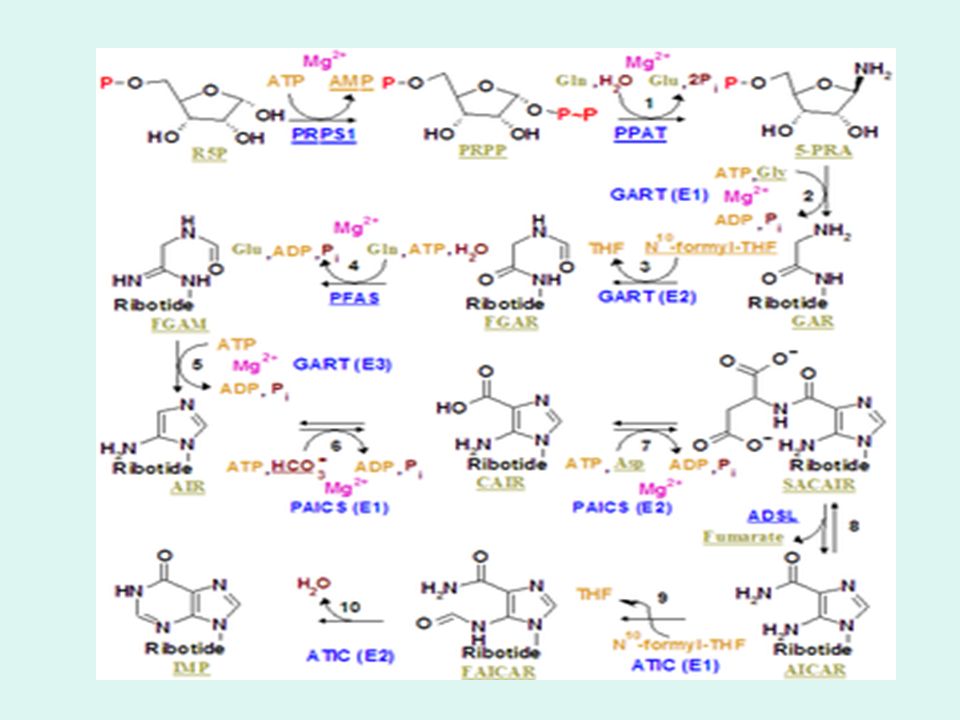
The whole glycine molecule, adds to the amino group to be atoms 4, 5, and 7 of the purine ring. This step uses ATP. The amino group of 5-phosphoribosyl amine becomes nitrogen 9 of the purine ring. 5, 10-Methenyl tetrahydrofolate supply the last atom to the 5-membered ring. the amide of glutamine adds to carbon 4 to start the six-membered ring portion. It becomes nitrogen 3. Then condensing of carbon 8 and nitrogen 9 to form the five-membered ring.

46
the addition of carboxyl group (from carbon dioxide) to form carbon 6 of the ring.
The amine group of aspartate adds to the carboxyl group with a subsequent removal of fumarate. The amino group is now nitrogen 1 of the final ring. The final atom of the purine ring, carbon 2, is supplied by 10-Formyl tetrahydrofolate. Ring closure produces the purine nucleotide, IMP (inosine monophosphate). IMP can then become either AMP or GMP via appropriate rxn Total 4 ATP are required for the whole process
. IMP can then become either AMP or GMP via appropriate rxn. Total 4 ATP are required for the whole process.")
47
Schematic presentation of purine synthesis
NAD + Gln ATP GMP Schematic presentation of purine synthesis

48
RDS- Adenylsuccinate lyse
Goes to TCA in muscle

50
Regulation of Purine Nucleotide Synthesis

51
Regulation of Purine Biosynthesis
1- Concentration of PRPP which depends on: Availability of ribose 5- phosphate. Activity of PRPP synthase. 2-Accumulation of purine nucleotides: The first limiting step, PRPP amidotransferase is synergistically inhibited by IMP & GMP binding to one allosteric site, and AMP binding to another. Adenylosuccinate synthetase & IMP dehydrogenase, the two enzymes at IMP branch point are also allosterically regulated. 51

52
AMP ADP + ADP Conversion of MPNucleotide TPNucleotide
- For ATP, 2 systems i- by a 2-step reactions a) ATP-dependent transphosphorylation of AMP into ADP AMP ADP + ADP b) oxidative phosphorylation of ADP into ATP ADP + Pi + O ATP + H2O this system is a compartmentalization type, i.e the enzymes catalysing the 2 rxn are linked into a complex .The ATP which is the phosphate donor for the transphosphorylation reaction is not the free ATP pre-existing in mitochondria, but the ATP produced by the complex itself; on the other hand, the ADP formed as a transient intermediate in the AMP-ATP conversion is immediately phosphorylated to give ATP without mixing with the free internal ADP. *** Formation of ATP - by phosphorylation of ADP or AMP ATP NADH NAD+
 ATP-dependent transphosphorylation of AMP into ADP. AMP ADP + ADP. b) oxidative phosphorylation of ADP into ATP. ADP + Pi + O2 ATP + H2O. this system is a compartmentalization type, i.e the enzymes catalysing the 2 rxn are linked into a complex .The ATP which is the phosphate donor for the transphosphorylation reaction is not the free ATP pre-existing in mitochondria, but the ATP produced by the complex itself; on the other hand, the ADP formed as a transient intermediate in the AMP-ATP conversion is immediately phosphorylated to give ATP without mixing with the free internal ADP. *** Formation of ATP. - by phosphorylation of ADP or AMP. ATP. NADH. NAD+")
53
ii- Conversion of mononucleotides to nucleotide di & triphosphates
Subsequent phosphorylation of AMP & GMP by ATP, leads to formation of di- & triphosphates catalyzed by kinases. 53

54
continuo 3-Energy sources: as seen in the reactions above, ATP is required to synthesize GMP from XMP, while GTP is required to synthesize AMP. Adenylosuccinate synthetase IMP dehydrogenase PRPP amidotransferase 54

55
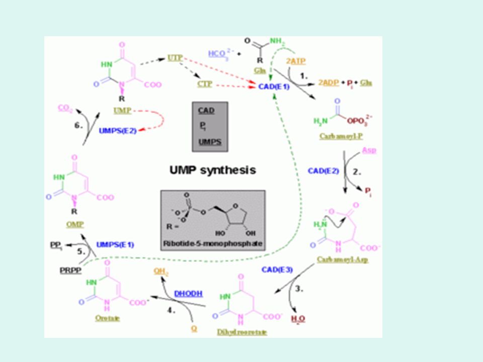
Synthesis of Pyrimidine Nucleotides
pyrimidine molecules are simpler than purines. their synthesis is simpler occurs in spleen, thymus, GI tract and testes

56
Glutamine's amide nitrogen and carbon dioxide provide atoms 2 and 3 of the pyrimidine ring via formation of cabamoyl-PO4.

57
The other four atoms of the ring are from aspartate, incorporated followed by dehydration forming orotate derivative

59
the sugar phosphate portion of the molecule is supplied by PRPP via formation of OMP (orotate monophosphate), and subsequent reaction leads to formation of UMP. UMP then acts as substrate for synthesis of other nucleotides

60
** Synthesis of nucleotide of pyrimidine differs from that of purine in that:
In purine synthesis, a nucleotide is formed first while pyrimidines are first synthesized as the free base. The control of pyrimidine nucleotide synthesis in man is exerted primarily at the level of cytoplasmic CPS II. UTP inhibits the enzyme, competitively with ATP. PRPP activates it .

61
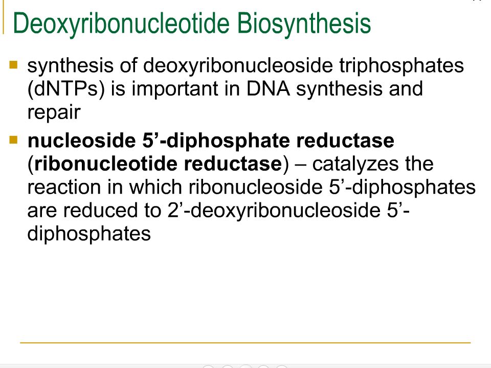
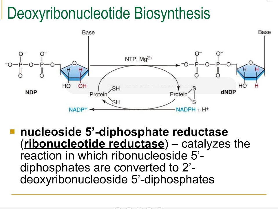
Synthesis of deoxyribonucleotides DNA requires deoxyribonucleotides.
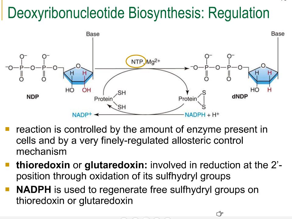
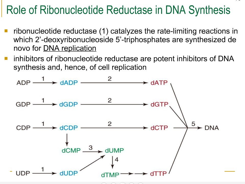
Conversion of purine & pyrimidine ribonucleotides to deoxyribonucleotides occurs only at nucleoside diphosphate level. Nucleoside Diphosphates [NDPs] are reduced by ribonucleotide reductase complex forming deoxyribonucleoside diphosphates [dNDPs]. This reduction requires thioredoxin (a protein cofactor), thioredoxin reductase (flavoproteins), & NADPH.H+ This enzyme complex is active only when cells are synthesizing DNA preparatory to cell division. 61
, thioredoxin reductase (flavoproteins), & NADPH.H+ This enzyme complex is active only when cells are synthesizing DNA preparatory to cell division. 61.")
70
GOOD LUCK

72
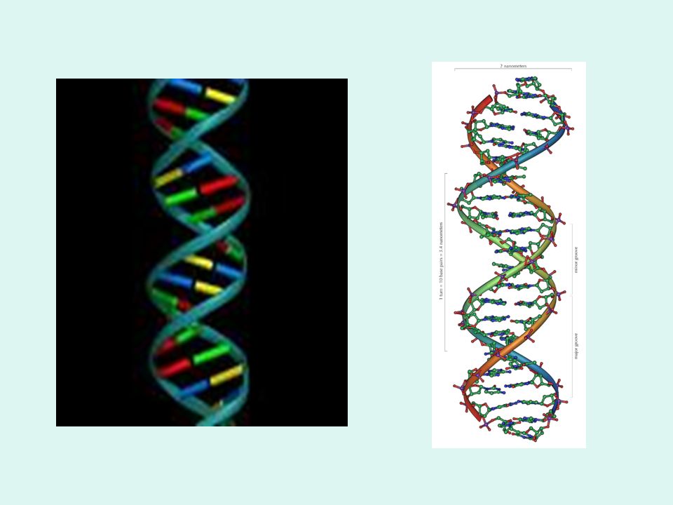
Deoxyribonucleic Acid (DNA)
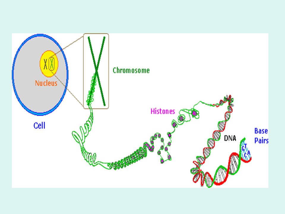
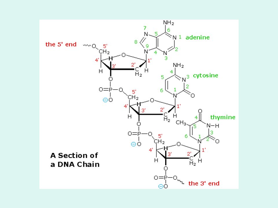
It has high molecular weight i.e molecule has high number of nucleotides. In euaryotic cells, it is found chiefly in the nuclei. It wrapped around small proteins known as histones forming bead-like structure then organized and folded into chromatin aggregates that make up the chromosomes and in procaryotic cells e.g bacteria, in the nucleoid regions. It contains two polynucleotide strands wound around each other through base-pairing i.e double helix The backbone of each strand consists of alternating deoxyribose and phosephate groups. The phosphate group bonded to the 5' carbon atom of one deoxyribose is covalently bonded to the 3' carbon of the next. The two strands are "antiparallel"; that is, one strand runs 5′ to 3′ while the other runs 3′ to 5′.

73
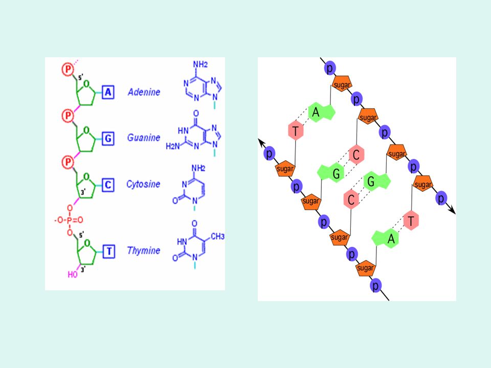
Base-pair adenine-thymine pair guanine-cytosine pair

75
The DNA strands are assembled in the 5′ to 3′ direction and, by convention, we "read" them the same way. The purine or pyrimidine attached to each deoxyribose projects in toward the axis of the helix. Has a cistron region i.e a sequence that contains information for a polypeptide and several signals that are required for ribosome function. Each base forms hydrogen bonds with the one directly opposite it, forming base pairs (also called nucleotide pairs). adenine-thymine base pair has 2 hydrogen bonds, and guanine-cytosine base pair has 3 hydrogen bonds guanine-cytosine base pair is stronger than the adenine-thymine base pair
. adenine-thymine base pair has 2 hydrogen bonds, and guanine-cytosine base pair has 3 hydrogen bonds. guanine-cytosine base pair is stronger than the adenine-thymine base pair.")
79
RIBONUCLEIC ACID (RNA)
Mostly is a single-stranded molecule which can coil back on itself and form unique and quite complex 3-D structure e.g hairpin, clover-shape. RNA is involved in the synthesis of proteins. "Information" is typically passed from DNA to RNA to the resulting proteins. There are 3 major species : ribosomal RNA (rRNA): 80-90%; transfer RNA (tRNA): 15 % messenger RNA (mRNA): 5% The size of the rRNA varies, but is generally less than a thousandth the size of DNA. It is a component of ribosomes ( rRNA + proteins). The secondary structure is extraordinary complex. Its size is designated by S (= sedimentation) value e.g 5 S, 5.8 S, 28 S.
: 80-90%; transfer RNA. (tRNA): 15 % messenger RNA (mRNA): 5% The size of the rRNA varies, but is generally less than a thousandth the size of DNA. It is a component of ribosomes. ( rRNA + proteins). The secondary structure is extraordinary complex. Its size is designated by S (= sedimentation) value e.g 5 S, 5.8 S, 28 S.")
80
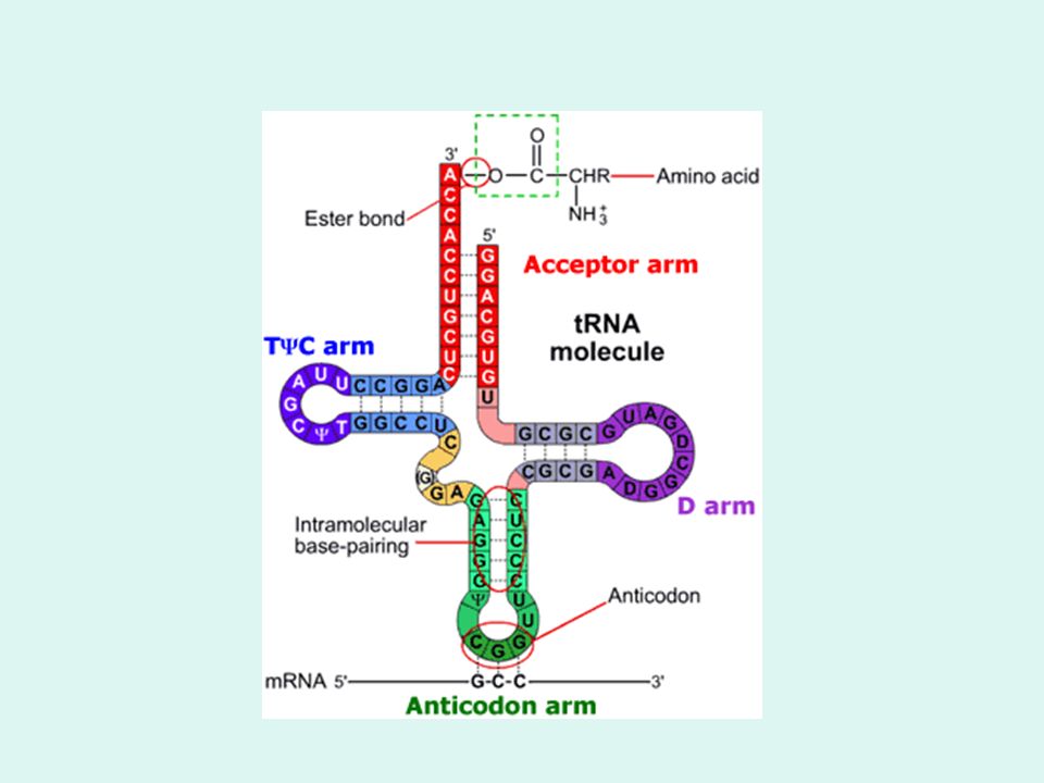
tRNA is a small molecule consists of nucleotides, function to carry activated a.a to the protein synthesis site, the ribosomes. It is a stable molecule but short-lived. There are at least 56 types in any cell. Each recognizes a different codon for an a.a. The different tRNA that accepts an a.a is called isoacceptor. Each carries only 1 a.a It has a ''cloverleaf '' structure i.e consisting of a stem and 3 loops.Intrachain base pairing at some point results in double helix portion . One of the loop is anticodon loop. This loop has ''Anticodon zone'' which is a triplet that base pair to mRNA during protein synthesis, and plays a role in specifying which a.a becomes attach to the tRNA .The stem ends in the sequence...CCA (3’end) which is the attachment site for the a.a. It contains other determinants of which a.a is to attach to the tRNA. An extra arm (variable loop) may also exist on the structure. tRNA made up 15% of cellular RNA. Contain modified bases e.g 4-thioUridine, dihydrouridine,
 which is the attachment site for the. a.a. It contains other determinants of which a.a is to attach to the tRNA. An extra arm (variable loop) may also exist on the structure. tRNA made up 15% of cellular RNA. Contain modified bases e.g 4-thioUridine, dihydrouridine,")
82
mRNA is the carrier of genetic information on
the primary structure of protein from DNA, has features allowing it to attach to ribosome and function in protein synthesis. It is of variable size depending on the protein size of which it codes and the cell type e.g E.coli mRNA compose of nucleotides. It is relatively short-lived, varies with protein species. 1 mRNA of eukaryotic codes one protein i.e it is monocistronic whereas that of prokaryotes are polycistronic i.e contain coding information for many polypeptide chains.

Similar presentations


 Recognize and apply the.>")

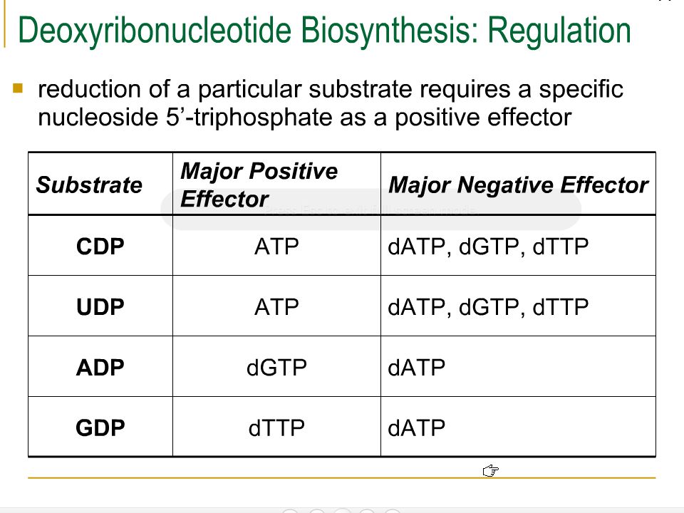
 ATP, are the sources of.>")


.>")


