Download presentation
Presentation is loading. Please wait.

1
ANATOMY OF CORONARY SINUS AND CLINICAL APPLICATION
Dr Gaurav Chaudhary MD,DM Cardiology Assistant Professsor Department of Cardiology

2
VENOUS DRAINAGE OF HEART
Coronary sinus Anterior cardiac vein Thebesian vein

4
60 % of venous blood of heart drains into right atrium via coronary sinus .
40 % remaining blood drains via anterior cardiac vein

5
Coronary sinus conveys blood from left coronary territory .
Anterior cardiac vein drains most of blood from right coronary artery

6
CORONARY SINUS Great cardiac vein
Oblique vein of left atrium [ vein of marshall ] Posterior vein of LV Middle cardiac vein Small cardiac vein

7
Coronary Venous System
CORONARY SINUS TRIBUTARIES

8
Great cardiac vein - anterior interventricular sulcus
Oblique vein of LA post surface of LA Post .vein of LV runs on diaphragmatic surface Middle cardiac vein - posterior IV groove . Small cardiac vein accompanies RCA

9
Developmental anatomy of the coronary sinus (CS) (26 weeks)—the right horn of the sinus venosus remains as the venous portion of the right atrium between the vena cava (light blue). Developmental anatomy of the coronary sinus (CS) (26 weeks)—the right horn of the sinus venosus remains as the venous portion of the right atrium between the vena cava (light blue). The left horn of the sinus venosus remains in adult life as the CS and portion of the great cardiac vein (dark blue). H
 (26 weeks)—the right horn of the sinus venosus remains as the venous portion of the right atrium between the vena cava (light blue). The left horn of the sinus venosus remains in adult life as the CS and portion of the great cardiac vein (dark blue). H.")
10
The CS has been dissected open along its long axis, CS musculature is seen in the proximal portion of the CS up to the orifice of the vein of Marshall. In this patient multiple posterior and posterolateral veins are also seen draining into the CS. The CS has been dissected open along its long axis, CS musculature is seen in the proximal portion of the CS up to the orifice of the vein of Marshall. In this patient multiple posterior and posterolateral veins are also seen draining into the CS.

11
The CS has been dissected open along its long axis
The CS has been dissected open along its long axis. Note the multiple ostia of various ventricular veins (hatched arrows). A remnant of the Thebesian valve that partly covers the ostium of the middle cardiac vein (MCV) is seen (arrow).
. A remnant of the Thebesian valve that partly covers the ostium of the middle cardiac vein (MCV) is seen (arrow).")
12
Valves can be found in the CS at various locations
Valves can be found in the CS at various locations. Most common are at the ostium of the CS (Thebesian valve) and at the ostium of the postural lateral vein at the junction of the Great Cardiac vein and CS (Vieussen's valve). These valves cover various extents of the area of the orifice.
 and at the ostium of the postural lateral vein at the junction of the Great Cardiac vein and CS (Vieussen s valve). These valves cover various extents of the area of the orifice.")
13
2-D ECHOCARDIOGRAPHIC IMAGING OF CORONARY SINUS

14
CORONARY SINUS -2D ECHO

15
ANAMOLIES OF CORONARY SINUS
Absent thebesian valve . Membranous thebesian valve . Absent tributaries of coronary sinus . Obstucted coronary sinus ostia Dilated coronary sinus

17
DILATED CORONARY SINUS

18
ABNORMAL CORONARY SINUS DRAINAGE

20
PERSISTENT LSVC

26
Figure 1. Transesophageal echocardiography revealed both atrial and right ventricular enlargement (left), a defect of the partial coronary sinus (middle), and shunt of the left atrium to the dilated coronary sinus (right) at the near longitudinal plane. AO indicates aorta; CS, coronary sinus; d, coronary sinus defect; LA, left atrium; LV, left ventricle; RA, right atrium; and RV, right ventricle. Figure 1. Transesophageal echocardiography revealed both atrial and right ventricular enlargement (left), a defect of the partial coronary sinus (middle), and shunt of the left atrium to the dilated coronary sinus (right) at the near longitudinal plane. Huang X Circulation 2007;116:e373-e373 Copyright © American Heart Association
, a defect of the partial coronary sinus (middle), and shunt of the left atrium to the dilated coronary sinus (right) at the near longitudinal plane. Huang X Circulation 2007;116:e373-e373. Copyright © American Heart Association.")
27
FLOUROSCOPIC IMAGING OF CORONARY SINUS

28
Figure 2. A partial coronary sinus defect beyond the range of the interatrial septum and an intact flap valve of the oval fossa with its muscular rims were revealed simultaneously at ≈140° section by transesophageal echocardiography (left). Figure 2. A partial coronary sinus defect beyond the range of the interatrial septum and an intact flap valve of the oval fossa with its muscular rims were revealed simultaneously at ≈140° section by transesophageal echocardiography (left). The shunt of the left atrium to the dilated coronary sinus was confirmed by the color Doppler flow image (right). Abbreviations as in Figure 1. Huang X Circulation 2007;116:e373-e373 Copyright © American Heart Association
. The shunt of the left atrium to the dilated coronary sinus was confirmed by the color Doppler flow image (right). Abbreviations as in Figure 1. Huang X Circulation 2007;116:e373-e373. Copyright © American Heart Association.")
29
AP Venogram

30
The MCV is a very consistent tributary of the CS present in nearly all hearts.
The MCV is a very consistent tributary of the CS present in nearly all hearts. In our study of 219 hearts the MCV was found in its usual location in the posterior interventricular sulcus draining to the ostium of the CS in 98% of these cases. Rarely, in 2.8%, the MCV has a separate ostium into the right atrium and in 18% of the cases the vein receives an immediate tributary from a posterior or posterolateral vein.

31
Coronary venous angiogram in the left anterior oblique (LAO) projection showing a near occlusive valve (arrow) in the region of the posterolateral vein (Vieussen's valve). Coronary venous angiogram in the left anterior oblique (LAO) projection showing a near occlusive valve (arrow) in the region of the posterolateral vein (Vieussen's valve).
 projection showing a near occlusive valve (arrow) in the region of the posterolateral vein (Vieussen s valve).")
32
CT ANGIO IMAGING OF CORONARY SINUS

33
Tops, L. F. et al. J Am Coll Cardiol Img 2008;1:94-106
Coronary Venous Anatomy and Relation Between Coronary Sinus and Mitral Annulus Tops, L. F. et al. J Am Coll Cardiol Img 2008;1:94-106 Copyright ©2008 American College of Cardiology Foundation. Restrictions may apply.

34
Reconstructed computed tomography image showing the coronary veins
Reconstructed computed tomography image showing the coronary veins. Note the complex origin of the MCV close to the ostium of the CS. In this patient there is a paucity of venous drainage of the posterolateral wall.

35
Case no. 15. (A) X-ray antero-posterior view during CS angiography; (B) CT reconstruction of CS and its branches, trachea and the two main bronchi, superior vena cava, and left ventricle; (C) fusion image of X-ray with CT reconstruction. RA, right atrium; RV, right ventricle; LA, left atrium; LV, left ventricle; CS, coronary sinus; VCS, vena cava superior.
 X-ray antero-posterior view during CS angiography; (B) CT reconstruction of CS and its branches, trachea and the two main bronchi, superior vena cava, and left ventricle; (C) fusion image of X-ray with CT reconstruction. RA, right atrium; RV, right ventricle; LA, left atrium; LV, left ventricle; CS, coronary sinus; VCS, vena cava superior..")
36
X-ray view of the phantom and fusion of CT reconstruction.
X-ray view of the phantom and fusion of CT reconstruction. (A–B) Antero-posterior (AP) view. (C–D) Left anterior oblique (LAO) 90° view. (E–F) Left anterior oblique (LAO) 30° view. (G–H) Right anterior oblique (RAO) 30° view. X-ray view of the phantom and fusion of CT reconstruction.
 Antero-posterior (AP) view. (C–D) Left anterior oblique (LAO) 90° view. (E–F) Left anterior oblique (LAO) 30° view. (G–H) Right anterior oblique (RAO) 30° view. X-ray view of the phantom and fusion of CT reconstruction.")
37
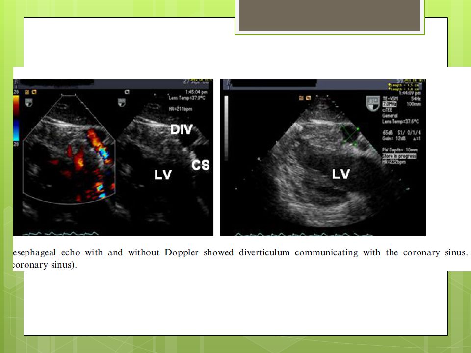
Coronary sinus diverticulum
Should be suspected in patient having WPW syndrome ,refractory to ablation Angiography reveals coronary diverticulum Associated with refractory posteroseptal pathway

41
Autopsy heart specimen depicting an absent Thebesian valve.
Autopsy heart specimen depicting an absent Thebesian valve. The lateral right atrium and lateral right ventricle have been cut open, displaying the septal leaflet of the tricuspid valve, the CS ostium (white arrow), and the fossa ovalis. Similar to this specimen, a Thebesian valve was absent in 20 of the 75 heart specimens in our study.
, and the fossa ovalis. Similar to this specimen, a Thebesian valve was absent in 20 of the 75 heart specimens in our study.")
42
Examples of membranous Thebesian valves with fenestrations.
Examples of membranous Thebesian valves with fenestrations. Two autopsy heart specimens are shown in which the right atrium and right ventricle are cut open along the direction of blood flow. (A) A ‘band-like’ Thebesian valve (white arrow) that is attached from 9 o'clock to 7 o'clock and from 1 o'clock to 4 o'clock of the ostial margin of the CS. The cranial and caudal aspect of the CS ostium is devoid of any valve attachment. (B) A ‘crescentic’ Thebesian valve (white arrow) with attachments along the entire margins of the CS ostium except at the cranial most aspect. The valves shown in (A and B) are thin and fenestrated, and likely should not impede the passage of a catheter through the ostium.
 A ‘band-like’ Thebesian valve (white arrow) that is attached from 9 o clock to 7 o clock and from 1 o clock to 4 o clock of the ostial margin of the CS. The cranial and caudal aspect of the CS ostium is devoid of any valve attachment. (B) A ‘crescentic’ Thebesian valve (white arrow) with attachments along the entire margins of the CS ostium except at the cranial most aspect. The valves shown in (A and B) are thin and fenestrated, and likely should not impede the passage of a catheter through the ostium.")
43
Examples of Thebesian valves that are fibromuscular, non-fenestrated, and occlusive.
Examples of Thebesian valves that are fibromuscular, non-fenestrated, and occlusive. Two autopsy heart specimens are shown in which the right atrium and right ventricle are cut open along the direction of blood flow. (A) A non-fenestrated Thebesian valve (white arrow) that is fibromuscular and is seen to occlude ∼80% of the ostium except for the cranial most aspect of the ostium. (B) A muscular, non-fenestrated Thebesian valve (white arrow) that occludes the entire CS ostium. To appreciate the CS ostium, the non-attached cranial most aspect of the valve has to be grasped with the forceps and peeled back. (A and B) Examples of Thebesian valves that potentially would interfere with CS cannulation (seen in 12/75—16% hearts).
 A non-fenestrated Thebesian valve (white arrow) that is fibromuscular and is seen to occlude ∼80% of the ostium except for the cranial most aspect of the ostium. (B) A muscular, non-fenestrated Thebesian valve (white arrow) that occludes the entire CS ostium. To appreciate the CS ostium, the non-attached cranial most aspect of the valve has to be grasped with the forceps and peeled back. (A and B) Examples of Thebesian valves that potentially would interfere with CS cannulation (seen in 12/75—16% hearts).")
Similar presentations


>")


