Download presentation
Presentation is loading. Please wait.

1
Prenatal development is divided into three periods called Two main periods of development: of development (defined as the first 8 weeks) and of development (from the start of the ninth week until birth)
 and of development (from the start of the ninth week until birth)")
2
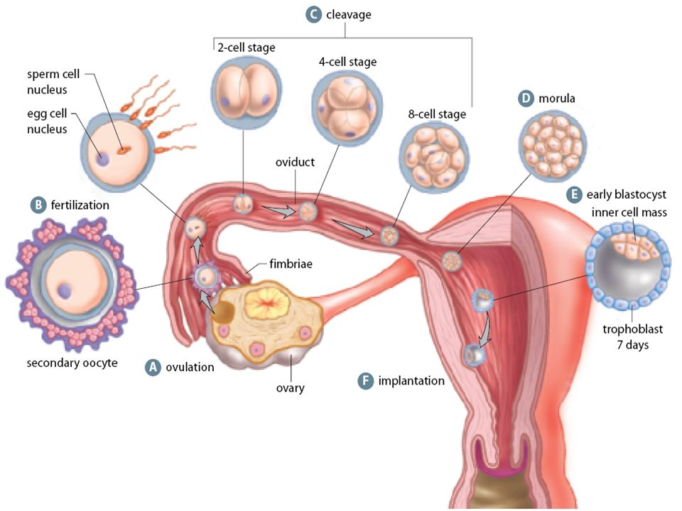
Fertilization Fertilization -

3
Fertilization An egg is released from the, gets swept into the, then moves toward the uterus by wave‐like muscular contractions as well as movement of cilia (it takes about to reach the uterus) If fertilization is to occur, it must happen within after release Once the sperm are released, they make their way through the, uterus then Fallopian Tube – most do not survive the trip!
 If fertilization is to occur, it must happen within after release Once the sperm are released, they make their way through the, uterus then Fallopian Tube – most do not survive the trip!")
4
The Layers of the Egg The egg has several layers around it: 1.The– a thin, clear layer of protein and 2. The– jelly‐like layers

5
When a sperm cell encounters the it releases from a structure called the These enzymes digest a “path” through these layers (many sperm are necessary to clear a pathway for the one eventual winner!) Once a sperm cell gains access to the egg, the egg’s plasma membrane – this prevents other sperm from entering Eventually the membranes around the sperm cell and egg cell disappear – there are now in one cell (fertilization is complete once this happens) The resulting structure is called a
 Once a sperm cell gains access to the egg, the egg’s plasma membrane – this prevents other sperm from entering Eventually the membranes around the sperm cell and egg cell disappear – there are now in one cell (fertilization is complete once this happens) The resulting structure is called a")
7
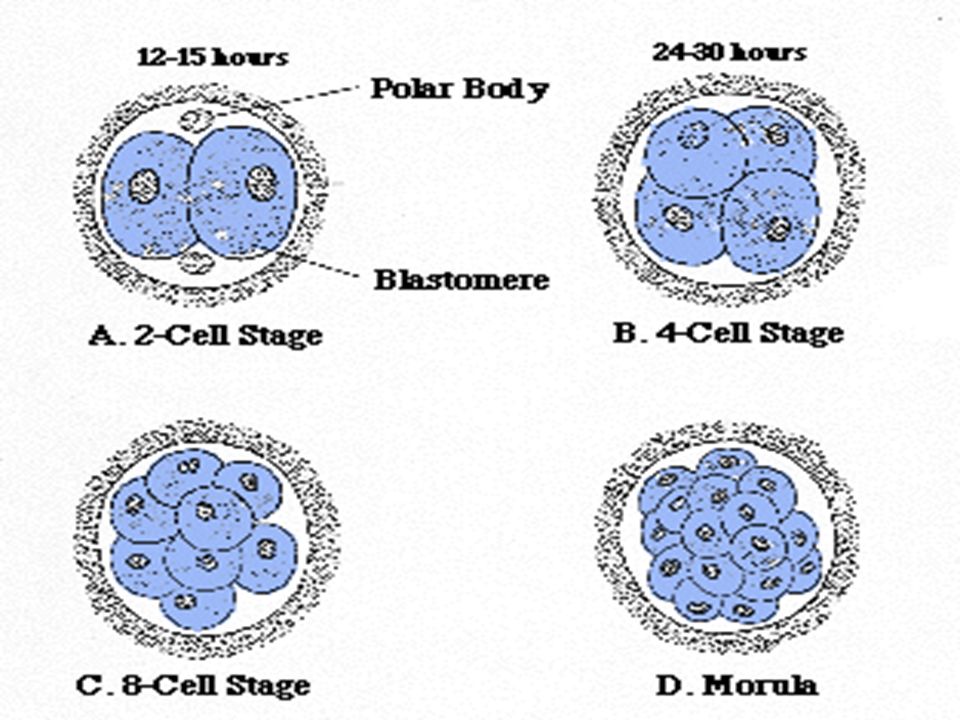
EARLY DEVELOPMENT The zygote (fertilized ovum before cleavage) while still in fallopian tube begins to divide () with double the number of cells produced each division (cleavage) (C) These divisions produce a solid ball of cells called the (D) Note that The zygote get any bigger – the cells get smaller = Refer to diagram
 while still in fallopian tube begins to divide () with double the number of cells produced each division (cleavage) (C) These divisions produce a solid ball of cells called the (D) Note that The zygote get any bigger – the cells get smaller = Refer to diagram")
10
EARLY DEVELOPMENT Further division results in the formation of a called the (~120 cells) (E) which will implant in the lining of the uterus (F).
 (E) which will implant in the lining of the uterus (F).")
11
The Blastocyst The blastocyst has 2 components: The which will become the The develops into the

12
Around 5‐7 days after fertilization, the blastocyst attaches to the The trophoblast enzymes that digest some of the tissues and blood vessels of the endometrium allowing the blastocyst to slowly sink into the uterine wall This process is called and is usually completed by the day

14
EARLY DEVELOPMENT Endometrium, under the influence of and from the, is prepared for the arrival of blastocyst -> menstruation stops

15
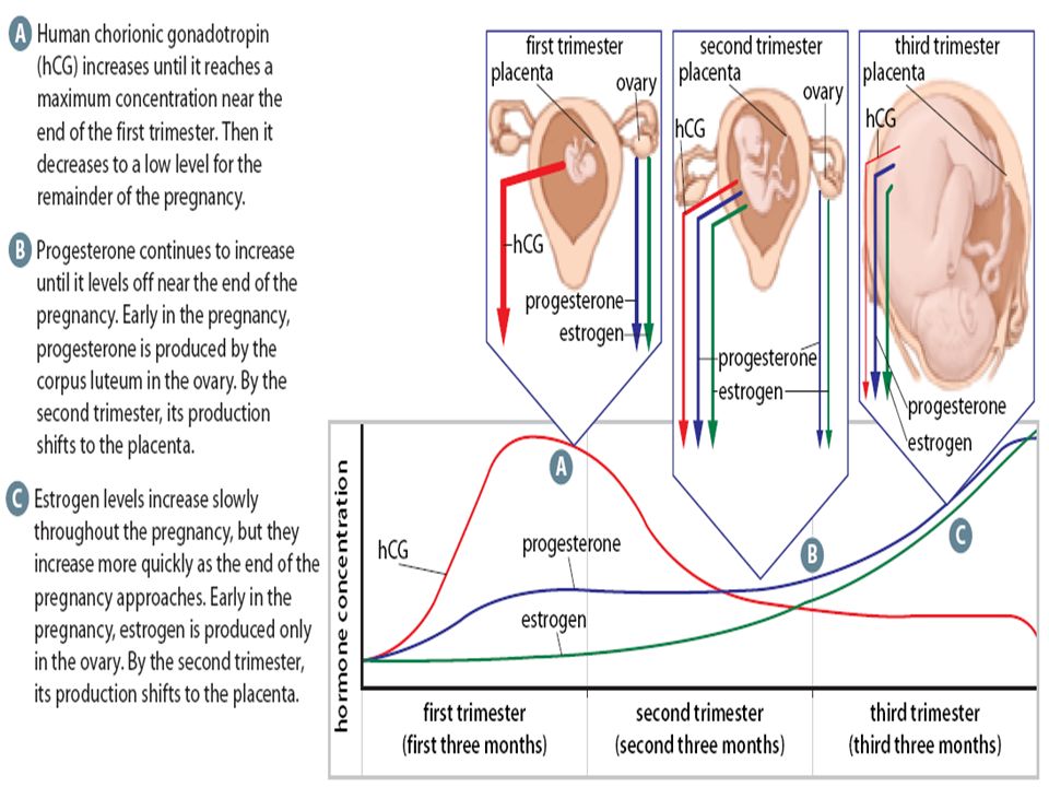
When implantation begins the trophoblast starts to secrete human chorionic gonadotropin hormone ( ) hCG has the same effects as so will maintain the corpus luteum The corpus luteum will secrete progesterone and estrogen to maintain the endometrium and prevent menstruation
 hCG has the same effects as so will maintain the corpus luteum The corpus luteum will secrete progesterone and estrogen to maintain the endometrium and prevent menstruation")
17
Gastrulation and Tissue Formation 2 nd week : a space forms between the inner cell mass and the trophoblast ( ) Fills with fluid and is where the fetus will develop Shortly after this the inner cell mass will form into three layers ()
 Fills with fluid and is where the fetus will develop Shortly after this the inner cell mass will form into three layers ()")
18
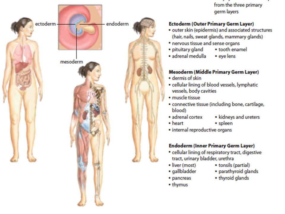
Gastrulation The blastocyst develops three layers ( ) and is now called the gastrula: Germ LayerPositionMajor Systems/Organs EctodermOutermost-Nervous system - Middle-Skeleton -Muscles -Reproductive Endoderm-Digestive -Respiratory -Endocrine glands
 and is now called the gastrula: Germ LayerPositionMajor Systems/Organs EctodermOutermost-Nervous system - Middle-Skeleton -Muscles -Reproductive Endoderm-Digestive -Respiratory -Endocrine glands")
20
Gastrulation marks the start of A series of events that form distinct structures of the organism Makes the different cells of the body () ()
 ()")
21
Differentiation Cleavage results in some cells becoming as a result of differential activation or of genes. Each cell contains a set of functioning genes Embryology

22
Neurulation Is part of in vertebrates and forms the

23
Neurulation and Organ Formation Between the 3 rd and 8 th weeks the form 3 rd week – band of cells start forming a The nervous system forms from the By day the heart starts beating

25
4 th week – growth, blood cells start to form and fill blood vessels, and kidneys take shape, is visible, arm and leg buds are visible 5 th week – head is, eyes open () 6 th week – brain continues development, limbs lengthen, start producing hormones
 6 th week – brain continues development, limbs lengthen, start producing hormones")
26
7 th & 8 th weeks –human characteristics, organs are formed, eyes are well developed, nostrils are developed By eight weeks the embryo is the size and mass of a paper clip but has approximately of all of its organs Embryo is now called a

27
At the same time as the internal structures are forming, a series of membrane structures are also forming: – The allantois, amnion, chorion and yolk sac These membranes and the embryo These membranes are after the baby is born – often called

28
Support structures (extra embryonic membranes) Amnion - thin sac filled with () protects developing fetus from
 Amnion - thin sac filled with () protects developing fetus from")
29
Amniocentesis Prenatal diagnosis of genetic or chromosomal disorders A sample of the amniotic fluid is collected and contained cells are tested for defect Can be carried out 16-20 weeks after the last period Video Clip

30
Chorion Develops into the fetal part of the

31
Chorionic Villus Sampling Prenatal diagnosis of genetic or chromosomal disorders A sample of the chorionic villi is collected and tested for defect Can be carried out 10- 13 weeks after the last period Video Clip

32
Support structures (extra embryonic membranes) Yolk sac - functions only in humans (NB in egg forming animals - nutrient yolk which is connected to embryos digestive tract)
 Yolk sac - functions only in humans (NB in egg forming animals - nutrient yolk which is connected to embryos digestive tract)")
33
Support Structures Allantois –forms the foundation of the

34
Support structures (extra embryonic membranes) Umbilical cord - connection between and Contains which transport blood from the fetus to the placenta Has which brings to the fetus
 Umbilical cord - connection between and Contains which transport blood from the fetus to the placenta Has which brings to the fetus")
36
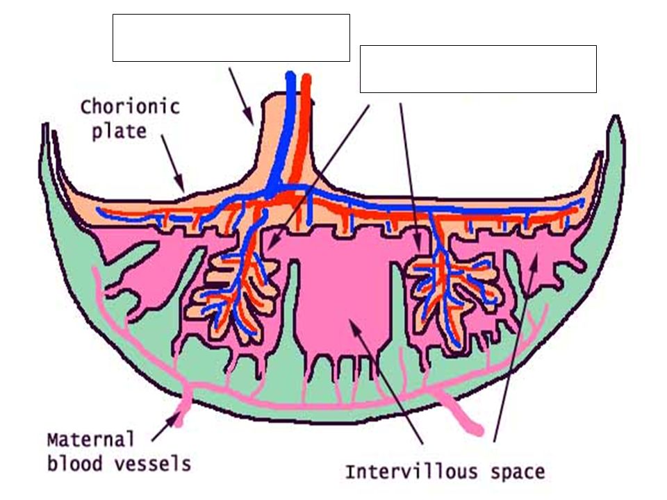
The Placenta Chorionic villi extend into the uterine wall within 2 weeks after Fertilization Cells from both the embryo and the mother make up the placenta It transports nutrients,, oxygen, and antibodies from the mother’s blood to the fetus’ blood and waste, carbon dioxide and hormones from the fetus to the mother It is fully developed by around the 10th week, and weighs about 600g It contains many blood vessels for

38
Placenta Produces a hormone called human chorionic gonadotropin (HCG) which stimulates the to continue producing hormones until the of pregnancy [presence of HCG is often used as an early pregnancy test] NOTE: EPT's are not always accurate! First Response EPT The Rabbit Died
![Placenta Produces a hormone called human chorionic gonadotropin (HCG) which stimulates the to continue producing hormones until the of pregnancy [presence of HCG is often used as an early pregnancy test] NOTE: EPT s are not always accurate.](http://images.slideplayer.com/35/10432639/slides/slide_38.jpg "First Response EPT The Rabbit Died.")
39
Keep in mind that there is of and blood! Nutrients diffuse from the mother’s blood vessels to the placenta, then from the placenta to the fetus’s blood vessels (wastes )
.")
40
Infectious diseases, alcohol, and some drugs cross the placental barrier and affect the developing fetus (FAS;SIDS)
")
41
In the placenta, chorionic villi that extend from the embryo are in contact with pools of blood from the mother. Nutrients and oxygen pass by diffusion from the maternal blood to the embryo, and wastes diffuse in the opposite direction.

42
Functions of the Placenta Nutritional functions transports nutrients (for example, glucose, amino acids, fatty acids, minerals, and vitamins) from the mother’s blood to the fetus’s blood stores nutrients, such as carbohydrates, proteins, iron, and calcium, in early pregnancy and releases them to the fetus later, when fetal demand is greater than the mother can absorb from her diet Excretory functions transports wastes (such as urea, ammonia, and creatinine) from the fetal blood to the mother’s blood Respiratory functions transports oxygen from the mother to the fetus and carbon dioxide from the fetus to the mother Endocrine functions secretes hormones, such as estrogen, progesterone, and human chorionic gonadotropin allows hormones from the fetus to diffuse into the mother’s blood and hormones from the mother to diffuse into the fetus’s blood Immune functions transports antibodies from the mother into the fetus’s blood to provide passive immunity
 from the mother’s blood to the fetus’s blood stores nutrients, such as carbohydrates, proteins, iron, and calcium, in early pregnancy and releases them to the fetus later, when fetal demand is greater than the mother can absorb from her diet Excretory functions transports wastes (such as urea, ammonia, and creatinine) from the fetal blood to the mother’s blood Respiratory functions transports oxygen from the mother to the fetus and carbon dioxide from the fetus to the mother Endocrine functions secretes hormones, such as estrogen, progesterone, and human chorionic gonadotropin allows hormones from the fetus to diffuse into the mother’s blood and hormones from the mother to diffuse into the fetus’s blood Immune functions transports antibodies from the mother into the fetus’s blood to provide passive immunity")
43
Hormones Secreted During Pregnancy Fig. 15.5

44
Gestation Total time embryo in uterus (in humans from beginning of last period) http://www.babycenter.com/2_inside-pregnancy-labor-and-birth_3658872.bc#videoplaylist
")
47
Twins

48
TERATOGENS Anything that causes a abnormality due to exposure during pregnancy The first seem to be the time – Examples include: – Thalidomide – Cigarette smoke – Alcohol – Drugs (including some prescription drugs)
")
49
Thalidomide Babies Powerful teratogen sold from 1957 to 1961 in almost 100 countries under at least 40 names – helped with morning sickness and sleep 1956 to 1962, approximately 10,000 children in Africa and Europe were born with severe malformations Impact in the United States was minimized by FDA blockage – only 17 children in the U.S. were born with the defects. Currently used as leprosy & cancer treatment (also for some mental illness)
.")
50
FASD – Fetal Alcohol Spectrum Disorder Alcohol causes in the developing baby. THERE IS OF ALCOHOL DURING PREGNANCY. There is also no safe TIME to drink alcohol during pregnancy Common problems include having trouble : – Adding, and handling money – Thinking things through / / remembering things – Learning from experience /understanding of their actions – Getting along with others.

52
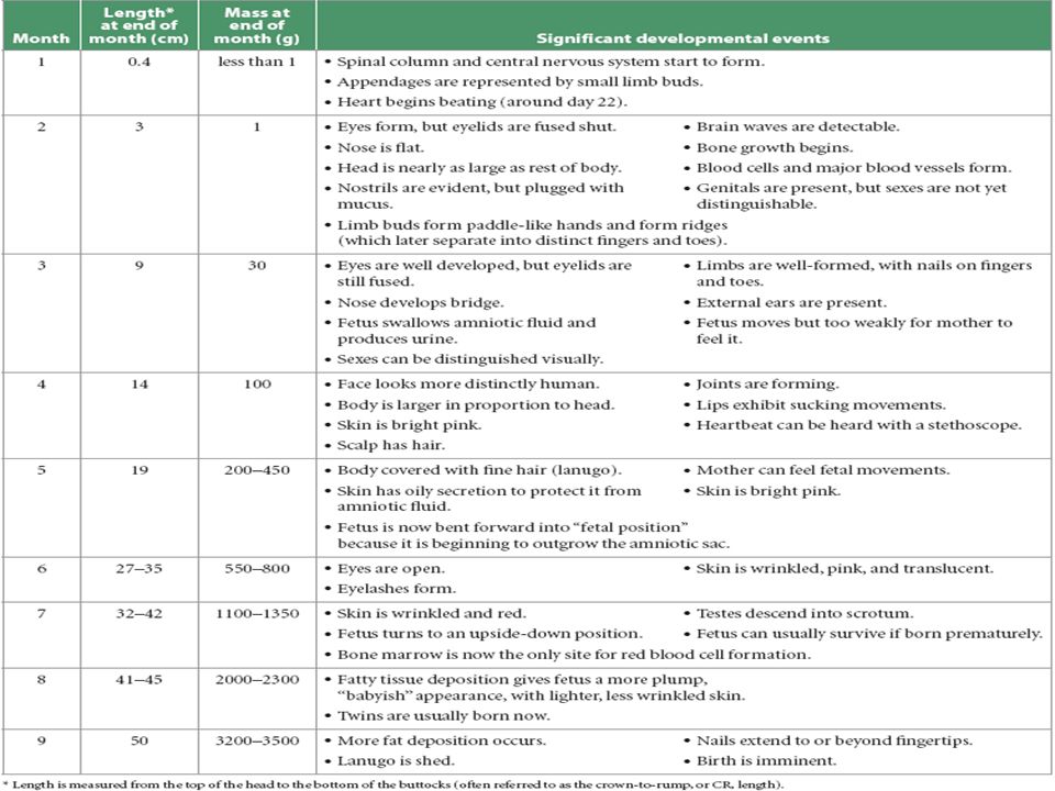
Critical Phases of Prenatal Development

53
Your Task Practice Question 1 on page 532 Practice Questions 2-3 on page 536 Complete a full response to the Statement Question on page 538-539

Similar presentations


>")


