Download presentation
Presentation is loading. Please wait.

1
Pleural Disease

2
Pneumothorax Classification Spontaneous Acquired PSP SSP Blunt trauma
Penetrating trauma Barotrauma Iatragenic injury

3
Pathophysiology

4
Primary Spontaneous Pneumothorax (PSP)
Young men (teens – mid 20s) Tall and thin Families Smokers Secondary Spontaneous Pneumothorax (SSP) Older people (45 – 64 years) Pre-existing lung disease Higher incidence of respiratory failure Higher mortality rate
 Tall and thin. Families. Smokers. Secondary Spontaneous Pneumothorax (SSP) Older people (45 – 64 years) Pre-existing lung disease. Higher incidence of respiratory failure. Higher mortality rate.")
5
Clinical presentation
1. Asymptomatic 2. Symptomatic a. Pain (PSP) b. Dyspnea (SSP) c. Orthopnea, hemoptysis, cough
 b. Dyspnea (SSP) c. Orthopnea, hemoptysis, cough.")
6
On examination Inspection: Dyspnea ± cyanosis
Decrease or absence chest wall movement Palpation: Apex shifted to the other side Trachea shifted to the other side Decreased chest wall expansion Decreased or absent tactile vocal fremitus Percussion: Hyper-resonance (tympanic) Auscultation: Decrease or absent breath sounds
 Auscultation: Decrease or absent breath sounds.")
7
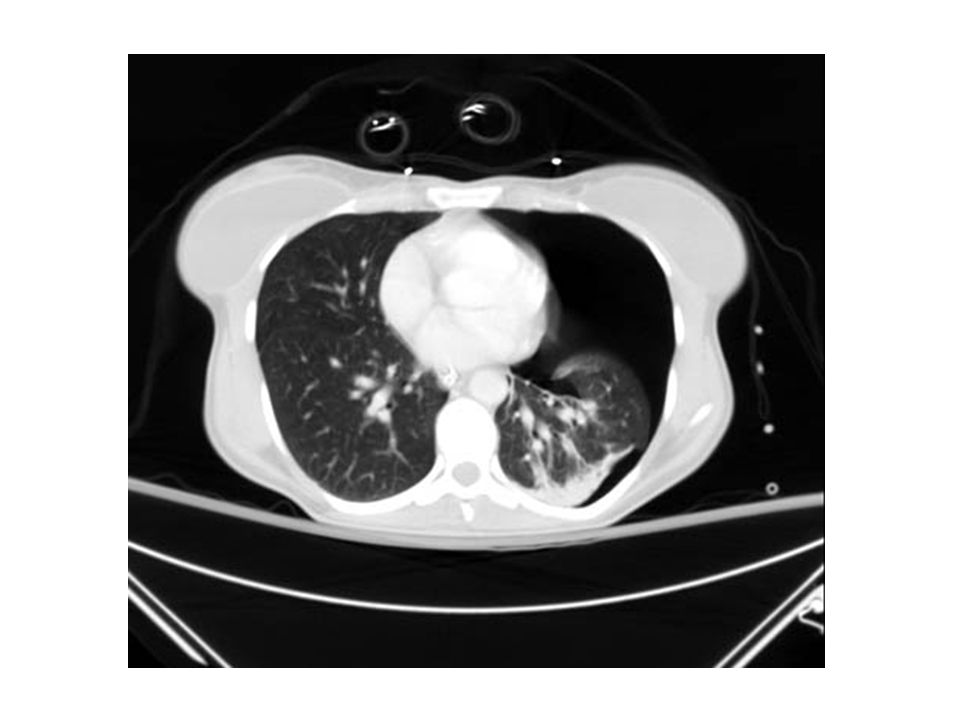
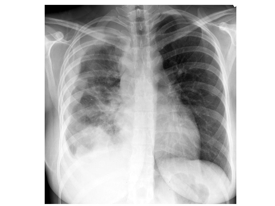
CXR: closed Pneumothorax

8
Tension Pnenmothorax

9
What is the difference??

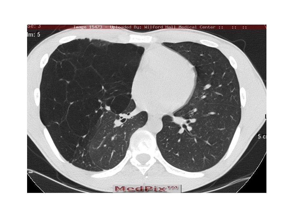
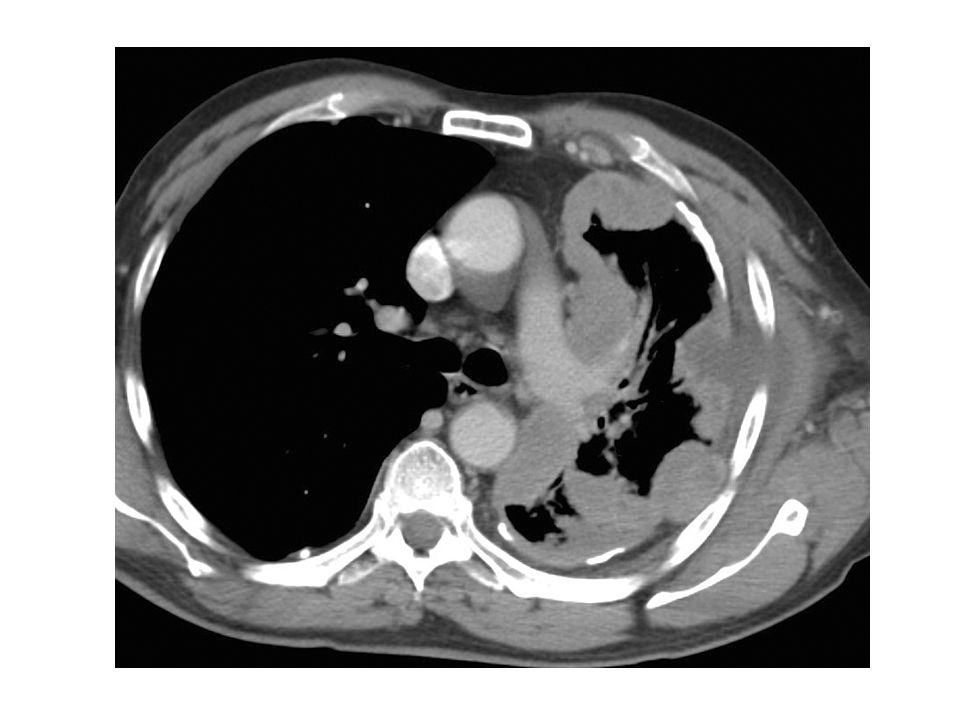
14
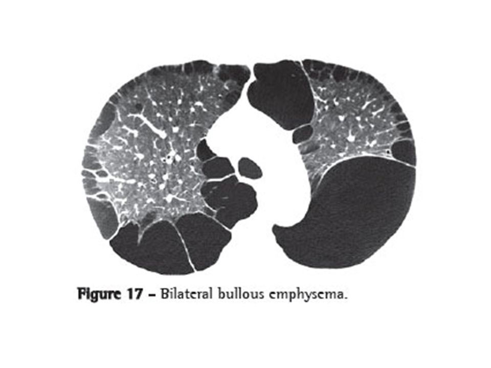
CT scan Bronchoscopy Emphysema Pneumothorax

15
Complications Pleural effusion
Hemothorax due to torn pleural adhesions Empyema Trapped lung (fibrothorax) due to failure of re-expansion Tension pneumothorax
 due to failure of re-expansion. Tension pneumothorax.")
16
Treatment Observation Pleurocenthesis or thoracocenthesis
Chest tube thoracostomy Surgery Massive air leak Persistent air leak Recurrent pneumothorax Bilateral pneumothorax Previous pneumonectomy Occupational hazard Pleurodesis

17
Malignant pleural effusion
Causes lung cancer pleural malignancy mediastinal LN malignancy Treatment of pleural effusion→physician or oncologist When to reffer to a surgeon →recurrent &/or a suspicion of being a malignant effusion Why reffer →biopsy + prevent reccurence of effusion

18
Biopsy: Prevent recurrence Cytology of pleural effusion Abram's needle
CT or U/S guided biopsy VATS biopsy Open biopsy Prevent recurrence Repeated thoracocenthesis Chemical pleurodesis Surgical pleurodesis/pleurectomy Pleuro-peritoneal shunt (Denver's shunt)
")
19
Denver’s Shunt

20
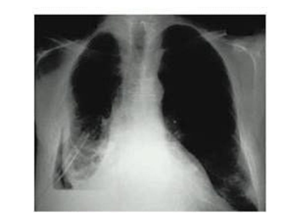
Whole lung atelectasis
What is the difference?? Whole lung atelectasis Massive effusion

21
Empyema Causes: Complication of pulmonary infection
Following chest trauma Extrapulmonary spread Complication of pneumothorax Non sterile aspiration of pleural fluid

22
Pathogenesis 1. Acute or exudative phase
Thin pus, Thin pleura, Expandable lungs Antibiotics and drainage 2. Transitional or fibrinopurulent phase Pus thicker, fibrin deposition, lung less expandable 3. Chronic or organization phase Thick pus, thick pleura with fibrous coat, non expandable lungs Surgery (decortication)
")
23
Clinical presentation
fever, maliase, anorexia, weight loss pleuretic chest pain dyspnea, cough, purulent sputum O/E: signs of infection + signs of pleural effusion Investigations: CXR Thoracocenthesis CT scan

24
CXR:

26
Treatment Objectives: Treatment line
Control infection Drain purulent material Restore lung function Treatment line Chest tube thoracostomy + physiotherapy Decortication / pleurectomy Open drainage e.g., Eloesser's flap

27
Eloesser’s flap Decortication

28
Chylothorax Etiology:
Disruption or tear in the thoracic duct during its coarse in the chest. Trauma including penetrating, blunt or iatrogenic injury Neoplasm with invasion of the thoracic duct Infection

29
Clinical presentation
Dyspnea, orthopnea, and cough Malnutrition Dehydration Decreased immunity Investigations: 1. Pleural fluid analysis: odorless milky white appearance with a creamy layer on standing. 2. Lymphangiography

30
Treatment 1. Conservative treatment a. Chest tube drainage
b. Correct dehydration c. Correct electrolyte imbalance d. Nutritional support by TPN or fat free oral diet 2. Surgical repair or ligation of thoracic duct 3. Denver's shunt (pleuro-peritoneal shunt)
")
31
Pleural malignancy Primary pleural tumors are rare
The most common primary tumor malignant mesothelioma (usually as a consequence of asbestos exposure). Poor prognosis Respiratory failure or symptoms of invasion of nearby organ. CXR: lung surrounded by thick irregular pleura with multiple nodules with extension to nearby structures. Curative treatment is surgery (extrapleural pneumonectomy). Both radiotherapy and chemotherapy are weakly effective.
. Poor prognosis. Respiratory failure or symptoms of invasion of nearby organ. CXR: lung surrounded by thick irregular pleura with multiple nodules with extension to nearby structures. Curative treatment is surgery (extrapleural pneumonectomy). Both radiotherapy and chemotherapy are weakly effective.")
34
Thank You

Similar presentations


, FCCP>")


. -Cytological tests (>")


