Download presentation
Presentation is loading. Please wait.

1
Sensory receptors are responsive to external and internal stimuli. Such sensory input is conveyed to integration centers where the sensory input is interpreted and associated with a response. Motor output is the conduction of signals from integration centers to effector cells. Effector cells (e.g. muscle, gland) carry out the body’s response to a stimulus. A. Nervous systems perform overlapping functions
 carry out the body’s response to a stimulus. A. Nervous systems perform overlapping functions.")
3
A Simple Nerve Circuit – the Reflex Arc. A reflex is an autonomic response.

4
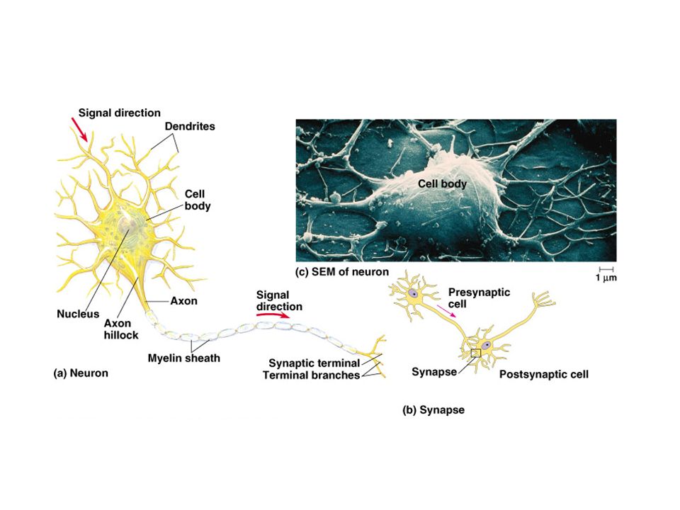
The neuron is the structural and functional unit of the nervous system. Nerve impulses are conducted along a neuron. Dentrite cell body axon Some axons are insulated by a myelin sheath. Axon endings are called synaptic terminals and contain neurotransmitters which conduct a signal across a synapse. A synapse is the junction between a presynaptic and postsynaptic neuron. B. Neuron Structure and Synapses

6
Neurons differ in terms of both function and shape.

7
Three Types of Neurons in Humans Sensory Neurons (part of PNS) from sensory receptor to CNS long dendrites and short axons Motor Neurons (part of PNS) from CNS to effector short dendrites and long axons Interneurons ( part of CNS) short dendrites and long or short axons
 from sensory receptor to CNS long dendrites and short axons Motor Neurons (part of PNS) from CNS to effector short dendrites and long axons Interneurons ( part of CNS) short dendrites and long or short axons")
8
Supporting Cells (Neuroglia) 1.Astrocytes are found within the CNS. Structural and metabolic support. By inducing the formation of tight junctions between capillary cells astrocytes help form the blood-brain barrier. Like neurons, astrocytes communicate with one another via chemical signals. 2.Oligodendrocytes are found within the CNS. Form a myelin sheath by insulating axons.

9
3.Schwann cells are found within the PNS. Form a myelin sheath by insulating axons.

10
A membrane potential is a localized electrical gradient across membrane. C. Every cell has a voltage, or membrane potential, across its plasma membrane An un-stimulated cell usually has a resting potential of -70mV.

11
1.How a Cell Maintains a Membrane Potential. K + the principal intracellular cation. Moves through channel proteins in neuron membrane Na + is the principal extracellular cation. Moves through channel proteins in neuron membrane Proteins, amino acids, sulfate, and phosphate are the principal intracellular anions. Too large to leave axoplasm of neuron Cl – is principal extracellular anion. Moves through channel proteins in neuron membrane

12
Ungated ion channels allow ions to diffuse across the plasma membrane. These channels are always open. This diffusion does not achieve an equilibrium since a sodium-potassium pump transports these ions against their concentration gradients.

13
Neurons have the ability to generate large changes in their membrane potentials. Gated K + and Na + ion channels open or close in response to stimuli. The subsequent diffusion of K + and Na + ions leads to a change in the membrane potential: the creation of the action potential 2. Changes in the membrane potential of a neuron give rise to nerve impulses

14
The Action Potential: All or Nothing If potentials received by each dendrite sum to - 55mV a threshold potential is achieved. This triggers the creation of an action potential of + 40 mV in the axons only.

15
Step 1: Resting Potential.

16
Step 2: Threshold Potential.

17
Step 3: Depolarization phase of the action potential.

18
Step 4: Repolarization phase of the action potential.

19
Step 5: Undershoot or Refractory Period.

20
During the undershoot or refractory period, the Na + gates are closed. At this time the neuron cannot depolarize in response to another stimulus The sodium-potassium pump is at work re- establishing the resting potential ion gradients

21
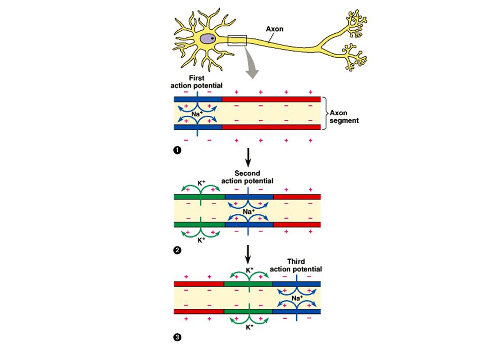
The action potential is repeatedly regenerated along the length of the axon. An action potential achieved at one region of the membrane is sufficient to depolarize a neighboring region above threshold. Thus triggering a new action potential. The refractory period assures that impulse conduction is unidirectional. 3. Nerve impulses propagate themselves along an axon

23
Saltatory conduction. In myelinated neurons only unmyelinated regions of the axon, called the nodes of Ranvier, depolarize. Thus, the impulse moves faster than in unmyelinated neurons.

24
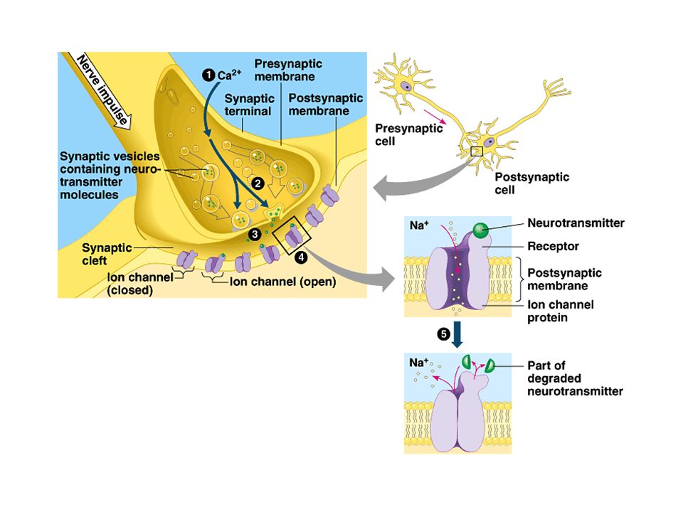
4. Chemical communication between cells occurs at synapses Postsynaptic chemically-gated channels exist for ions such as Na +, K +, and Cl -. Depending on which gates open, an influx of ions into the postsynaptic neuron can cause it depolarize,

26
Excitatory postsynaptic potentials (EPSP) depolarize the postsynaptic neuron. The binding of neurotransmitter to postsynaptic receptors open gated channels that allow Na + to diffuse into and K + to diffuse out of the cell. Inhibitory postsynaptic potential (IPSP) hyperpolarize the postsynaptic neuron. The binding of neurotransmitter to postsynaptic receptors open gated channels that allow K+ to diffuse out of the cell and/or Cl- to diffuse into the cell. 5. Neural integration occurs at the cellular level
 hyperpolarize the postsynaptic neuron. The binding of neurotransmitter to postsynaptic receptors open gated channels that allow K+ to diffuse out of the cell and/or Cl- to diffuse into the cell. 5. Neural integration occurs at the cellular level.")
27
Summation: potentials (EPSPs and IPSPs) are summed to either depolarize or hyperpolarize a postsynaptic neuron.
 are summed to either depolarize or hyperpolarize a postsynaptic neuron.")
28
1.Acetylcholine. Excitatory to skeletal muscle. Inhibitory to cardiac muscle. Secreted by the CNS, PNS, and at vertebrate neuromuscular junctions. 6. The same neurotransmitter can produce different effects on different types of cells

29
2.Epinephrine and norepinephrine. Can have excitatory or inhibitory effects. Secreted by the CNS and PNS. Secreted by the adrenal glands.

30
3.Dopamine Generally excitatory; may be inhibitory at some sites. Widespread in the brain. Affects sleep, mood, attention, and learning. Secreted by the CNS and PNS. A lack of dopamine in the brain is associated with Parkinson’s disease. Excessive dopamine is linked to schizophrenia.

31
4.Serotonin. Generally inhibitory. Widespread in the brain. Affects sleep, mood, attention, and learning Secreted by the CNS.

32
C. Vertebrate nervous systems have central and peripheral components Central nervous system (CNS). Brain and spinal cord. Both contain fluid-filled spaces which contain cerebrospinal fluid (CSF). The central canal of the spinal cord is continuous with the ventricles of the brain. White matter is composed of bundles of myelinated axons Gray matter consists of unmyelinated axons, nuclei, and dendrites. Peripheral nervous system. Everything outside the CNS.
. The central canal of the spinal cord is continuous with the ventricles of the brain. White matter is composed of bundles of myelinated axons Gray matter consists of unmyelinated axons, nuclei, and dendrites. Peripheral nervous system. Everything outside the CNS..")
33
Structural composition of the PNS. Paired cranial nerves that originate in the brain and innervate the head and upper body. Paired spinal nerves that originate in the spinal cord and innervate the entire body. Ganglia associated with the cranial and spinal nerves. The divisions of the peripheral nervous system interact in maintaining homeostasis

34
Functional composition of the PNS.

35
A closer look at the divisions of the autonomic nervous system (ANS).
.")
37
1.Medulla oblongata. Control autonomic homeostatic functions. Breathing. Heart and blood vessel activity. Swallowing. Vomiting. Digestion. Relays information to and from higher brain centers. Structures of the Brain

38
2.Pons. Involved in the regulation of breathing. Relays information to and from higher brain centers. 3.The Midbrain. Involved in the integration of sensory information. Relays information to and from higher brain centers.

39
4.The Cerebellum. Functions to error-check and coordinate motor activities, and perceptual and cognitive factors. Relays sensory information about joints, muscles, sight, and sound to the cerebrum. Coordinates motor commands issued by the cerebrum.

40
5.Thalamus. Relays all sensory information to the cerebrum. Relays motor information from the cerebrum. Receives input from the cerebrum. Receives input from brain centers involved in the regulation of emotion and arousal.

41
6.Hypothalamus. Regulates autonomic activity. Involved in thermoregulation, hunger, thirst, sexual and mating behavior, aggression, etc. Regulates the pituitary gland.

42
7. The cerebrum is the most highly evolved structure of the mammalian brain

43
The cerebrum is divided into left and right cerebrum hemispheres. The corpus callosum is the major connection between the two hemispheres. The left hemisphere is primarily responsible for the right side of the body. The right hemisphere is primarily responsible for the left side of the body. Cerebral cortex: outer covering of gray matter. Neocortex: region unique to mammals. The more convoluted the surface of the neocortex the more surface area the more neurons.

44
Lateralization of Brain Function. The left hemisphere. Specializes in language, math, logic operations, and the processing of serial sequences of information, and visual and auditory details. Specializes in detailed activities required for motor control. The right hemisphere. Specializes in pattern recognition, spatial relationships, nonverbal ideation, emotional processing, and the parallel processing of information.

45
The cerebrum is divided into frontal, temporal, occipital, and parietal lobes. Regions of the cerebrum are specialized for different functions

Similar presentations


. Brain and spinal cord. Both contain fluid-filled spaces which contain cerebrospinal fluid (CSF). The central canal of the.>")
 Nervous system functions Structure of a neuron Sensory, motor, inter- neurons Membrane potential Sodium.>")


