Download presentation
Presentation is loading. Please wait.

1
Female reproduction

5
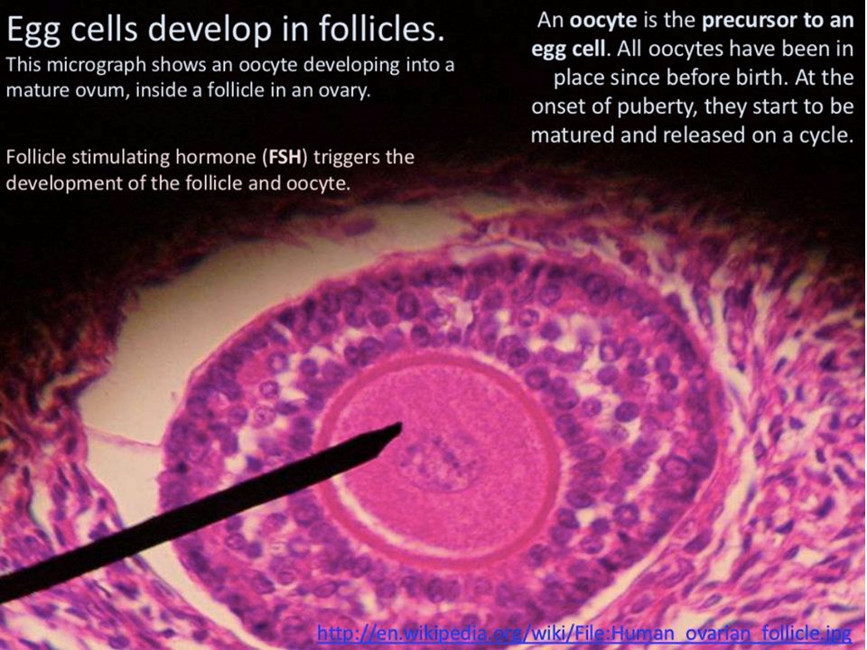
BIGPICTUREEDUCATION.COM Egg cell in follicle Light microscopy image of a transverse cross-section of an immature egg cell (oocyte) in a maturing follicle. Once a month, during the female menstrual cycle, an oocyte matures in one of the many ovarian follicles. As the follicle matures it increases in size, and different cell types are recruited to the developing follicle to support the oocyte before ovulation. Credit: Dr Ivor Mason, King’s College London, Wellcome Images.

6
A human secondary oocyte (which will later become an egg cell) during in vitro fertilisation, viewed with a light microscope using Nomarski optics, a technique used to highlight cell structure. The small cell at the bottom left is a polar body. Credit: Spike Walker, Wellcome Images. BIGPICTUREEDUCATION.COM Secondary oocyte during in vitro fertilisation

8
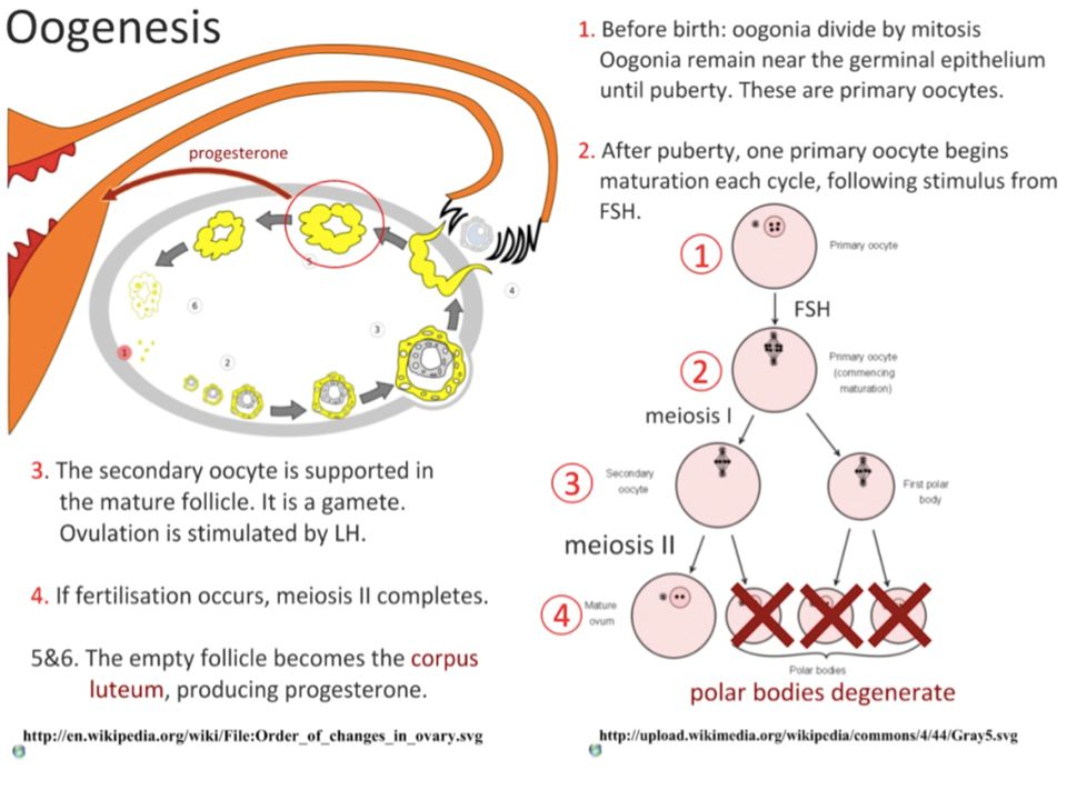
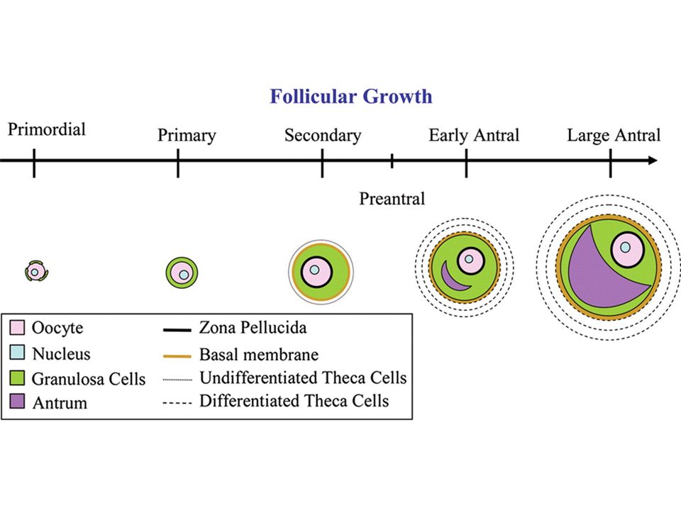
Oogenesis is quite different from spermatogenesis Oogenesis describes the production of female gametes (ova) within the ovary The process begins during foetal development, when a large number of cells (oogonia) are formed by mitosis before undergoing a period of growth These cells begin meiosis but are arrested in prophase I until puberty At puberty, some follicles continue to develop each month in response to FSH secretion These follicles complete the first meiotic division to form two cells of unequal size The cell with less cytoplasm is a polar body (which degenerates), while the larger cell forms a secondary oocyte The secondary oocyte begins the second meiotic division but is arrested in prophase II (until fertilisation) It is released from the ovary (ruptured follicle develops into corpus luteum) and, only if fertilisation occurs, will complete meiosis The second meiotic division will produce an ovum and a second polar body
 within the ovary The process begins during foetal development, when a large number of cells (oogonia) are formed by mitosis before undergoing a period of growth These cells begin meiosis but are arrested in prophase I until puberty At puberty, some follicles continue to develop each month in response to FSH secretion These follicles complete the first meiotic division to form two cells of unequal size The cell with less cytoplasm is a polar body (which degenerates), while the larger cell forms a secondary oocyte The secondary oocyte begins the second meiotic division but is arrested in prophase II (until fertilisation) It is released from the ovary (ruptured follicle develops into corpus luteum) and, only if fertilisation occurs, will complete meiosis The second meiotic division will produce an ovum and a second polar body")
13
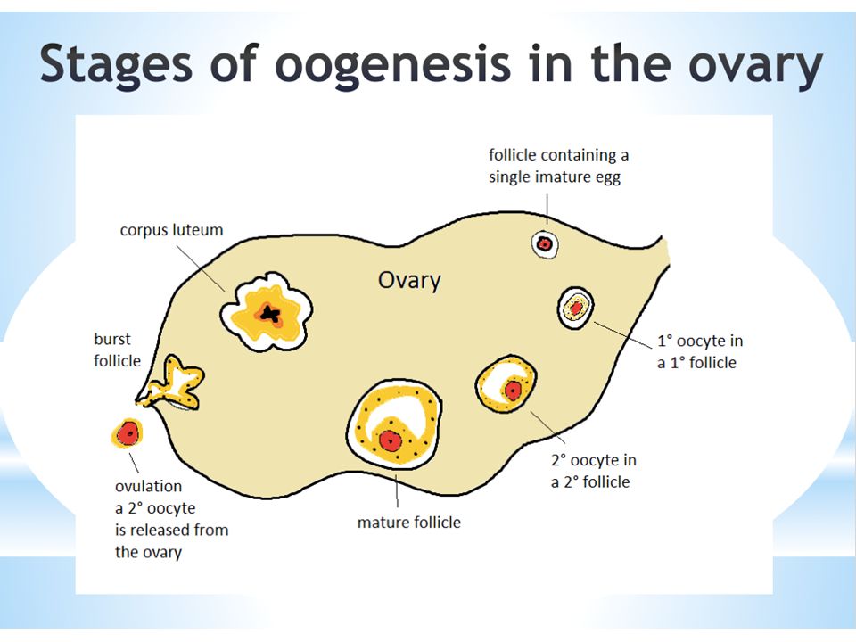
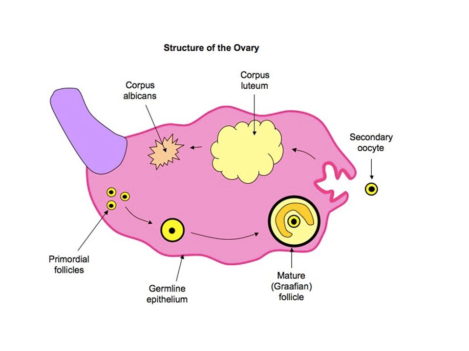
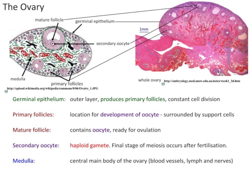
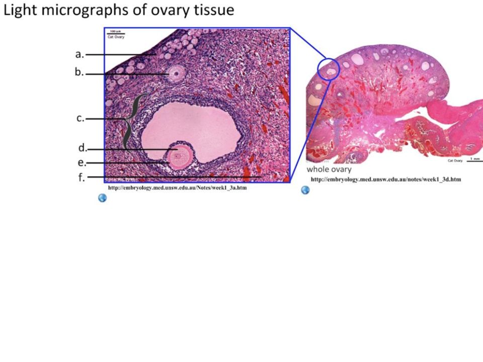
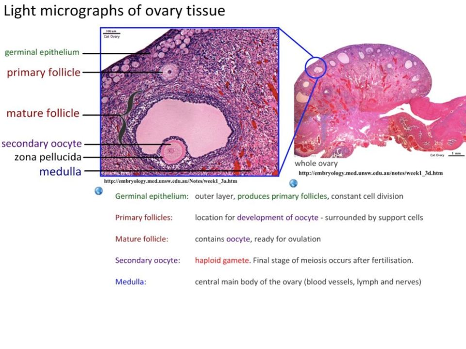
The ovary contains follicles in various stages of development Egg cells within primordial follicles have been arrested in prophase I of meiosis and have yet to undergo meiotic division Egg cells within mature follicles have begun meiotic division and are released from the ovary as secondary oocytes (arrested in prophase II) The ruptured follicle develops into a corpus luteum that will, in time, degenerate into a corpus albicans The germline epithelium functions as an epithelial layer separating ovarian tissue from the rest of the body - it is not involved in oocyte development
 The ruptured follicle develops into a corpus luteum that will, in time, degenerate into a corpus albicans The germline epithelium functions as an epithelial layer separating ovarian tissue from the rest of the body - it is not involved in oocyte development")
18
Structure of a mature oocyte

19
Light microscopy image showing sperm and an egg cell (or ovum) at the moment of conception during in vitro fertilisation. The egg is surrounded by protective cumulus cells around the outside surface, coloured yellow. The sperm need to penetrate these cells and the membrane surrounding the egg, called the zona pellucida, if successful fertilisation is to occur. Credit: Spike Walker, Wellcome Images. BIGPICTUREEDUCATION.COM Egg and sperm

20
Digital artwork showing intracytoplasmic sperm injection (ICSI). ICSI is a method of in vitro fertilisation (IVF) that is used to treat infertile couples when standard IVF techniques are not likely to be successful. ICSI is the process of injecting a single sperm cell directly into the egg; it is normally used when the male has a low sperm count or sperm motility is low and fertilisation is unlikely to occur naturally. This illustration shows the egg cell (ovum) being held at the end of a micropipette. The egg is surrounded by cumulus cells, which provide nutrients to the egg. Credit: Maurizio De Angelis, Wellcome Images. BIGPICTUREEDUCATION.COM Intracytoplasmic sperm injection
 that is used to treat infertile couples when standard IVF techniques are not likely to be successful. ICSI is the process of injecting a single sperm cell directly into the egg; it is normally used when the male has a low sperm count or sperm motility is low and fertilisation is unlikely to occur naturally. This illustration shows the egg cell (ovum) being held at the end of a micropipette. The egg is surrounded by cumulus cells, which provide nutrients to the egg. Credit: Maurizio De Angelis, Wellcome Images. BIGPICTUREEDUCATION.COM Intracytoplasmic sperm injection.")
21
A false-colour scanning electron micrograph of a human egg cell (gold) surrounded by cumulus cells (orange). Cumulus cells are specialised cells that nourish the large egg cell while it grows in the ovarian follicle. Credit: Yorgos Nikas, Wellcome Images Human egg

22
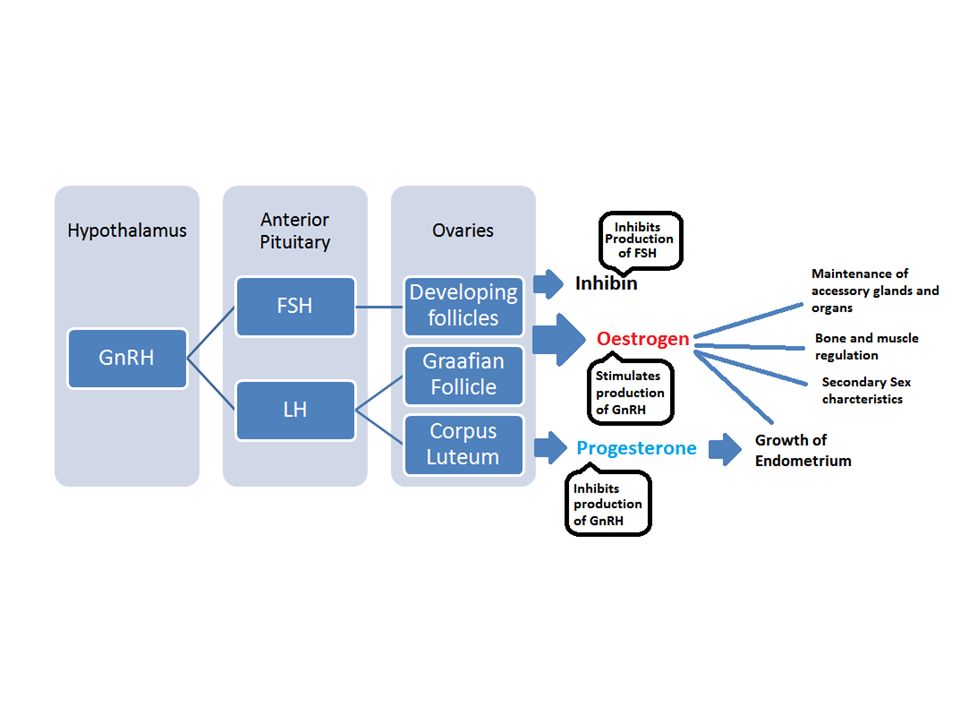
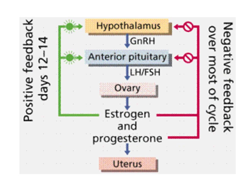
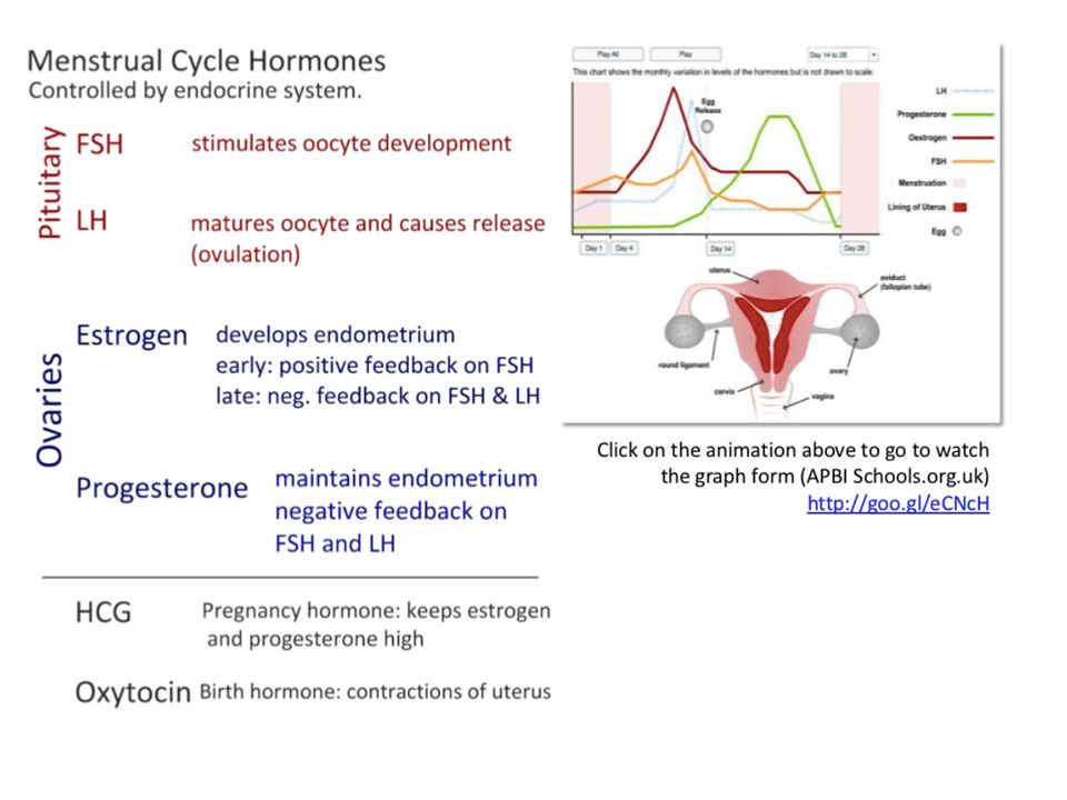
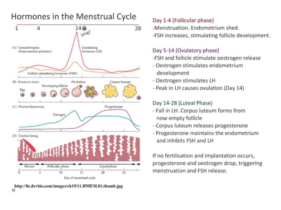
Female hormones

Similar presentations


 Gametogenesis Process of gamete formation with the reduction by half.>")


 produce sperm. Female gonads (ovaries) produce eggs.>")

