Download presentation
1
Integumentary System Part 2 Accessory Organs of the Skin

2
Nails Protective coverings on the ends of the fingers and toes The nail bed is covered with a nail plate Lunula-base of each nail; most active growing region Specialized epidermal cells that are keratinized make up nails The keratin of nails is harder than the skin’s epidermal cells The thumbnail grows the slowest and the middle nail grows the fastest. Fg. 6.4 page 117

3
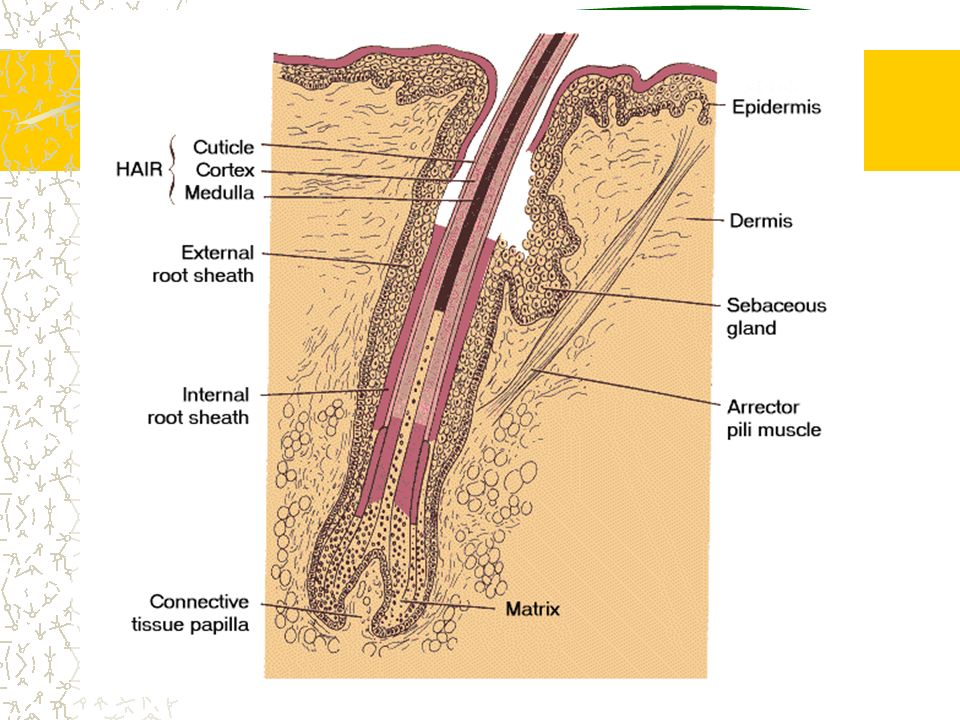
Hair Follicles Areas with no hair: palms, soles, lips, nipples, parts of the external reproductive organs Each hair develops from epidermal cells at the base of a tube-like hair follicle. Hair root is at the base: these cells are nourished by BV’s in the dermis As newly formed cells develop and grow, older cells are pushed toward the surface and undergo keratinization. Hair shaft is dead epidermal cells

5
Hair Color Genes determine hair color by directing the type and amount of pigment that epidermal melanocytes produce. Melanocytes are at the base of the follicle Lot of pigment-dark hair Intermediate amount of pigment-blond hair No pigment-white hair Trichosiderin pigment is produced in people with red hair Gray hair is a mixture of pigmented and unpigmented hair

6
“Goose Bumps” Arrector pili muscles attach to each hair follicle Nerve impulses cause the muscles to contract When muscles contract the hair stands up

7
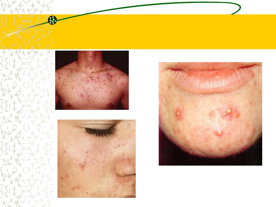
Sebaceous Glands Specialized epithelial cells associated with hair follicles Secrete sebum: a mixture of fatty material and cellular debris Helps keep hair soft, pliable, and waterproof Acne-disorder of sebaceous glands

10
Sweat Glands (sudoriferous glands) Coiled tube in the subcutaneous layer-has a long tube that extends to a pore on the surface of skin Lined with sweat secreting epithelial cells Eccrine glands: most numerous sweat glands; respond to temp. and physical exercise; common on the forehead, neck and back Apocrine glands: active at puberty; respond to being emotionally upset, frightened, or in pain; numerous in the axillary and groin area Sweat is mostly water, but has salt and wastes (urea and uric acid) also
 also.")
11
Modified Sweat Glands Ceruminous glands of ear secrete wax Mammary glands secrete milk

12
Regulation of Body Temperature Vital because heat affects the rates of metabolic reactions Normal body temp. 98.6 When body temp. rises above the normal set point, dermal BV’s dilate, and sweat glands secrete sweat If body temp. drops below normal, dermal bv’s constrict, sweat glands become inactive Extensive heat loss stimulates skeletal muscles to contract involuntarily 80% of body heat is lost through the head

13
Skin Color Genetic Factors All people have the same number of melanocytes (dark skin just produces more melain) Environmental Factors: Sunlight, UV, X-rays (stimulate melanocytes to produce more melanin) Physiological Factors: Blood in dermal BV’s (oxygenated vs deoxygenated) Dilation or constriction of BV’s Presence of pigments (carotene-jaundice)
 Environmental Factors: Sunlight, UV, X-rays (stimulate melanocytes to produce more melanin) Physiological Factors: Blood in dermal BV’s (oxygenated vs deoxygenated) Dilation or constriction of BV’s Presence of pigments (carotene-jaundice)")
14
Healing of Wounds Skin injuries trigger inflammation. The affected area becomes red, warm, swollen and tender Fg. 6.13 page 186 Dividing epithelial cells fill in shallow cuts in the epidermis. Clots close deeper cuts, sometimes leaving a scar where CT replaces the skin (scab forms) Granulations form in large, open wounds as part of the healing process
 Granulations form in large, open wounds as part of the healing process.")
15
Burns First degree: minor sunburn, skin warm and reddened; dermal bv’s dilate; heals in a few days to 2 weeks; no scarring Second degree: destroys some epidermis and some dermis; blisters form Third degree: destroys epidermis, dermis, and accessory organs; sometimes requires skin grafts

16
“Rule of Nines” Fg. 6.14 page 187 Skin’s surface is divided into regions (each accounts for 9% or some multiple of 9% of the total surface area Estimate is important-how much body fluid and electrolytes to replace from injured tissues

17
Types of Membranes p. 162 Serous-line body cavities that lack openings to the outside; secrete serous fluid which lubricates membrane surfaces Mucous membranes-line cavities and tubes that open to the outside; secrete mucus Cutaneous membrane-the skin Synovial membrane-lines joints

18
Skin Cancer Skin cancer cells develop from nonpigmented epithelial cells Cutaneous carcinomas are most commonly caused by exposure to UV light People most likely to develop cutaneous carcinomas: over 40, light skin, work and play in sunlight Cutaneous melanomas are commonly caused by short exposure to high intensity sunlight

19
A,B,C,D’s of Skin Cancer A=assemetry B=border C=color D=diameter

20
Things to read in your text: Pressure ulcers (p. 171) Psoriasis (p. 173) Sunburn (p. 174) Skin Cancer (p. 175) Contact Dermatitis (p. 176) Tatto (p. 176) Alopecia (p. 178) Life Span Changes (p. 187-188)
 Sunburn (p. 174) Skin Cancer (p. 175) Contact Dermatitis (p. 176) Tatto (p. 176) Alopecia (p. 178) Life Span Changes (p ).")


>")

 Structure: Hair follicle- organs producing.>")


 1. Physical protection 2. Temperature Regulation 3. Protects against water loss 4. Excretion 5. Synthesis.>")

