Download presentation
Presentation is loading. Please wait.

1
Cytology of Body Fluid Pleural peritoneal pericardial
Visceral and parietal Mesothelial cells Tissue culture medium Normal: minimal fluid

3
Pathology of Body Cavity
Air; - pneumothorax - pneumoperitoneum - pneumopericardium Effusion; - pleural effusion - peritoneal effusion (ascites) - pericardial effusion
 - pericardial effusion.")
4
Pathology of Body Cavity
Transudate (여출액); Filtration of serum due to ↑osmotic pressure of circulating blood (hypoproteinemia, LC, kidney and heart dis. …) Exudate (삼출액); Active loss of blood component due to ↑vascular permeability of damaged capillaries (inflammation, cancers …)
; Filtration of serum due to ↑osmotic pressure of circulating blood. (hypoproteinemia, LC, kidney and heart dis. …) Exudate (삼출액); Active loss of blood component due to. ↑vascular permeability of damaged capillaries. (inflammation, cancers …)")
5
Transudate Exudate Gross Clear Turbid Specific gravity < 1.015 > 1.015 Protein < 3g/dl > 3g/dl LDH < serum > serum Fibrin clot Absent Present Cell Few; Mesothelial Many; Mesothelial, WBC, RBC, tumor cells…

6
Transudate Exudate Exudate

7
NL Cell Components in Effusion
Mesothelial cells Macrophages Leukocytes

8
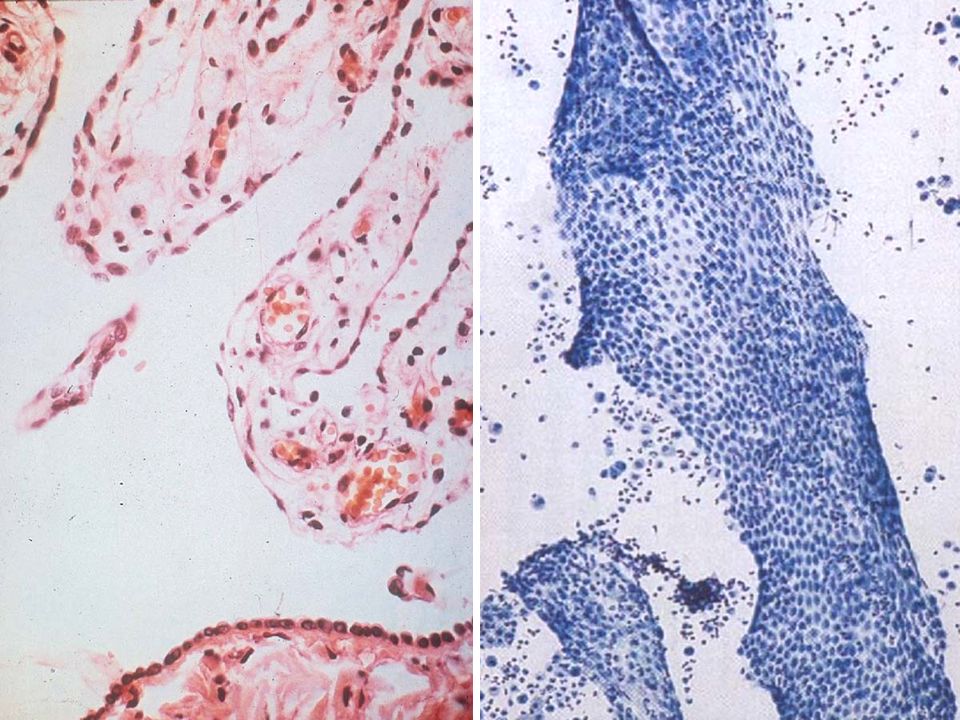
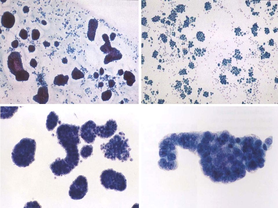
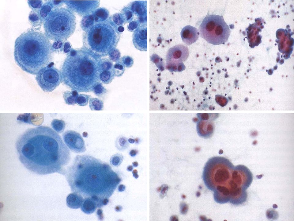
1. Mesothelial Cells Almost absent in normal state
Sheets composed of polygonal cells when scraped from the surface Mesothelial reaction → mesothelial hyperplasia Mesothelial cells (about 20㎛); - well-demarcated cytoplasm - Single or aggregated - Rounded cells with large round & central nuclei - Chromatin: finely granular Reactive mesothelial cells; coarse, irregular chromatin with prominent nucleoli
; - well-demarcated cytoplasm. - Single or aggregated. - Rounded cells with large round & central nuclei. - Chromatin: finely granular. Reactive mesothelial cells; coarse, irregular chromatin with prominent nucleoli.")
10
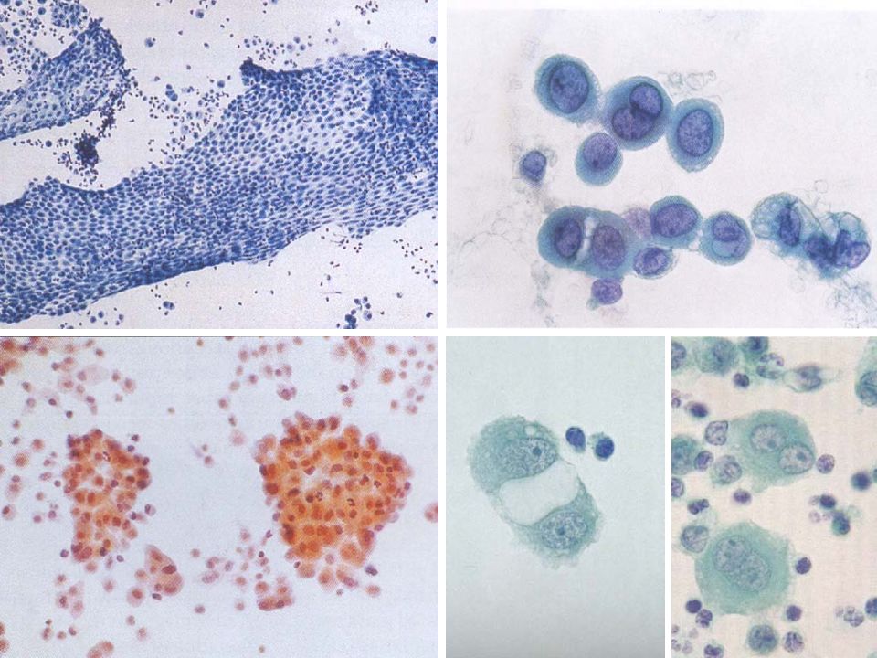
Peripheral foaminess Vacuolated mesothelial cells

14
Characteristics of Mesothelial Cell Clusters
Presence of intercellular windows (cytoplasmic molding) Absence of common cell borders; irregular margins of cell clusters with individual cell borders Pseudo-acinus
 Absence of common cell borders; irregular margins of cell clusters with individual cell borders. Pseudo-acinus.")
15
Mesothelial cells Adenocarcinoma

16
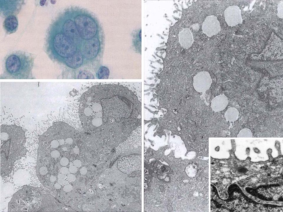
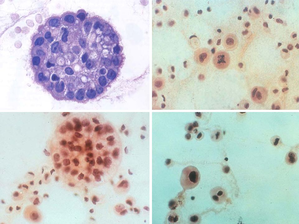
2. Macrophages Cell size similar to mesothelial cells (10~20 ㎛)
Single or loose aggregates, but no cytoplasmic molding Foamy cytoplasm Indistinct cytoplasmic borders Eccentric nuclei with frequent kidney shape and multi-nucleated

17
Cytokeratin MØ Cytokeratin CD-68

18
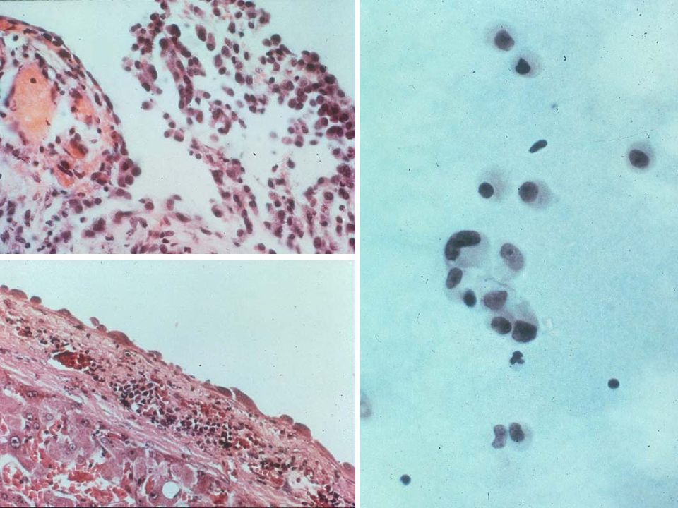
3. Leukocytes Lymphocytes; present frequently, numerous lymphocytes in lymphoma, leukemia, and Tbc PMN cells; indicates inflammatory reaction Eosinophils; allergic disease, asthma, cancer, pneumothorax, idiopathic in 40% of cases Plasma cells; chronic inflammation, multiple myeloma, Hodgkin’s disease

19
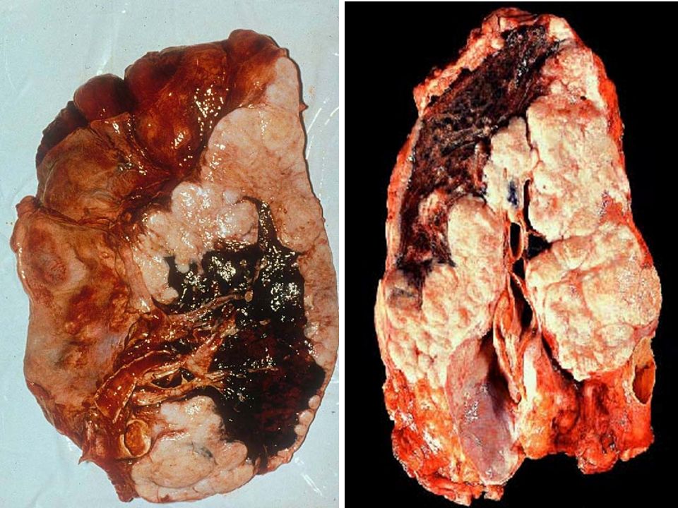
Mesothelioma Most common in pleura Asbestos exposure Carcinomatous,
Fibrosarcomatous, and mixed (biphasic)
")
21
Calretinin

22
Mesothelioma Cytology; Profuseness of cells in body cavity smear
Mesothelial cell clusters, frequent Larger and irregular clusters than reactive Full spectrum of mesothelial cells from malignant mesothelial cells to reactive and normal mesothelial cells

26
Criteria for Mesothelial Differentiation
Lack of a ‘foreign’ population Structure of cell aggregates Cytoplasmic characteristics Cell-to-cell relationships Vacuoles Collagen basement membrane Glycogen

27
Structure of Cell Aggregates
Mesothelioma, cuboidal cells with rounded nuclei Adenocarcinoma, columnar cells with elongated nuclei

28
Cytoplasmic Characteristics
Usually abundant, dense and basophilic Often shows a tinctorial gradation from green at the periphery to reddish orange at the peri-nuclear zone

29
Cell-to-Cell relationships
Brush-like border of microvilli “Window” effect between adjacent cells Cell engulfment

30
Characteristics of Mesothelial Cell Clusters
Presence of intercellular windows (cytoplasmic molding) Absence of common cell borders; irregular margins of cell clusters with individual cell borders Pseudo-acinus → Cell culture effect
 Absence of common cell borders; irregular margins of cell clusters with individual cell borders. Pseudo-acinus. → Cell culture effect.")
32
Immuno-profiles of Malignant Mesothelioma
Cytokeratin Calretinin WT1 TTF-1

33
Metastatic Adenocarcinoma
Lung Carcinoma CEA TTF-1 Cytokeratin Calretinin

34
Adenocarcinoma, CEA Mesothelioma, CEA EMA Calretinin CK 5/6

35
Metastatic Cancer Primary Sites of Metastatic Cancer of Body Cavities in Adults Body cavity Male Female Pleural Lung GI tract Lymphoma Breast Ovary Peritoneal Pericardial

36
Pleural, adenocarcinoma
forming cell clumps, lung primary Ascites, serous adenocarcinoma, ovary primary

37
Pleural fluid; adenocarcinoma, breast primary, ductal type

38
Ascites; adenocarcinoma, breast primary, lobular type

39
Metastatic Ca from breast

40
Malignant Lymphoma; typically do not form clumps, karyorrhectic nuclei (mercury-drop appearance), often need immunohistochemistry for definite diagnosis

Similar presentations


 Dr. Essam H. Jiffri.>")

 The fluid is a plasma filtrate.>")


 and intercellular.>")

