Download presentation
Presentation is loading. Please wait.

1
Exam 1 Section 2 ATHT 205

3
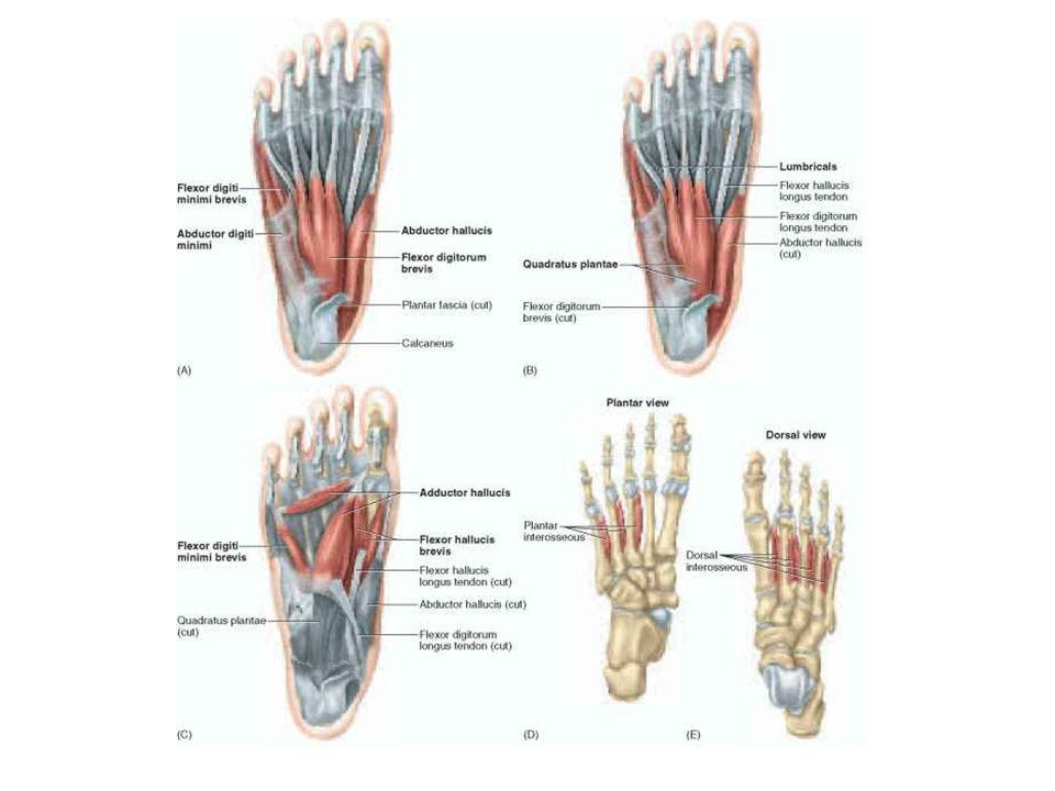
Layers of muscles 1-Superficial – abduct 1 st toe, abduct 5 th toe, flex toes 2-5 2- middle- changes angle of pull for flexor digitorium longus and and lumbricals 3 rd - deep- secondary flexors of 1 st and 5 th toes 4 th - interosseous abduct and adduct middle 3 toes

4
First layer MuscleProximal Attachment (origin) Distal Attachment (insertion) ActionNerve Abductor HallucisMedial process of calcaneal tuberosity Medial side of base of proximal phalax of great toe (hallux) Abducts and flexes great toe Medial plantar (S1, S2) Flexor digitorum brevis Medial process of calcaneal tuberosity Both sides of middle phalanges of lateral 4 digits Flexes lateral 4 digitsMedial plantar (S1, S2) Abductor digiti minimiMedial and lateral processes of calcaneal tuberosity Lateral side of the base of proximal phalax of 5 th digit Abducts and flexes 5 th digit Lateral plantar nerve (S2, S3) All extend from posterior part of calcaneous to phalanges; act as elastic spring to support arches and maintain foot concavity
 Distal Attachment (insertion) ActionNerve Abductor HallucisMedial process of calcaneal tuberosity Medial side of base of proximal phalax of great toe (hallux) Abducts and flexes great toe Medial plantar (S1, S2) Flexor digitorum brevis Medial process of calcaneal tuberosity Both sides of middle phalanges of lateral 4 digits Flexes lateral 4 digitsMedial plantar (S1, S2) Abductor digiti minimiMedial and lateral processes of calcaneal tuberosity Lateral side of the base of proximal phalax of 5 th digit Abducts and flexes 5 th digit Lateral plantar nerve (S2, S3) All extend from posterior part of calcaneous to phalanges; act as elastic spring to support arches and maintain foot concavity")
5
2 nd layer MuscleProximal Attachment (origin) Distal Attachment (insertion) ActionNerve Quadratus plantaeMedial surface and lateral margin of plantar surface of calcaneus (2 heads separated by long plantar ligament) Posterolateral margin of tendon of flexor digitorium longus Assists flexor digitorium longus in flexing lateral 4 digits Lateral plantar nerve (S2, S3) LumbricalsTendons of flexor digitorium longus Medial side of bases of proximal phalanges of lateral 4 digits and extensor expansions of tendons of flexor digitorium longus Flex proximal phalanges and extend middle and distal phalanges of lateral 4 digits Medial #2: medial plantar nerve (S2, S3) Lateral #3, 4, 5 Lateral plantar nerve (S2, S3)
 Distal Attachment (insertion) ActionNerve Quadratus plantaeMedial surface and lateral margin of plantar surface of calcaneus (2 heads separated by long plantar ligament) Posterolateral margin of tendon of flexor digitorium longus Assists flexor digitorium longus in flexing lateral 4 digits Lateral plantar nerve (S2, S3) LumbricalsTendons of flexor digitorium longus Medial side of bases of proximal phalanges of lateral 4 digits and extensor expansions of tendons of flexor digitorium longus Flex proximal phalanges and extend middle and distal phalanges of lateral 4 digits Medial #2: medial plantar nerve (S2, S3) Lateral #3, 4, 5 Lateral plantar nerve (S2, S3)")
6
3 rd Layer MuscleProximal Attachment (origin) Distal Attachment (insertion) ActionNerve Flexor Hallucis BrevisPlantar surfaces of cuboid and lateral cuneiforms Both sides of base of proximal phalanx of 1 st digit Flexes proximal phalanx of 1 st digit Medial plantar (S2,S3) Adductor hallucisOblique head: base of metatarsals 2-4 Transverse head: plantar ligaments of matatarsophalangeal joints Tendons of both heads attach to lateral side of base of proximal phalanx of 1 st digit Adducts 1 st digit; assists in maintaining transverse arch Deep branch of lateral plantar nerve (S2, S3) Flexor digiti minimiBase of 5 th metatarsal Base of proximal phalanx of 5 th digit Flexes proximal phalanx of 5 th Superficial branch of lateral plantar nerve (S2-S3)
 Distal Attachment (insertion) ActionNerve Flexor Hallucis BrevisPlantar surfaces of cuboid and lateral cuneiforms Both sides of base of proximal phalanx of 1 st digit Flexes proximal phalanx of 1 st digit Medial plantar (S2,S3) Adductor hallucisOblique head: base of metatarsals 2-4 Transverse head: plantar ligaments of matatarsophalangeal joints Tendons of both heads attach to lateral side of base of proximal phalanx of 1 st digit Adducts 1 st digit; assists in maintaining transverse arch Deep branch of lateral plantar nerve (S2, S3) Flexor digiti minimiBase of 5 th metatarsal Base of proximal phalanx of 5 th digit Flexes proximal phalanx of 5 th Superficial branch of lateral plantar nerve (S2-S3)")
7
4 th layer MuscleProximal Attachment (origin) Distal Attachment (insertion) ActionNerve Plantar interossei (PADs) (3 muscles) Bases and medial sides of metatarsals 3-5 Medial sides of bases of proximal phalanges of digits 3- 5 Adduct digits 3-5Lateral plantar nerve (S2, S3) Dorsal interossei (DABs) (4 muscles) Adjacent sides of metatarsals 1-5 1 st - medial side of proximal phalanx of 2 nd digit 2-4- lateral sides of 2-4 digits Abduct digits 2-4Lateral plantar nerve (S2, S3) Approximate bones during WB to maintain integrity of forefoot
 Distal Attachment (insertion) ActionNerve Plantar interossei (PADs) (3 muscles) Bases and medial sides of metatarsals 3-5 Medial sides of bases of proximal phalanges of digits 3- 5 Adduct digits 3-5Lateral plantar nerve (S2, S3) Dorsal interossei (DABs) (4 muscles) Adjacent sides of metatarsals st - medial side of proximal phalanx of 2 nd digit 2-4- lateral sides of 2-4 digits Abduct digits 2-4Lateral plantar nerve (S2, S3) Approximate bones during WB to maintain integrity of forefoot")
8
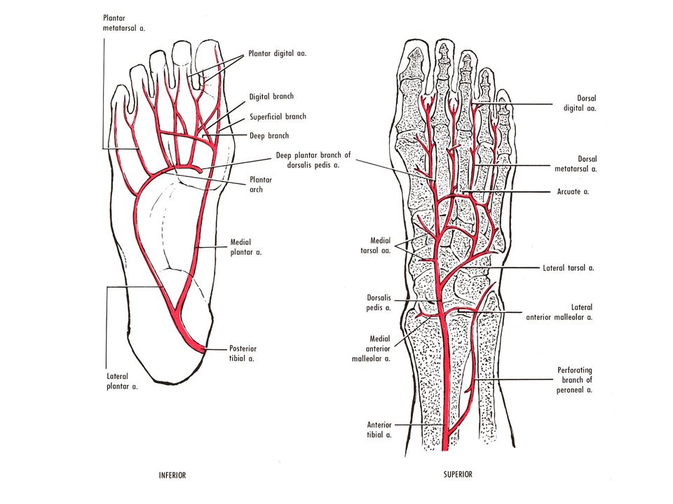
Clinical Anatomy Arteries Dorsal pedis artery –Lateral tarsal artery Arcuate artery –First dorsal MT artery Posterior tibial artery –Plantar structures –Medial tibial artery –Deep plantar artery –Plantar arch Veins Medial marginal vein –Great saphenous vein Lateral marginal vein –Small saphenous vein

10
Muscles Leg divided into compartments –Anterior, lateral, superficial posterior, deep posterior –Surrounded by fascia –Accumulation of fluids can compress nerves and blood vessels within

11
Anterior Compartment MuscleProximal Attachment Distal AttachmentActionNerve Tibialis AnteriorLateral condyle and superior half half of lateral surface of tibia Medial and inferior surfaces of medial cuneiform and base of 1 st metatarsal Dorsiflexes ankle and inverts foot Deep peroneal L4- L5 Extensor Hallicus Longus Middle part of anterior surface of fibula and interossous membrane Dorsal aspect of base of distal phalanx of great toe (hallux) Extends great toe and dorsiflex ankle Deep peroneal L5- S1 Extensor Digitorium Longus Lateral condyle of tibia and superior three fourths of anterior surface of interosseous membrane Middle and distal phalanges of lateral 4 digits Extends lateral 4 digits and dorsiflex ankle Deep peroneal L5- S1 Peroneal TertiusInferior third of anterior surface of fibula and interosseus membrane Dorsum of base of 5 th metatarsal Dorsiflex ankle and aids eversion of foot Deep peroneal L5- S1
 Extends great toe and dorsiflex ankle Deep peroneal L5- S1 Extensor Digitorium Longus Lateral condyle of tibia and superior three fourths of anterior surface of interosseous membrane Middle and distal phalanges of lateral 4 digits Extends lateral 4 digits and dorsiflex ankle Deep peroneal L5- S1 Peroneal TertiusInferior third of anterior surface of fibula and interosseus membrane Dorsum of base of 5 th metatarsal Dorsiflex ankle and aids eversion of foot Deep peroneal L5- S1")
12
Anterior Compartment Extensor retinaculum Common Peroneal Nerve Deep Peroneal Nerve Anterior Tibial Artery Dorsalis Pedis Artery

13
Lateral Compartment MuscleProximal Attachment Distal AttachmentActionNerve Peroneus longusHead and superior two thirds of lateral surface of fibula Base of 1 st metatarsal medial cuneiform Evert foot and weakly plantarflex ankle Superficial peroneal nerve L5, S1, S2 Peroneus brevisInferior two thirds of lateral surface of fibula Dorsal surface of tuberosity on lateral side of base of 5 th metatarsal Evert foot and weakly plantarflex ankle Superficial peroneal nerve L5, S1, S2

14
Lateral Compartment Superior peroneal retinaculum Inferior peroneal retinaculum Superficial peroneal nerve Posterior Tibial Artery

15
Superficial Posterior Compartment MuscleProximal Attachment Distal AttachmentActionNerve GastrocnemiusLateral head: lateral aspect of lateral condyle of femur Medial head: popliteal surface of femur, superior to medial condyle Posterior surface of calcaneus with calcaneal tendon (tendocalcaneus) Plantarflexes ankle when knee is extended; raises heel during walking, and flexes leg at knee joint Tibial Nerve (S1 and S2) SoleusPosterior aspect of head of fibula, superior fourth of posterior surface of fibula, soleal line and medial border of tibia Posterior surface of calcaneus with calcaneal tendon (tendocalcaneus) Plantarflexes ankle (independent of knee position) and steadies leg on foot Tibial Nerve (S1 and S2) PlantarisInferior end of lateral supracondylar line of femur and oblique popliteal ligament Posterior surface of calcaneus with calcaneal tendon (tendocalcaneus Weakly assists gastrocnemius in plantarflexing ankle and flexing knee Tibial Nerve (S1 and S2)
 Plantarflexes ankle when knee is extended; raises heel during walking, and flexes leg at knee joint Tibial Nerve (S1 and S2) SoleusPosterior aspect of head of fibula, superior fourth of posterior surface of fibula, soleal line and medial border of tibia Posterior surface of calcaneus with calcaneal tendon (tendocalcaneus) Plantarflexes ankle (independent of knee position) and steadies leg on foot Tibial Nerve (S1 and S2) PlantarisInferior end of lateral supracondylar line of femur and oblique popliteal ligament Posterior surface of calcaneus with calcaneal tendon (tendocalcaneus Weakly assists gastrocnemius in plantarflexing ankle and flexing knee Tibial Nerve (S1 and S2)")
16
Superficial Posterior Compartment Tibial nerve Posterior tibial artery Plantaris absent in 7- 20% of people

17
Deep Posterior Compartment MuscleProximal Attachment Posterior Attachment ActionNerve PopliteusLateral surface of lateral condyle of femur and lateral meniscus Posterior surface of tibia, superior to soleal line Unlocks fully extended knee (laterally rotates femur 5 degrees on planted tibia); weakly flexes knee Tibial Nerve (L4,L5, and S1) Flexor hallucis longus Inferior two thirds of posterior surface of fibula and inferior part of interosseous membrane Base of distal phalanx of great toe (hallux) Flexes great toe at all joints and plantarflexes ankle; supports medial longitudinal arch of foot Tibial Nerve (L4,L5, and S1) Flexor digitorum longus Medial part of posterior surface of tibia inferior to soleal line, and by a broad tendon to fibula Bases of distal phalanges of lateral four toes Flexes lateral four digits and plantarflexes ankle; supports longitudinal arches of foot Tibial nerve (S2 and S3) Tibialis PosteriorInterosseous membrane, posterior surface of tibia inferior to soleal line and posterior surface of fibula Tuberosity of navicular, cuneiform, and cuboid bases of metatarsals 2-4 Plantarflexes ankle and inverts foot Tibial Nerve (L4 and L5)
; weakly flexes knee Tibial Nerve (L4,L5, and S1) Flexor hallucis longus Inferior two thirds of posterior surface of fibula and inferior part of interosseous membrane Base of distal phalanx of great toe (hallux) Flexes great toe at all joints and plantarflexes ankle; supports medial longitudinal arch of foot Tibial Nerve (L4,L5, and S1) Flexor digitorum longus Medial part of posterior surface of tibia inferior to soleal line, and by a broad tendon to fibula Bases of distal phalanges of lateral four toes Flexes lateral four digits and plantarflexes ankle; supports longitudinal arches of foot Tibial nerve (S2 and S3) Tibialis PosteriorInterosseous membrane, posterior surface of tibia inferior to soleal line and posterior surface of fibula Tuberosity of navicular, cuneiform, and cuboid bases of metatarsals 2-4 Plantarflexes ankle and inverts foot Tibial Nerve (L4 and L5)")
19
Bursae Subtendinous calcaneal (retrocalcaneal) –Between Achilles and calcaneus Subcutaneous calcaneal bursa –Between posterior Achilles and skin
 –Between Achilles and calcaneus Subcutaneous calcaneal bursa –Between posterior Achilles and skin")
Similar presentations