Download presentation
Presentation is loading. Please wait.

1
LEHNINGER PRINCIPLES OF BIOCHEMISTRY
David L. Nelson and Michael M. Cox LEHNINGER PRINCIPLES OF BIOCHEMISTRY Fifth Edition CHAPTER 17 Fatty Acid Catabolism © 2008 W. H. Freeman and Company

2
Introduction The oxidation of long-chain fatty acids to acetyl-CoA as a central energy-yielding pathway- in mammalian heart and liver; it provide 80% of energetic need Fatty acid are converted into acetyl-CoA by repetitive four-step processes, b-oxidation Advantage of fatty acid as storage fuel - highly reduced structure with high energy - do not raise the osmolarity - no undesired chemical reaction with other cellular constituents 4. Fatty acid oxidation in mitochondria

3
Dietary Fats are absorbed in the small intestine
Three sources: in the diet, in cells as lipid droplet, synthesized in one organ (liver) Dietary Fats are absorbed in the small intestine FIGURE 17-1 Processing of dietary lipids in vertebrates. Digestion and absorption of dietary lipids occur in the small intestine, and the fatty acids released from triacylglycerols are packaged and delivered to muscle and adipose tissues. The eight steps are discussed in the text.
 Dietary Fats are absorbed in the small intestine. FIGURE 17-1 Processing of dietary lipids in vertebrates. Digestion and absorption of dietary lipids occur in the small intestine, and the fatty acids released from triacylglycerols are packaged and delivered to muscle and adipose tissues. The eight steps are discussed in the text.")
4
FIGURE 17-2 Molecular structure of a chylomicron
FIGURE 17-2 Molecular structure of a chylomicron. The surface is a layer of phospholipids, with head groups facing the aqueous phase. Triacylglycerols sequestered in the interior (yellow) make up more than 80% of the mass. Several apolipoproteins that protrude from the surface (B-48, C-III, C-II) act as signals in the uptake and metabolism of chylomicron contents. The diameter of chylomicrons ranges from about 100 to 500 nm.
 make up more than 80% of the mass. Several apolipoproteins that protrude from the surface (B-48, C-III, C-II) act as signals in the uptake and metabolism of chylomicron contents. The diameter of chylomicrons ranges from about 100 to 500 nm.")
5
When low levels of glucose in the blood trigger the release of glucagon, 1 the hormone binds its receptor in the adipocyte membrane and thus 2 stimulates adenylyl cyclase, via a G protein, to produce cAMP. This activates PKA, which phosphorylates 3 the hormone-sensitive lipase and 4 perilipin molecules on the surface of the lipid droplet. Phosphorylation of perilipin permits hormone-sensitive lipase access to the surface of the lipid droplet, where 5 it hydrolyzes triacylglycerols to free fatty acids. 6 Fatty acids leave the adipocyte, bind serum albumin in the blood, and are carried in the blood; they are released from the albumin and 7 enter a myocyte via a specific fatty acid transporter. 8 In the myocyte, fatty acids are oxidized to CO2, and the energy of oxidation is conserved in ATP, which fuels muscle contraction and other energy-requiring metabolism in the myocyte. FIGURE 17-3 Mobilization of triacylglycerols stored in adipose tissue. When low levels of glucose in the blood trigger the release of glucagon, 1 the hormone binds its receptor in the adipocyte membrane and thus 2 stimulates adenylyl cyclase, via a G protein, to produce cAMP. This activates PKA, which phosphorylates 3 the hormone-sensitive lipase and 4 perilipin molecules on the surface of the lipid droplet. Phosphorylation of perilipin permits hormone-sensitive lipase access to the surface of the lipid droplet, where 5 it hydrolyzes triacylglycerols to free fatty acids. 6 Fatty acids leave the adipocyte, bind serum albumin in the blood, and are carried in the blood; they are released from the albumin and 7 enter a myocyte via a specific fatty acid transporter. 8 In the myocyte, fatty acids are oxidized to CO2, and the energy of oxidation is conserved in ATP, which fuels muscle contraction and other energy-requiring metabolism in the myocyte.

6
FIGURE 17-4 Entry of glycerol into the glycolytic pathway.

7
Fatty acid are activated.
The enzymes of fatty acid oxidation in animal cells are located in the mitochondrial matrix. Fatty acid are activated. MECHANISM FIGURE 17-5 Conversion of a fatty acid to a fatty acyl–CoA. The conversion is catalyzed by fatty acyl–CoA synthetase and inorganic pyrophosphatase. Fatty acid activation by formation of the fatty acyl–CoA derivative occurs in two steps. The overall reaction is highly exergonic.

8
Fatty acid are transported into mitochondria
FIGURE 17-6 Fatty acid entry into mitochondria via the acyl-carnitine/carnitine transporter. After fatty acyl–carnitine is formed at the outer membrane or in the intermembrane space, it moves into the matrix by facilitated diffusion through the transporter in the inner membrane. In the matrix, the acyl group is transferred to mitochondrial coenzyme A, freeing carnitine to return to the intermembrane space through the same transporter. Acyltransferase I is inhibited by malonyl-CoA, the first intermediate in fatty acid synthesis (see Figure 21-1). This inhibition prevents the simultaneous synthesis and degradation of fatty acids.
. This inhibition prevents the simultaneous synthesis and degradation of fatty acids.")
9
Aceyl-CoA are oxidized to CO2 in the citric acid cycle
Three stages Fatty acids undergo oxidative removal of successive two-carbon units in the form of acetyl –CoA, starting from the carboxyl end of the fatty acyl chain Aceyl-CoA are oxidized to CO2 in the citric acid cycle Electrons from first and second stage are transferred to ETS chain, passing to oxygen with the formation of ATP. FIGURE 17-7 Stages of fatty acid oxidation. Stage 1: A long-chain fatty acid is oxidized to yield acetyl residues in the form of acetyl-CoA. This process is called β oxidation. Stage 2: The acetyl groups are oxidized to CO2 via the citric acid cycle. Stage 3: Electrons derived from the oxidations of stages 1 and 2 pass to O2 via the mitochondrial respiratory chain, providing the energy for ATP synthesis by oxidative phosphorylation.

10
b-Hydroyacyl-CoA deghydrogenase
Acyl-CoA dehydrogenase: dehydrogenation of fatty-acyl CoA produces a double bond between a and b carbon, FAD is a prosthetic group. Similar to succinate dehydrogenation system (bound inner membrane of Mito.; transfer electron to ETS) Enoyl-CoA hydratase: water is added to the double bond of the trans-enoyl-CoA, analogous to the fumarase reaction b-Hydroyacyl-CoA deghydrogenase NADH formed and then transfer electorn to NADH dehydrogenase in ETS. Analogous to the malate dehydrogenase Thiolase thiolysis reaction FIGURE 17-8 The β-oxidation pathway. (a) In each pass through this four-step sequence, one acetyl residue (shaded in pink) is removed in the form of acetyl-CoA from the carboxyl end of the fatty acyl chain—in this example palmitate (C16), which enters as palmitoyl-CoA. (b) Six more passes through the pathway yield seven more molecules of acetyl-CoA, the seventh arising from the last two carbon atoms of the 16-carbon chain. Eight molecules of acetyl-CoA are formed in all.
 Enoyl-CoA hydratase: water is added to the double bond of the trans-enoyl-CoA, analogous to the fumarase reaction. b-Hydroyacyl-CoA deghydrogenase. NADH formed and then transfer electorn to NADH dehydrogenase in ETS. Analogous to the malate dehydrogenase. Thiolase. thiolysis reaction. FIGURE 17-8 The β-oxidation pathway. (a) In each pass through this four-step sequence, one acetyl residue (shaded in pink) is removed in the form of acetyl-CoA from the carboxyl end of the fatty acyl chain—in this example palmitate (C16), which enters as palmitoyl-CoA. (b) Six more passes through the pathway yield seven more molecules of acetyl-CoA, the seventh arising from the last two carbon atoms of the 16-carbon chain. Eight molecules of acetyl-CoA are formed in all.")
11
C>12, trifunctional protein (TFP(a4b4)), multienzyme complex associated with inner membrane, catalyzed the reaction a subunit: enoyl-CoA hadratase and b-hydroxyacyl-CoA dehydrogenase activities b-subunit: thiolase substrate channeling C<12, catalyzed by four soluble enzyme 5. Methylene group (-CH2-) in fatty acid is relatively stable first three step create a much less stable C-C bond, in which the a carbon is bonded to two carbonyl carbon then b-carbon makes it a good target for nucleophilic attack by the –SH of CoA 6. The four b-oxidation steps are repeated to yield Acetyl-CoA and ATP palmitoyl-CoA + 7CoA + 7 FAD + 7NAD + 7H2O - 8acetyl-CoA + 7FADH2 + 7NADH + 7H+ -8acetyl-CoA + 28 ATP + 7H+ 7. Acetyl-CoA can be further oxidized in the citric acid cycle - 10 ATP/1turn 80 ATP -108 ATP/palmitoryl-CoA - for activation of palmitoryl, 2 phosphoanhydride bond was used - 106 ATP/palmitate
 in fatty acid is relatively stable. first three step create a much less stable C-C bond, in which the a carbon is bonded to two carbonyl carbon. then b-carbon makes it a good target for nucleophilic attack by the –SH of CoA. 6. The four b-oxidation steps are repeated to yield Acetyl-CoA and ATP. palmitoyl-CoA + 7CoA + 7 FAD + 7NAD + 7H2O - 8acetyl-CoA + 7FADH2 + 7NADH + 7H+ -8acetyl-CoA + 28 ATP + 7H+ 7. Acetyl-CoA can be further oxidized in the citric acid cycle. - 10 ATP/1turn 80 ATP. -108 ATP/palmitoryl-CoA. - for activation of palmitoryl, 2 phosphoanhydride bond was used - 106 ATP/palmitate.")
12
Oxidation of unsaturated fatty acid
FIGURE 17-9 Oxidation of a monounsaturated fatty acid. Oleic acid, as oleoyl-CoA (Δ9), is the example used here. Oxidation requires an additional enzyme, enoyl-CoA isomerase, to reposition the double bond, converting the cis isomer to a trans isomer, a normal intermediate in β oxidation.
, is the example used here. Oxidation requires an additional enzyme, enoyl-CoA isomerase, to reposition the double bond, converting the cis isomer to a trans isomer, a normal intermediate in β oxidation.")
13
Oxidation of unsaturated fatty acid
FIGURE Oxidation of a polyunsaturated fatty acid. The example here is linoleic acid, as linoleoyl-CoA (Δ9,12). Oxidation requires a second auxiliary enzyme in addition to enoyl-CoA isomerase: NADPH-dependent 2,4-dienoyl-CoA reductase. The combined action of these two enzymes converts a trans-Δ2,cis-Δ4-dienoyl-CoA intermediate to the trans-Δ2-enoyl-CoA substrate necessary for β oxidation.
. Oxidation requires a second auxiliary enzyme in addition to enoyl-CoA isomerase: NADPH-dependent 2,4-dienoyl-CoA reductase. The combined action of these two enzymes converts a trans-Δ2,cis-Δ4-dienoyl-CoA intermediate to the trans-Δ2-enoyl-CoA substrate necessary for β oxidation.")
14
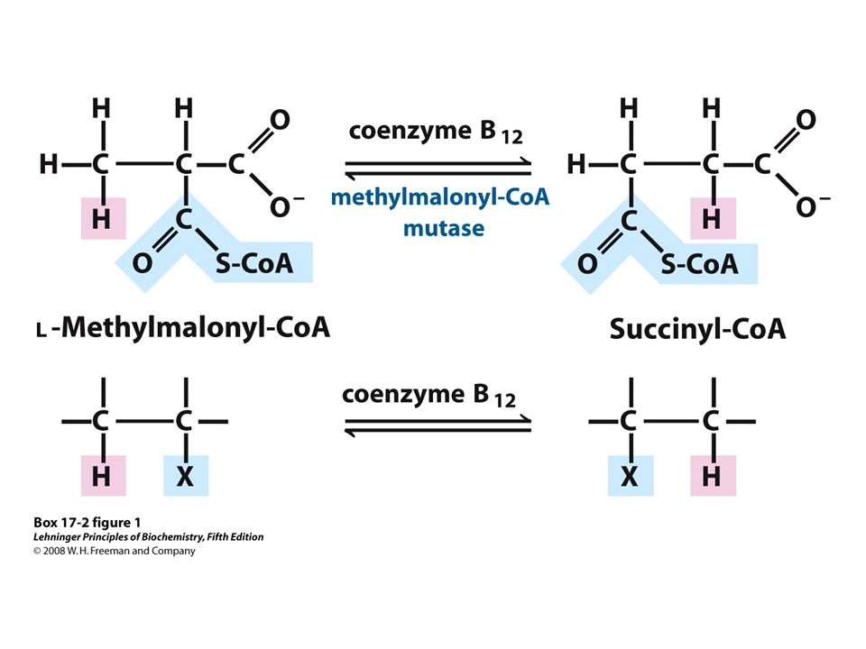
Complete oxidation of odd number fatty acids requires three extra reactions
FIGURE Oxidation of propionyl-CoA produced by β oxidation of odd-number fatty acids. The sequence involves the carboxylation of propionyl-CoA to D-methylmalonyl-CoA and conversion of the latter to succinyl-CoA. This conversion requires epimerization of D- to L-methylmalonyl-CoA, followed by a remarkable reaction in which substituents on adjacent carbon atoms exchange positions (see Box 17-2).
.")
18
Fatty acid oxidation is tightly regulated
The three step process (carnitine shuttle) is rate-limiting for fatty acid oxidation and is an important point of regulation Malonyl-CoA, the first intermediate in the cytosolic biosynthesis of fatty acid from acetyl-CoA, inhibits carnitine acyltransferase I. [NADH]/[NAD+] ration is high, b-hydroxyl-CoA dehydrogenase is inhibited. High concentrations of aceyl-CoA inhibit thiolase. [AMP] is high, AMPK is activated and phosphorylated acetyl-CoA carboxylase, which catalyze the formation of malonyl-CoA, thereby decreasing the malonyl-CoA concentration. allowing b-oxidation 6. Transcription factors turn on the synthesis of proteins for lipid catabolism: - PPARa acts in muscle, adipose tissue, and liver to turn on a set of genes essential for fatty acid oxidation. - Glucagon [cAMP] CREB turn on a set of genes essential for fatty acid.
 is rate-limiting for fatty acid oxidation and is an important point of regulation. Malonyl-CoA, the first intermediate in the cytosolic biosynthesis of fatty acid from acetyl-CoA, inhibits carnitine acyltransferase I. [NADH]/[NAD+] ration is high, b-hydroxyl-CoA dehydrogenase is inhibited. High concentrations of aceyl-CoA inhibit thiolase. [AMP] is high, AMPK is activated and phosphorylated acetyl-CoA carboxylase, which catalyze the formation of malonyl-CoA, thereby decreasing the malonyl-CoA concentration. allowing b-oxidation. 6. Transcription factors turn on the synthesis of proteins for lipid catabolism: - PPARa acts in muscle, adipose tissue, and liver to turn on a set of genes essential for fatty acid oxidation. - Glucagon [cAMP] CREB turn on a set of genes essential for fatty acid.")
19
Coordinate regulation of fatty acid synthesis and breakdown
Two enzymes are key to the coordination of fatty acid metabolism: acetyl-CoA carboxylase (ACC), the first enzyme in the synthesis of fatty acids, and carnitine acyltransferase I, which limits the transport of fatty acids into the mitochondrial matrix for b-oxidation. Ingestion of a high-carbohydrate meal raises the blood glucose level and thus 1 triggers the release of insulin. 2 Insulin-dependent protein phosphatase dephosphorylates ACC, activating it. 3 ACC catalyzes the formation of malonyl-CoA (the first intermediate of fatty acid synthesis), and 4 malonyl-CoA inhibits carnitine acyltransferase I, thereby preventing fatty acid entry into the mitochondrial matrix . When blood glucose levels drop between meals, 5 glucagon release activates cAMP-dependent protein kinase (PKA), which 6 phosphorylates and inactivates ACC. The concentration of malonyl-CoA falls, the inhibition of fatty acid entry into mitochondria is relieved, and 7 fatty acids enter the mitochondrial matrix and 8 become the major fuel. Because glucagon also triggers the mobilization of fatty acids in adipose tissue, a supply of fatty acids begins arriving in the blood. FIGURE Coordinated regulation of fatty acid synthesis and breakdown. When the diet provides a ready source of carbohydrate as fuel, β oxidation of fatty acids is unnecessary and is therefore downregulated. Two enzymes are key to the coordination of fatty acid metabolism: acetyl-CoA carboxylase (ACC), the first enzyme in the synthesis of fatty acids (see Figure 21-1), and carnitine acyltransferase I, which limits the transport of fatty acids into the mitochondrial matrix for β oxidation (see Figure 17-6). Ingestion of a high-carbohydrate meal raises the blood glucose level and thus 1 triggers the release of insulin. 2 Insulin-dependent protein phosphatase dephosphorylates ACC, activating it. 3 ACC catalyzes the formation of malonyl-CoA (the first intermediate of fatty acid synthesis), and 4 malonyl-CoA inhibits carnitine acyltransferase I, thereby preventing fatty acid entry into the mitochondrial matrix. When blood glucose levels drop between meals, 5 glucagon release activates cAMP-dependent protein kinase (PKA), which 6 phosphorylates and inactivates ACC. The concentration of malonyl-CoA falls, the inhibition of fatty acid entry into mitochondria is relieved, and 7 fatty acids enter the mitochondrial matrix and 8 become the major fuel. Because glucagon also triggers the mobilization of fatty acids in adipose tissue, a supply of fatty acids begins arriving in the blood.
, the first enzyme in the synthesis of fatty acids, and carnitine acyltransferase I, which limits the transport of fatty acids into the mitochondrial matrix for b-oxidation. Ingestion of a high-carbohydrate meal raises the blood glucose level and thus 1 triggers the release of insulin. 2 Insulin-dependent protein phosphatase dephosphorylates ACC, activating it. 3 ACC catalyzes the formation of malonyl-CoA (the first intermediate of fatty acid synthesis), and 4 malonyl-CoA inhibits carnitine acyltransferase I, thereby preventing fatty acid entry into the mitochondrial matrix . When blood glucose levels drop between meals, 5 glucagon release activates cAMP-dependent protein kinase (PKA), which 6 phosphorylates and inactivates ACC. The concentration of malonyl-CoA falls, the inhibition of fatty acid entry into mitochondria is relieved, and 7 fatty acids enter the mitochondrial matrix and 8 become the major fuel. Because glucagon also triggers the mobilization of fatty acids in adipose tissue, a supply of fatty acids begins arriving in the blood. FIGURE Coordinated regulation of fatty acid synthesis and breakdown. When the diet provides a ready source of carbohydrate as fuel, β oxidation of fatty acids is unnecessary and is therefore downregulated. Two enzymes are key to the coordination of fatty acid metabolism: acetyl-CoA carboxylase (ACC), the first enzyme in the synthesis of fatty acids (see Figure 21-1), and carnitine acyltransferase I, which limits the transport of fatty acids into the mitochondrial matrix for β oxidation (see Figure 17-6). Ingestion of a high-carbohydrate meal raises the blood glucose level and thus 1 triggers the release of insulin. 2 Insulin-dependent protein phosphatase dephosphorylates ACC, activating it. 3 ACC catalyzes the formation of malonyl-CoA (the first intermediate of fatty acid synthesis), and 4 malonyl-CoA inhibits carnitine acyltransferase I, thereby preventing fatty acid entry into the mitochondrial matrix. When blood glucose levels drop between meals, 5 glucagon release activates cAMP-dependent protein kinase (PKA), which 6 phosphorylates and inactivates ACC. The concentration of malonyl-CoA falls, the inhibition of fatty acid entry into mitochondria is relieved, and 7 fatty acids enter the mitochondrial matrix and 8 become the major fuel. Because glucagon also triggers the mobilization of fatty acids in adipose tissue, a supply of fatty acids begins arriving in the blood.")
20
Peroxisome also carry out b oxidation
1. Peroxisome: membrane-inclosed organelles of animal and plant cells. 2. Dehydrogenation, addition of water, oxidation of b-hydroxyacyl-CoA to a ketone, thiolytic cleavage by conezyme A. 3. Difference between peroxisome and mitochondria. - In peroxisome, the flavoprotein acyl-CoA oxidase passes electrons directly to O2, producing H2O2 and cleaved to H2O and O2 by catalase - Specificity for fatty acyl-CoA: much more active on very-long cahin fatty acid such as hexacosanoic acid (26:0). 4. In mammal, high concentration of fats in the diet result in increase synthesis of the enzymes of peroxisomal b-oxidation in liver. long fatty acids are catabolized to shorter-chain products and then are exported to mitochondria. FIGURE Comparison of β oxidation in mitochondria and in peroxisomes and glyoxysomes. The peroxisomal/glyoxysomal system differs from the mitochondrial system in two respects: (1) in the first oxidative step electrons pass directly to O2, generating H2O2, and (2) the NADH formed in the second oxidative step cannot be reoxidized in the peroxisome or glyoxysome, so reducing equivalents are exported to the cytosol, eventually entering mitochondria. The acetyl-CoA produced by peroxisomes and glyoxysomes is also exported; the acetate from glyoxysomes (organelles found only in germinating seeds) serves as a biosynthetic precursor (see Figure 17-14). Acetyl-CoA produced in mitochondria is further oxidized in the citric acid cycle.
. 4. In mammal, high concentration of fats in the diet result in increase synthesis of the enzymes of peroxisomal b-oxidation in liver. long fatty acids are catabolized to shorter-chain products and then are exported to mitochondria. FIGURE Comparison of β oxidation in mitochondria and in peroxisomes and glyoxysomes. The peroxisomal/glyoxysomal system differs from the mitochondrial system in two respects: (1) in the first oxidative step electrons pass directly to O2, generating H2O2, and (2) the NADH formed in the second oxidative step cannot be reoxidized in the peroxisome or glyoxysome, so reducing equivalents are exported to the cytosol, eventually entering mitochondria. The acetyl-CoA produced by peroxisomes and glyoxysomes is also exported; the acetate from glyoxysomes (organelles found only in germinating seeds) serves as a biosynthetic precursor (see Figure 17-14). Acetyl-CoA produced in mitochondria is further oxidized in the citric acid cycle.")
21
Plant peroxisome and glyoxysome use acetyl-CoA from b-oxation as a biosynthetic precursor
In plant, fatty acid oxidation does not occur primarily in mitochondria but in the peroxisomes of leaf tissue and in the glyoxysomes of germinating seeds The biological role of b-oxidation is to use stored lipid primarily to provide biosynthetic precursors. Glyoxylate cycle to four-carbon precursors for gluconeogenesis. Glyoxysomes contain high concentrations of catalase. FIGURE Triacylglycerols as glucose source in seeds. β Oxidation is one stage in a pathway that converts stored triacylglycerols to glucose in germinating seeds. For more detail, see Figure

22
Omega-oxidation of fatty acids occurs in the endoplasmic reticulum
There is another pathway in some species, including vertebrates, that involves oxidation of the omega carbon. The enzymes unique to omega oxidation are located the ER of liver and kidney The preferred substrate are fatty acid of 10 to 12 carbon. The first step introduces a hydroxyl group on to omega carbon by mixed function oxidase involved in cytochrome p450 and NADPH Produce a carboxyl group at each end. In each pass through the b-oxidation pathway, the double-ended fattyacid yield dicarboxylic acid such as succinic acid, which can enter citric acid cycle. FIGURE The β oxidation of fatty acids in the endoplasmic reticulum. This alternative to β oxidation begins with oxidation of the carbon most distant from the α carbon—the ω (omega) carbon. The substrate is usually a medium-chain fatty acid; shown here is lauric acid (laurate). This pathway is generally not the major route for oxidative catabolism of fatty acids.
 carbon. The substrate is usually a medium-chain fatty acid; shown here is lauric acid (laurate). This pathway is generally not the major route for oxidative catabolism of fatty acids.")
23
Ketone bodies In human and most other mammals, acetyl-CoA formed in the liver during fatty acid oxidation can either enter the citric acid cycle or undergo conversion to the ketone bodies. Acetoacetate and b-hydorxybutyrate are transported by the blood to extrahepatic tissues, where they converted to acetyl-CoA and oxidized in citric acid cycle. The brain can adapt to the use for acetoacetate or b-hydorxybutyrate under starvation condition.

24
FIGURE 17-18 Formation of ketone bodies from acetyl-CoA
FIGURE Formation of ketone bodies from acetyl-CoA. Healthy, well-nourished individuals produce ketone bodies at a relatively low rate. When acetyl-CoA accumulates (as in starvation or untreated diabetes, for example), thiolase catalyzes the condensation of two acetyl-CoA molecules to acetoacetyl-CoA, the parent compound of the three ketone bodies. The reactions of ketone body formation occur in the matrix of liver mitochondria. The six-carbon compound β-hydroxy-β-methylglutaryl-CoA (HMG-CoA) is also an intermediate of sterol biosynthesis, but the enzyme that forms HMG-CoA in that pathway is cytosolic. HMG-CoA lyase is present only in the mitochondrial matrix.
, thiolase catalyzes the condensation of two acetyl-CoA molecules to acetoacetyl-CoA, the parent compound of the three ketone bodies. The reactions of ketone body formation occur in the matrix of liver mitochondria. The six-carbon compound β-hydroxy-β-methylglutaryl-CoA (HMG-CoA) is also an intermediate of sterol biosynthesis, but the enzyme that forms HMG-CoA in that pathway is cytosolic. HMG-CoA lyase is present only in the mitochondrial matrix.")
25
FIGURE 17-18 (part 1) Formation of ketone bodies from acetyl-CoA
FIGURE (part 1) Formation of ketone bodies from acetyl-CoA. Healthy, well-nourished individuals produce ketone bodies at a relatively low rate. When acetyl-CoA accumulates (as in starvation or untreated diabetes, for example), thiolase catalyzes the condensation of two acetyl-CoA molecules to acetoacetyl-CoA, the parent compound of the three ketone bodies. The reactions of ketone body formation occur in the matrix of liver mitochondria. The six-carbon compound β-hydroxy-β-methylglutaryl-CoA (HMG-CoA) is also an intermediate of sterol biosynthesis, but the enzyme that forms HMG-CoA in that pathway is cytosolic. HMG-CoA lyase is present only in the mitochondrial matrix.
 Formation of ketone bodies from acetyl-CoA. Healthy, well-nourished individuals produce ketone bodies at a relatively low rate. When acetyl-CoA accumulates (as in starvation or untreated diabetes, for example), thiolase catalyzes the condensation of two acetyl-CoA molecules to acetoacetyl-CoA, the parent compound of the three ketone bodies. The reactions of ketone body formation occur in the matrix of liver mitochondria. The six-carbon compound β-hydroxy-β-methylglutaryl-CoA (HMG-CoA) is also an intermediate of sterol biosynthesis, but the enzyme that forms HMG-CoA in that pathway is cytosolic. HMG-CoA lyase is present only in the mitochondrial matrix.")
26
FIGURE 17-18 (part 2) Formation of ketone bodies from acetyl-CoA
FIGURE (part 2) Formation of ketone bodies from acetyl-CoA. Healthy, well-nourished individuals produce ketone bodies at a relatively low rate. When acetyl-CoA accumulates (as in starvation or untreated diabetes, for example), thiolase catalyzes the condensation of two acetyl-CoA molecules to acetoacetyl-CoA, the parent compound of the three ketone bodies. The reactions of ketone body formation occur in the matrix of liver mitochondria. The six-carbon compound β-hydroxy-β-methylglutaryl-CoA (HMG-CoA) is also an intermediate of sterol biosynthesis, but the enzyme that forms HMG-CoA in that pathway is cytosolic. HMG-CoA lyase is present only in the mitochondrial matrix.
 Formation of ketone bodies from acetyl-CoA. Healthy, well-nourished individuals produce ketone bodies at a relatively low rate. When acetyl-CoA accumulates (as in starvation or untreated diabetes, for example), thiolase catalyzes the condensation of two acetyl-CoA molecules to acetoacetyl-CoA, the parent compound of the three ketone bodies. The reactions of ketone body formation occur in the matrix of liver mitochondria. The six-carbon compound β-hydroxy-β-methylglutaryl-CoA (HMG-CoA) is also an intermediate of sterol biosynthesis, but the enzyme that forms HMG-CoA in that pathway is cytosolic. HMG-CoA lyase is present only in the mitochondrial matrix.")
27
FIGURE 17-19 D-β-Hydroxybutyrate as a fuel
FIGURE D-β-Hydroxybutyrate as a fuel. D-β-Hydroxybutyrate, synthesized in the liver, passes into the blood and thus to other tissues, where it is converted in three steps to acetyl-CoA. It is first oxidized to acetoacetate, which is activated with coenzyme A donated from succinyl-CoA, then split by thiolase. The acetyl-CoA thus formed is used for energy production.

28
Ketone bodies are overproduced in diabetes and starvation
During starvation, gluconeogenesis depletes citric acid cycle intermediates, diverting acetyl-CoA to ketone body production. Untreated DM insulin down cannot uptake glucose malonyl-Co A fall fatty acid oxidation increase ketone body acidosis (ketosis) Individual on very low-calorie diets, using the fats stored in adipose tissue as their major energy source, also have increased level of ketone bodies in their blood and urine. FIGURE Ketone body formation and export from the liver. Conditions that promote gluconeogenesis (untreated diabetes, severely reduced food intake) slow the citric acid cycle (by drawing off oxaloacetate) and enhance the conversion of acetyl-CoA to acetoacetate. The released coenzyme A allows continued β oxidation of fatty acids.
 Individual on very low-calorie diets, using the fats stored in adipose tissue as their major energy source, also have increased level of ketone bodies in their blood and urine. FIGURE Ketone body formation and export from the liver. Conditions that promote gluconeogenesis (untreated diabetes, severely reduced food intake) slow the citric acid cycle (by drawing off oxaloacetate) and enhance the conversion of acetyl-CoA to acetoacetate. The released coenzyme A allows continued β oxidation of fatty acids.")
Similar presentations


 ~15%>")
>")


 Fatty acid Catabolism ( -oxidation)>")

>")


 is amphibolic (both catabolic and anabolic) The cycle is involved in.>")

 Acylcarnitine.>")
