Download presentation
Presentation is loading. Please wait.

1
Flexible Fiberoptic Workshop: Advanced Course
March 31, 2016 Orlando, FL Flexible Fiberoptic Workshop: Advanced Course Marti Felder, PA-C Updated 12/08/2015

2
Flexible Fiberoptic Workshop: Advanced Course
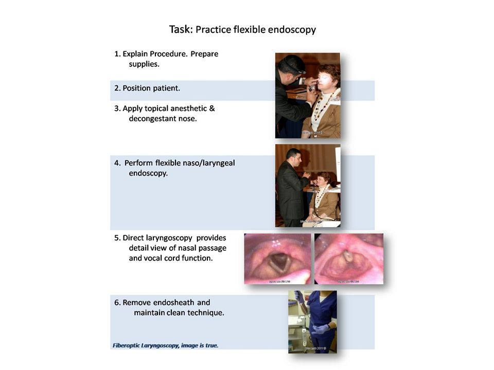
Basic Instruction Demonstration Hands-On Practice Learn by doing Identify abnormal pathology Perform flexible endoscopy adult Perform flexible endoscopy child/infant Perform rigid endoscopy

3
Introduction There are multiple methods and techniques available to successfully complete all the topics presented in this workshop. Some are based on patient request, available equipment or supervising physician’s preference. The goal of this workshop is to correctly demonstrate the most common methods and give participants time for hands on training.

4
Objectives Learning Objectives
Identify normal anatomy, normal variants and abnormal findings visible via flexible fiberoptic nasopharyngoscopy. Understand indications and perform flexible and rigid scope examination adult. Understand indications and perform flexible scope examination child/infant. Perform intranasal culture and sinus debridement using rigid scope adult.

5
Indications for Fiberoptic Endoscopy (FOE)
Dysphonia Presbylarynx VC paralysis VC Nodules LPR Neoplasms Dysphagia Cadidiasis Odynophagia Symptoms of aspiration Laryngomalecia Angioedema Strong gag reflex* Failed mirror exam* Nasal obstruction Foreign Body Septal deviation Adenoid hypertrophy Nasal mass Unilateral otitis media Polyps Sinusitis Chronic throat pain Chronic cough *Documentation of a strong gag reflex and failed mirror exam should be included in note to justify procedure for billing purposes.

6
Contraindications Epiglottitis (by inexperienced) Relative:
Coagulopathy Craniofacial trauma

7
Know Your Anatomy

8
Laryngeal Anatomy (Mirror*)
Images from aboutcancer.com 4. Aryepiglottic folds 5. Arytenoids 6. Pyriform sinuses 7. Base of Tongue True vocal cords False cords Epiglottis *Mirror Laryngoscopy, image is inverted.

9
Laryngeal Anatomy (FOE*)
True Vocal Cords abducted True Vocal Cords adducted *Fiberoptic endoscopy, image is true.

10
Review Tips For Starting the Exam
Patient informed of the procedure (obtain consent) Proper positioning Sniffing, head supported, use non-dominant hand to steady the pts. Head Choose the most patent of the nares Appropriate equipment Adult vs Pedi Decongestant/anesthetic Gloves Chair Photographic/video accessories Biopsy materials if needed Lubricant +/-
 Proper positioning. Sniffing, head supported, use non-dominant hand to steady the pts. Head. Choose the most patent of the nares. Appropriate equipment. Adult vs Pedi. Decongestant/anesthetic. Gloves. Chair. Photographic/video accessories. Biopsy materials if needed. Lubricant +/-")
11
Photo Courtesy Bernadine Sonnier 2011
Preparation for FOE May want patient to blow nose. Assess most patent of nares. Antifogging solution. Apply topical decongestant 0.05% Oxymetazoline 0.25% -2 % Phenylephrine Apply topical anesthetic 4% Lidocaine Pontacaine Photo Courtesy Bernadine Sonnier 2011

12
Video Courtesy J. Mercado
Normal FOE Click on video to begin play. Essentially normal flexible endoscopy on ASYMPTOMATIC patient. Note color of mucous membranes. Clear post nasal secretions noted. Nasopharynx shows white lines consistent with history of previous adenotonsillectomy. Also note left eustachian tube opening. No gross abnormalities. Emphasize how exam should de directed to evaluate symptoms. Video Video Courtesy J. Mercado

13
Causes of Nasal Obstruction
Nasal Foreign Body Septal deviation Synechiae Turbinate hypertrophy Sinusitis Adenoid hypertropy Nasopharyngeal masses Nasal polyps These are some of the most common causes of nasal obstruction that a clinician should be able to distinguish on physical examination.

14
Septal Deviation/ Turbinate Hypertrophy
Septal deviation with evidence of septal spur and mild turbinate hypertrophy.

15
Synechiae Fused intranasal tissue is called an adhesion or synechiae.
Adhesions are a common, usually minor complication of nasal or sinus surgery and nasal packing. They also may develop because of trauma (nose-picking or cocaine use) and such conditions as syphilis, tuberculosis, lupus, or sarcoidosis.
 and such conditions as syphilis, tuberculosis, lupus, or sarcoidosis.")
16
Video Courtesy J. Mercado
Nasal Polyps Click on video to begin play. Video of 34 year old female complaining of a 3 week history of right sided facial pain and nasal congestion that had failed to resolve with OTC medication. Video demonstrates right nasal polyp with thick yellow mucous. Symptoms resolved with 10 day course of penicillin based antibiotics, isotonic sinus rinse and topical nasal steriod spray. Video Courtesy J. Mercado Video

17
Nasal Polyps Nasal polyps are common, noncancerous, teardrop-shaped growths that form in the nose or sinuses, usually around the area where the sinuses open into the nasal cavity. Mature nasal polyps look like seedless, peeled grapes Top image polyps viewed via nasal speculum. Bottom image polyps viewed via fiberoptic intranasal exam. Mercado 2011 © Image on left, transnasal view of nasal polyp. Image on right shows several nasal polyps.

18
Video Courtesy J.Mercado
Sinusitis Click on video to begin play. 27 year old female, 32 weeks pregnant with a 4 week history of facial pressure. Symptoms resolved with 10 day course of amoxicilan and isotonic sinus rinse. Video Video Courtesy J.Mercado

19
Sinusitis Note thick nasal secretions, yellow to greenish in color. Discuss duration and severity of symptoms as predictors of viral vs. bacterial etiology. Treatment of sinusitis discussed more in detail during lectures. Mercado 2013 © Mercado 2013 ©

20
Sinusitis Sinusitis is diagnoses by history and physical examination.
Fiberoptic endoscopy (flexible or rigid) is ideal for evaluating the osteomeatal complex Image of fungal sinusitis. Note thick debris vs purulent secretions in previous images.
 is ideal for evaluating the osteomeatal complex. Image of fungal sinusitis. Note thick debris vs purulent secretions in previous images.")
21
Video Courtesy J. Mercado
Adenoid Hypertrophy Click on video to begin play. Examination of nasal obstruction in a 7 year old female with complaints of snoring. Copious nasal secretions make examination difficult. End of scope is brushed against nasopharynx when it becomes too foggy to see to clear scope. Note amount of nasal obstruction in terms of percentage. Roughly 70% obstruction. Patient was started on topical nasal steroid spray as well as environmental control measures for dust and mold avoidance with excellent improvement of symptoms. Video Video Courtesy J. Mercado

22
Adenoid Hypertrophy Adenoids are described based on % of obstruction
No obstruction Partial (percentage) obstruction Complete obstruction Endoscopic view of the nasopharynx showing adenoidal obstruction of the choana. Mercado 2011 © Mercado 2013 ©
 obstruction. Complete obstruction. Endoscopic view of the nasopharynx showing adenoidal obstruction of the choana. Mercado 2011 © Mercado 2013 ©")
23
Video Courtesy J. Mercado
Angiofibroma Click on video to begin play. Episodes of recurrent nosebleeds in a 15 year old Hispanic male with complaints of nasal congestion and difficulty breathing x 4 years. Born full term NSVD without complications. No pets at home and not exposed to second hand smoke. No obvious bleeding in keisslebach’s plexus prompted fiberoptic examination Video Video Courtesy J. Mercado

24
Juvenile Angiofibroma
Juvenile angiofibroma – benign, highly vascular invasive mass that occurs in the posterior nasal cavity in less than 0.5% of head and neck tumors. Almost always found in adolescent boys. Presents with epistaxis and nasal congestion or both. CT Scan w/contrast confirmed nasopharyngeal mass measuring 4.0 cm x 3.8 cm consistent with a juvenile angiofibroma.

25
Tornwaldt Cyst A Tornwaldt cyst is a midline pouch within the posterior roof of the nasopharnx which is caused by an adherence of the notochord to the pharyngeal ectoderm during development, resulting in the outpouching of ectoderm into the pharyngobasilar fascia. They are typically asymptomatic but may present with middle ear symptoms (eustachian tube obstruction), halitosis (when associated with a leaking sinus tract), foul taste in mouth, and, rarely, occipital headaches. These lesions will demonstrate low to increased signal on T1-weighted images depending on the protein content of the mucus. They will be high signal on T2-weighted images and will not enhance.
, halitosis (when associated with a leaking sinus tract), foul taste in mouth, and, rarely, occipital headaches. These lesions will demonstrate low to increased signal on T1-weighted images depending on the protein content of the mucus. They will be high signal on T2-weighted images and will not enhance.")
26
Unilateral Otitis Media
Unilateral Otitis Media in adults should raise suspicion of Nasopharyngeal mass blocking Eustachian Tube orifice which can cause unilateral middle ear effusion. 1.5:100,000 patients, more common in Asian and Alaskan people 20:100,000 Top image normal Torus Tubarius Bottom image opening covered with mucosal tissue (squamous cell carcinoma). Mercado 2011 © Bottom image was a 65 year old Asian female with a 6 week history of left, unilateral otitis media that failed to resolved with oral antibiotics. She complained of clogged sensation with hearing loss. Fiberoptic nasal endoscopy revealed left torus tubarius was blocked with mucosal tissue. Biopsy confirmed squamous cell carcinoma. Mercado 2011 ©
. Mercado 2011 © Bottom image was a 65 year old Asian female with a 6 week history of left, unilateral otitis media that failed to resolved with oral antibiotics. She complained of clogged sensation with hearing loss. Fiberoptic nasal endoscopy revealed left torus tubarius was blocked with mucosal tissue. Biopsy confirmed squamous cell carcinoma. Mercado 2011 ©")
27
Voice Change Voice overuse/misuse Vocal cord nodules/polyps Layngopharyngeal Reflux Vocal Cord Paralys Presbylarynx Laryngeal neoplasms

28
Video Courtesy M. Mitrani
Vocal Cord Paralysis Click on video to begin play. Video is courtesy of Dr. M. Mitrani. 68 year old male with previous history chronic cough, previous smoker, presented with complaints of 3 week history of hoarseness of voice. Denied dysphagia or odynophagia. Video Video Courtesy M. Mitrani

29
Video Courtesy J. Mercado
Vocal Cord Nodules, LPR Click on video to begin play. 33 year old male presented with complaints of foreign body throat sensation (globus), chronic throat clearing. Symptoms were worse in the morning and after eating. He is a salesman, non-smoker, who drinks 2-3 cups of regular coffee, 2 expresso (Cuban Coffee) and 1-2 diet colas daily. He has dinner around 9pm. He denies fever, SOB, weight changes or odynophagia. Video Video Courtesy J. Mercado
, chronic throat clearing. Symptoms were worse in the morning and after eating. He is a salesman, non-smoker, who drinks 2-3 cups of regular coffee, 2 expresso (Cuban Coffee) and 1-2 diet colas daily. He has dinner around 9pm. He denies fever, SOB, weight changes or odynophagia. Video. Video Courtesy J. Mercado.")
30
Video Courtesy J. Mercado
Laryngeal Cancer Click on video to begin play. 49 year old male that presented with clogged left ear. Initial impression was hoarseness of voice. Heavy smoker. Cerumen impaction cleared and fiberoptic laryngoscopy revealed large pendunculated left vocal cord exophytic lesion. Biospsy confirmed poorly differentiated squamous cell carcinoma. Video Video Courtesy J. Mercado

31
Video Courtesy J. Mercado
Cervical Osteophyte 76 year old nursing home patient wheelchair bound was referred for sore throat with “burning sensation” and difficulty swallowing. She denied weight loss or symptoms of aspiration. Video Video Courtesy J. Mercado

32
Mercado 2014 © Mercado 2014 © Cervical osteophytes are bone spurs that grow on any of the seven vertebrae in the cervical spine (C1 - C7 vertebrae). More than half of people over the age of 60 have osteophytes somewhere in their bodies. Osteophytes in the spine are a normal sign of aging and usually doe not cause symptoms. However, neurological symptoms or pain may occur if the osteophytes encroach upon the individual spinal nerves, the spinal cord itself, the vertebral discs, or the blood vessels in the region of the cervical vertebral column. The inflamed or damaged tissue that stimulates cervical osteophyte growth is often caused by cervical osteoarthritis, a degradation in the neck joints that occurs in many older people. These joints include the disc spaces themselves (a modified joint) and the facet joints, and this condition of cervical osteophyte formation is referred to as cervical spondylosis. CT scan of neck confirmed prominent anterior osteophyte at C3-C4. This patient had resolution of her symptoms with dietary modifications and a proton pump inhibitor.
 and the facet joints, and this condition of cervical osteophyte formation is referred to as cervical spondylosis. CT scan of neck confirmed prominent anterior osteophyte at C3-C4. This patient had resolution of her symptoms with dietary modifications and a proton pump inhibitor.")
33
Adult Mannequin Mercado 2013 © Mercado 2013 © Advanced airway and custom mannequins available to practice flexible fiberoptic endoscopy technique.

35
Respiratory Complications
Children Must rule out foreign body aspiration Choanal Atresia Laryngomalecia Subglotticstenosis Adults Angioedema

36
Flexible fiberoptic exam on children.
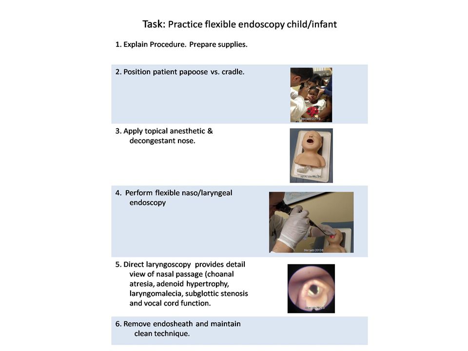
Generally well tolerated by children. Explain procedure in detail. Secure patient (papoose vs. cradle). Anesthesia vs. decongestant? Give adequate time for anesthesia/decongestant. Provides better visualization Mercado 2013 © Mercado 2013 ©
. Anesthesia vs. decongestant Give adequate time for anesthesia/decongestant. Provides better visualization. Mercado 2013 © Mercado 2013 ©")
37
Flexible fiberoptic exam on infants.
Generally well tolerated by infants. Explain procedure in detail. Secure patient (papoose vs. cradle) Anesthesia vs. decongestant? Give adequate time for anesthesia/decongestant. Provides better visualization In infants and neonates, decongestant alone is often better than combination anesthetic/decongestant. Mercado 2013 © Mercado 2013 © Mercado 2013 ©
 Anesthesia vs. decongestant Give adequate time for anesthesia/decongestant. Provides better visualization. In infants and neonates, decongestant alone is often better than combination anesthetic/decongestant. Mercado 2013 © Mercado 2013 © Mercado 2013 ©")
38
Flexible fiberoptic exam on infants and children
Evaluating pediatric airway via flexible fiberoptic Assess nares/choanae (choanal atresia) Assess adenoid and lingual tonsil (hypertrophy) Assess Airway Epiglottis (laryngomalacia) TVC mobility (paralyzed vocal cords) Assess laryngeal structures (stenosis) Foreign bodies A child's airway differs from that of an adult in that the child's tongue is proportionately larger in the oropharynx compered to that of an adult. Also, a child's airway is smaller and softer and more prone to foreign body obstruction. An infant's airway is smaller and softer still and the trachea is usually about the diameter of a pencil
 Assess adenoid and lingual tonsil (hypertrophy) Assess Airway. Epiglottis (laryngomalacia) TVC mobility (paralyzed vocal cords) Assess laryngeal structures (stenosis) Foreign bodies. A child s airway differs from that of an adult in that the child s tongue is proportionately larger in the oropharynx compered to that of an adult. Also, a child s airway is smaller and softer and more prone to foreign body obstruction. An infant s airway is smaller and softer still and the trachea is usually about the diameter of a pencil.")
39
Flexible fiberoptic exam on infants and children
A child's airway differs from that of an adult in that the child's tongue is proportionately larger in the oropharynx compared to that of an adult. A child's airway is smaller, softer and more prone to foreign body obstruction. The trachea is usually about the diameter of a pencil.

40
Flexible fiberoptic exam on infants and children
Difference Pediatric vs Adult Airway Relatively larger tongue Obstructs airway Obligate nasal breathers Difficult to visualize larynx Angled vocal cords Infant’s vocal cords have more angled attachment to trachea, whereas adult vocal cords are more perpendicular Important to note thicker tongue in children vs. adult. Also shorter neck in children often allows the epiglottis to be visualized in children intra-orally. Image from:

41
Video Courtesy J. Mercado
Laryngomalacia Click on video to begin play. 4 month old female with inspiratory stridor and noisy respirations. No shortness of breathe or cyanosis. On Nutramagen formula with mild reflux eating 4-5oz every 2 to 3 hours. Video Video Courtesy J. Mercado

42
Laryngomalacia Most common congenital abnormality of the larynx.
Most prominent SX – inspiratory stridor. Immature development results in soft laryngeal walls that close airway. Child outgrows, Usually, No treatment necessary, Omega Shaped epliglottis Mercado 2011 ©

43
Subglottic Stenosis Class I Class II Class III
Narrowing of subglottis can be congenital or acquired in etiology. Its nature can be membranous, cartilaginous, or mixed, with or without combination of glottic or upper tracheal stenosis. The lower limit of normal subglottis dimension in full term infant is 4.0 mm and in premature infant 3.5 mm. Circumferential edema of 1mm reduces its cross-sectional area by 60%. Myer-Cotton grading system is a useful classification for mature circumferential subglottic stenosis. It is divided into 4 grade as below:: Grade I - Obstruction of 0-50% of the lumen obstruction Grade II - Obstruction of 51-70% of the lumen Grade III - Obstruction of 71-99% of the lumen Grade IV - Obstruction of 100% of the lumen (ie, no detectable lumen) Class I Class II Class III
 Class I. Class II. Class III.")
44
Infant/Child Mannequin
Mercado 2013 © Mercado 2013 © Attendees can practice on infant mannequin. Solicit pearls from audience. Personally, secure patient, work quickly but make sure you get a good look. Mercado 2013 © Practice mannequins available to practice flexible fiberoptic endoscopy technique.

46
Angioedema Vascular leakage beneath the dermis and subcutanis. Response is mediated by vasoactive mediators, i.e., histamine, serotonin, and kinins (eg, bradykinins), which cause the arterioles to dilate while inducing a brief episode of vascular leakage in the venules, where the junction between the endothelial cells appears looser than in the capillaries and arterioles. Angioedema with or without urticaria, is classified as allergic, hereditary, or idiopathic. Complications range from dysphonia or dysphagia to respiratory distress, complete airway obstruction, and death. Symptoms - Severe facial/oral edema, Urticaria – (hives) food allergy, erythema. Most common cause - ACE Inhibitor sensitivity, Food allergies such as fresh berries, shellfish, fish, nuts, tomatoes, eggs, milk, chocolate, food additives, and preservatives Treatment - H 1 (antihistamine), H 2 (antacid), Steroids. Fresh Frozen Plasma, Protect airway. Mercado 2011 © Patient on top was admitted with chest pain and elevated blood pressure. He developed dysphagia and lip swelling 2 days after starting lisinopril. Patient on bottom suffers from hereditary angioedema and has recurrent episodes. Hereditary angioedema (HAE) is caused by low levels or improper function of a protein called C1 inhibitor. Mercado 2011 ©
, which cause the arterioles to dilate while inducing a brief episode of vascular leakage in the venules, where the junction between the endothelial cells appears looser than in the capillaries and arterioles. Angioedema with or without urticaria, is classified as allergic, hereditary, or idiopathic. Complications range from dysphonia or dysphagia to respiratory distress, complete airway obstruction, and death. Symptoms - Severe facial/oral edema, Urticaria – (hives) food allergy, erythema. Most common cause - ACE Inhibitor sensitivity, Food allergies such as fresh berries, shellfish, fish, nuts, tomatoes, eggs, milk, chocolate, food additives, and preservatives. Treatment - H 1 (antihistamine), H 2 (antacid), Steroids. Fresh Frozen Plasma, Protect airway. Mercado 2011 © Patient on top was admitted with chest pain and elevated blood pressure. He developed dysphagia and lip swelling 2 days after starting lisinopril. Patient on bottom suffers from hereditary angioedema and has recurrent episodes. Hereditary angioedema (HAE) is caused by low levels or improper function of a protein called C1 inhibitor. Mercado 2011 ©")
47
Angioedema Edema uvula Edema floor of mouth Edema epiglottis
Mercado 2011 © Edema uvula Edema floor of mouth Slide from Henry Ford from “Laryngoscopy 101” by Mausumi Syamal, MD, MS, Glendon Gardner, MD, Bob Standring, MD, and Werner Roennecke, MD from Henry Ford Health Systems. Patient on right was seen in ER with a 3 week history of sore throat that got worse over last 6 days. He had been started on accupril 1 week ago. Edema epiglottis Edema arytenoid

48
Rigid Scope The goal here is to provide familiarization of the rigid scope and discuss two specific uses; obtaining culture from sinuses and post operative FESS debridement. Attendees will have to practice on mannequins NOT each other.

49
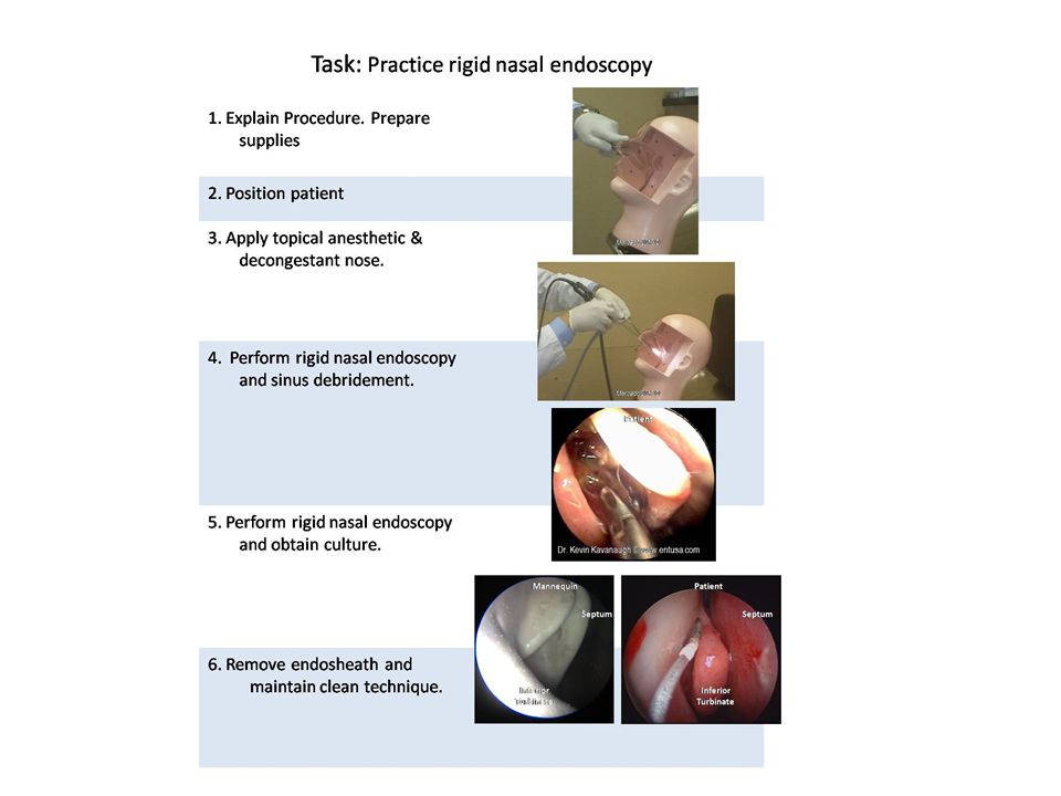
Rigid Scope The rigid endoscope provides superior image clarity, facilitates culture and tissue sampling, controls epistaxis, and affords the endoscopist the ability to perform surgery. Rigid endoscopes for the nose come in diameters of mm and have tips of different angles (generally 0-70º), allowing the clinician to visualize various sinuses and areas within the nasal cavity. This facilitates culture of the sinuses and dedribement postoperatively.
, allowing the clinician to visualize various sinuses and areas within the nasal cavity. This facilitates culture of the sinuses and dedribement postoperatively.")
50
Advise patient not to eat or drink anything 1 hour after procedure.
Complications Tearing Epistaxis Coughing Laryngospasm – rare Bleeding Advise patient not to eat or drink anything 1 hour after procedure.

51
Rhinosinusitis, Fifth Edition
Rigid Scope Nasal Septum When empiric treatment of sinusitis fails or symptoms persist consider obtaining a culture directly from the offending sinus. Direct visualization with a rigid endoscope will reduce chance of contamination. Inferior Turbinate Mercado 2011 © Nasal endoscopic examination and culture for definitive diagnosis best accomplished with rigid scope. Small culturette used to obtain mucous & pus sample from hiatus semilunaris. Be sure not to touch other tissue as this may contaminate specimen. Copyright 2006 The American Academy of Otolaryngology--Head and Neck Surgery Foundation

52
Rhinosinusitis, Fifth Edition
Nasal Cultures Nasal cultures are not routinely indicated in first- line management of Acute Rhinosinusitis Endoscopically guided microswab or suction aspiration culture of a draining sinus ostium are a strong consideration in Chronic Rhinosinusitis, especially when poorly responsive to prior antibiotics. Middle Turbinate Microswab Rhinosinusitis : Cultures Photo depicts a “microswab” obtaining a specimen from the middle meatus under direct vision via nasal endoscopy. Such culture, in the presence of visible pus draining into the hiatus semilunaris and then into the nasal cavity, yields organism(s) that correspond to those cultured directly from the affected sinus (e.g., from the maxillary sinus via direct needle puncture) about 75-85% of the time. Culture is not indicated for the average community acquired acute RS, or even some cases of CRS. However, for severe RS cases, those not responding per expectation to antibiotics, or when the patient is immunocompromised or has been on multiple prior courses of antibiotics, culture is prudent. References Benninger M, Appelbaum, Denneny J, Osguthorpe JD. Maxillary Sinus Puncture and Culture in the Diagnosis of Acute Rhinosinusitis: The Case for Pursuing Alternative Culture. Otolaryngol Head Neck Surg 2002;127:7-12. Benninger M, Ferguson B, Hadley J, Hamilos D, Jacobs M, Kennedy D, Lanza D, Marple B, Osguthorpe J, Stankiewicz J, Anon J, Denneny J, Emanuel I, Levine H. Adult Chronic Rhinosinusitis: Definitions, Diagnosis, Epidemiology, and Pathophysiology. Otolaryngol Head Neck Surg 2003;129(suppl 3) S1-32. Copyright 2006 The American Academy of Otolaryngology--Head and Neck Surgery Foundation
 that correspond to those cultured directly from the affected sinus (e.g., from the maxillary sinus via direct needle puncture) about 75-85% of the time. Culture is not indicated for the average community acquired acute RS, or even some cases of CRS. However, for severe RS cases, those not responding per expectation to antibiotics, or when the patient is immunocompromised or has been on multiple prior courses of antibiotics, culture is prudent. References. Benninger M, Appelbaum, Denneny J, Osguthorpe JD. Maxillary Sinus Puncture and Culture in the Diagnosis of Acute Rhinosinusitis: The Case for Pursuing Alternative Culture. Otolaryngol Head Neck Surg 2002;127:7-12. Benninger M, Ferguson B, Hadley J, Hamilos D, Jacobs M, Kennedy D, Lanza D, Marple B, Osguthorpe J, Stankiewicz J, Anon J, Denneny J, Emanuel I, Levine H. Adult Chronic Rhinosinusitis: Definitions, Diagnosis, Epidemiology, and Pathophysiology. Otolaryngol Head Neck Surg 2003;129(suppl 3) S1-32. Copyright 2006 The American Academy of Otolaryngology--Head and Neck Surgery Foundation.")
53
Dr. Kevin Kavanaugh © www.entusa.com
FOE After FESS Clinicians should know exactly what procedure patient underwent as well as surgical course and complications if any. In most cases debridement is indicated to restore airway, minimize obstruction, improve patient comfort, evaluate for bleeding and prevent infectious complications. Mercado 2011 © Dr. Kevin Kavanaugh © Post operative debridement following FESS is best accomplished with rigid scope. Rigid scope allows clinician to visualize area and suction or use forceps.

54
Interactive, live demonstration of rigid fiberoptic endoscopy
Rigid Endoscopy Interactive, live demonstration of rigid fiberoptic endoscopy Demonstrate technique on simulated patient (live person) then demonstrate obtaining culture, suction and debridement on mannequin.
 then demonstrate obtaining culture, suction and debridement on mannequin.")
55
Rigid Scope Practice DEMONSTRATE RIGID TECHNIQUE. Cut-away view of model allows instructor to give feedback regarding technique and anatomy. Picture on right is view of mannequin from rigid endoscope. Mercado 2013 © Mercado 2013 © Demonstration of Rigid Endoscopy technique. Custom made models available to practice Post-FESS Debridement and C/S of sinus

57
Care of Scope Briefly REVIEW scope care. This was covered during Basic course.

58
Pull endosheath from distal end
Endosheaths Loosen endosheath Pull endosheath from distal end To remove endosheath, loosen from tip and pull from distal end. Do not push from hub adapter as sheath will become wedged and may damage scope. Mercado 2011 © Mercado 2011 ©

59
Cold Sterilization Flexible scopes are non-autoclavable.
Rigid scopes can be autoclaved. Clean length of scope with an enzymatic detergent solution like ENZOL® to remove debris and reduce bacterial burden before instruments are disinfected or sterilized. DO NOT ALLOW debris to dry. Soak scope in a glutaraldehyde solution like Cidex ® which provides quick high-level disinfection. Noncorrosive solution reduces instrument damage and associated repair costs. Soaking times vary by product. Steam is the most common and least expensive method of sterilization. However, many lensed endoscopic instruments cannot be steam sterilized. Even instruments and telescopes marketed as "autoclavable" will last longer if processed by alternative methods. Mercado 2011 ©

60
Leak Testing Routine leak testing in accordance with specific manufacture depending on volume of use. Introduce air pressure via attached bulb (DO NOT overinflate) and submerge looking for leaks. Leaks can slowly damage fiberoptics and internal parts causing expensive yet preventable damage. Mercado 2011 © Mercado 2011 © Mercado 2011 ©
 and submerge looking for leaks. Leaks can slowly damage fiberoptics and internal parts causing expensive yet preventable damage. Mercado 2011 © Mercado 2011 © Mercado 2011 ©")
61
Review of Scope Care Avoid bending scope in tight angles.
Clean lens with lens cleaner/paper. Pre-clean with enzymatic cleaner. Soak only for required period depending on brand and manufacture. Store in dry safe place. Perform regular leak testing to avoid damage.

62
Flexible Fiberoptic Workshop: Advanced
Room Set Up Station 5 Video Tower Adult examination Screen Station 4 Video Tower Rigid Station 1 Pediatric Projector Speaker Station 3 Video Tower Rigid Station 2 Adult examination Proctors

63
Flexible Fiberoptic Workshop: Advanced Evaluation
Score cards will be used for admission to workshops and attendance. Credit will only be awarded for completed score cards. Name Date On scale of 1through 5 with 5 being most likey Scale 1-5 1. Were learning objectives met? 2. Was instruction free of commercial bias? 3. Was there adequate instruction before practice? 4. Was there adequate supervision during practice? 5. Were training aids useful/realistic in learning skill? 6. How likely are you to perform these skills in future 7. Did this training improve your skills? Comments:

64
Flexible Fiberoptic Workshop: Advanced Score Card
Rotate and complete each station. “Go/No Go” for internal use only. Completion of workshop is NOT contingent on pass/fail. Task Go No Go Perform flexible endoscopy child mannequin. Perform flexible endoscopy infant mannequin. Perform flexible fiberoptic endoscopy on adult mannequin. Perform flexible fiberoptic endoscopy on simulated patient. Perform rigid endoscopy (culture collection) Perform rigid endoscopy debridement. Comments
 Perform rigid endoscopy debridement. Comments.")
65
Functional Voice Disorders
Resources On-Line New England Journal of Medicine Video Excellent pictures and videos by Dr. Kevin Kavenaugh Dr Rahmat Omar Direct Laryngoscopy video Functional Voice Disorders Neil N Chheda, MD

66
Recommend Reading Examination of the Larynx and Pharynx
n engl j med 358;3 january 17, 2008 Laryngeal Evaluation by Kendall & Leonard Publication Date: August pp, 309 illustrations hardcover & video ISBN (Americas): ISBN (EUR, Asia, Africa, AUS):
: ISBN (EUR, Asia, Africa, AUS):")
67
Documentation Effective January 1, 2014, any payer requesting documentation for a scope procedure could either deny the service or reduce the payment if documentation doesn’t show that all areas were examined during the endoscopy and the findings noted. According to Medicare, documentation should identify specific anatomical landmarks. “form over substance” . CPT® guidelines state, “For endoscopic procedures, report appropriate endoscopy of each anatomic site examined. 31575 Flexible Fiberoptic Laryngoscopy, Laryngoscopy, Indirect, 31515, Laryngoscopy, Direct, includes examination of the tongue base, larynx, and hypopharynx. If using operating microscope, telescope, or both, use the applicable code only once per operative session. 31231 Nasal Endoscopy (diagnostic), and Debridement Endoscopy, Nasal should include mention of the superior turbinate, superior meatus and sphenoethmoid recess Clinicians need to clearly document each area’s examination and whatever findings were observed. This information stems from increased number of procedure denials and reduced payments as well as from audits from Zone Program Integrity Contractors (ZPICs) on behalf of Medicare ( Otolaryngology Coding Alert, The Coding Institute, February 2015, Vol. 17, No. 2 (Pages 9-16) Coding Corner, AAO-HNSF
, and Debridement Endoscopy, Nasal should include mention of the superior turbinate, superior meatus and sphenoethmoid recess. Clinicians need to clearly document each area’s examination and whatever findings were observed. This information stems from increased number of procedure denials and reduced payments as well as from audits from Zone Program Integrity Contractors (ZPICs) on behalf of Medicare ( s=ent) Otolaryngology Coding Alert, The Coding Institute, February 2015, Vol. 17, No. 2 (Pages 9-16) Coding Corner, AAO-HNSF")
68
Sample Templates Template should also include description of positioning of patient, application of topical anesthetic and any decongestants. If consent was obtained, verbal vs. written. Document indication for procedure. At completion of procedure note if patient tolerated procedure and if there were any complications. Fiberoptic Laryngoscopy Findings; Nasopharnyx -WNL Oropharynx -WNL Base of Tongue -WNL Vallecula –WNL* Lateral Pharyngeal Wall -WNL Posterior Pharyngeal Wall -WNL Epiglottis -WNL Aryepiglottic Folds –WNL* Pyriform Sinuses -WNL Interarytenoid -WNL Tracheal Rings -WNL Vocal Cords –WNL* False Cords –WNL* Ventricle –WNL* Fiberoptic Nasal Endoscopy Findings Septum Right Side Inferior Turbinates -WNL Middle Turbinates -WNL Middle Meatus -WNL Mucosa -WNL Mucous -WNL Polyps - None Left Side Nasopharynx -patent Sphenoid and ethmoid cavities-WNL Template should also include description of positioning of patient, application of topical anesthetic and any decongestants. If consent was obtained, verbal vs. written. Document indication for procedure. At completion of procedure note if patient tolerated procedure and if there were any complications. * Denotes laterally i.e. Left side, Right side

Similar presentations


 Root Ala Dorsum>")


