Download presentation
Presentation is loading. Please wait.

1
TRAUMATIC DELIVERY Guslihan Dasa Tjipta Division of Perinatology Department of Child Health Medical School University of Sumatera Utara

2
Predisposing Factors Maternal factors: Primigravida
Cephalopelvic disproportion, small maternal stature maternal pelvic anomalies Prolonged or rapid labor Dystocia Oligohydramnios

3
Predisposing factors Fetal factors: Abnormal presentation
Breech, face VLBW or extreme prematurity Fetal macrosomia Large fetal head Fetal anomalies

4
Predisposing Factors Obstetrical Interventions:
Use of mid-cavity forceps or vacuum extraction Versions and extractions

5
Types of Injury Soft tissue injuries Head and Skull Face
Musculoskeletal injuries Intra-abdominal injuries Peripheral nerve injuries

6
Soft Tissue Injuries Erythema & Abrasions - Forceps, Dystocia
Petechiae head/neck/chest/back - Cord around neck /breech - thrombocytopenia Ecchymoses breech/prematurity

8
Soft Tissue Injuries Lacerations scalp, buttocks, thighs
(Fetal scalp electrodes, surgeons knife!) Infection a risk, but most heal uneventfully Management: careful cleaning, application of antibiotic ointment, and observation Bring edges together using Steri-Strips Lacerations occasionally require suturing
 Infection a risk, but most heal uneventfully. Management: careful cleaning, application of antibiotic ointment, and observation. Bring edges together using Steri-Strips. Lacerations occasionally require suturing.")
9
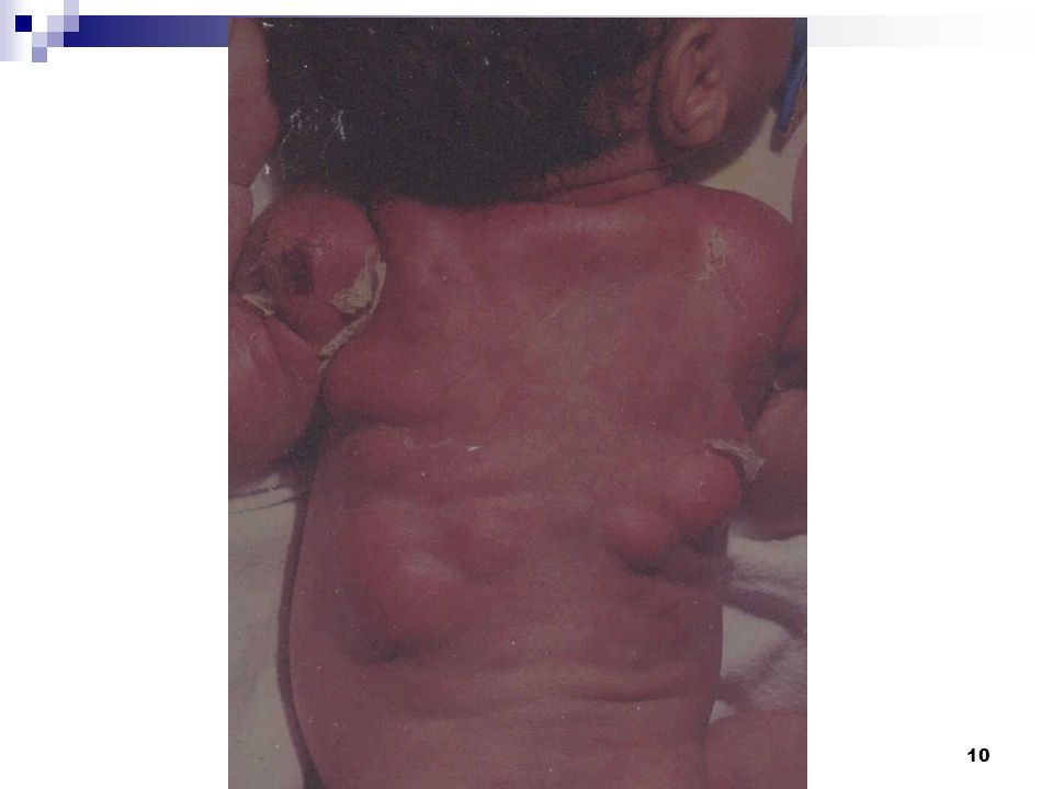
Soft Tissue Injuries Subcutaneous fat Necrosis (SFN)
Not usually detected at birth Irregular, hard, non-pitting, subcutaneous plaques with overlying dusky red-purple discoloration on the extremities, face, trunk, or buttocks May be caused by pressure during delivery Hypothermia/ischemia/asphyxia days 6-8 wk/atrophy Sometimes calcifies

11
Soft Tissue Injuries SFN: Treatment
Treat symptomatic hypercalcemia aggressively increased fluid intake low calcium/ vit. D diet furosemide -calcium-wasting diuretic Steroids-inhibit metabolism of vit. D Biphosphonates-reduce bone resorption

12
Injuries to the Head Caput Succedaneum most frequently observed lesion
pressure on the scalp against cervix subcutaneous, extraperiosteal accumulation of blood/serum presenting part involved overlying bruising/Petechiae crosses suture lines resolves within days

13
Injuries to the Head Cephalhematoma 0.4%-2.5% of all live births
sub-periosteal hemorrhage from rupture of blood vessels between the skull and the periosteum buffeting of fetal head against the pelvis no extension across suture lines most commonly parietal, may occasionally be observed over the occipital bone

14
Injuries to the Head Cephalhematoma increases in size with time
15% bilateral 18% associated skull fracture Forceps Vacuum

15
Injuries to the Head Subgaleal Hemorrhage
Diagnosis is generally clinical: fluctuant boggy mass developing over the scalp (especially over the occiput) develops gradually hours after delivery hematoma spreads across the whole calvarium Usually insidious and may not be recognized for hours swelling may obscure the fontanelle and cross suture lines (distinguishing it from cephalhematoma)
 develops gradually hours after delivery. hematoma spreads across the whole calvarium. Usually insidious and may not be recognized for hours. swelling may obscure the fontanelle and cross suture lines (distinguishing it from cephalhematoma)")
16
Injuries to the Head Subgaleal Hemorrhage
Rx if signs of substantial volume loss: compression wrap restore blood volume surgical drainage 25% mortality

18
Caput Succedaneum Cephalhematoma Subgaleal hemorrhage with skull fracture

19
extradural hemorrhage
Skin Caput Cephalhematoma Epicranial aponeuroses Subgaleal hemorrhage extradural hemorrhage Periosteum Skull Dura Lesion External swelling ↑ after birth Crosses suture lines ↑↑↑acute blood loss Caput succedaneum Soft, pitting No Yes Cephalhematoma Firm, tense Subgaleal hematoma Firm, fluctuant Extracranial hemorrhages: Caput succedaneum: Subdermal edema, secondary to compression of uterus/cervix against the presenting parts Occurs % of deliveries with vacuum extraction Resolves gradually, no intervention is recommended Cephalhematoma A circumscribed region of hemorrhages overlying the skull and confined by cranial sutures Caused by mechanical force No intervention is recommended Subgaleal hematoma Hemorrhage beneath the aponeurosis covering the scalp and connecting the fromtal and occipital components of the occipito-frontalis muscle A strong association with vacuum delivery Observe closely for acute massive blood loss and signs of DIC

20
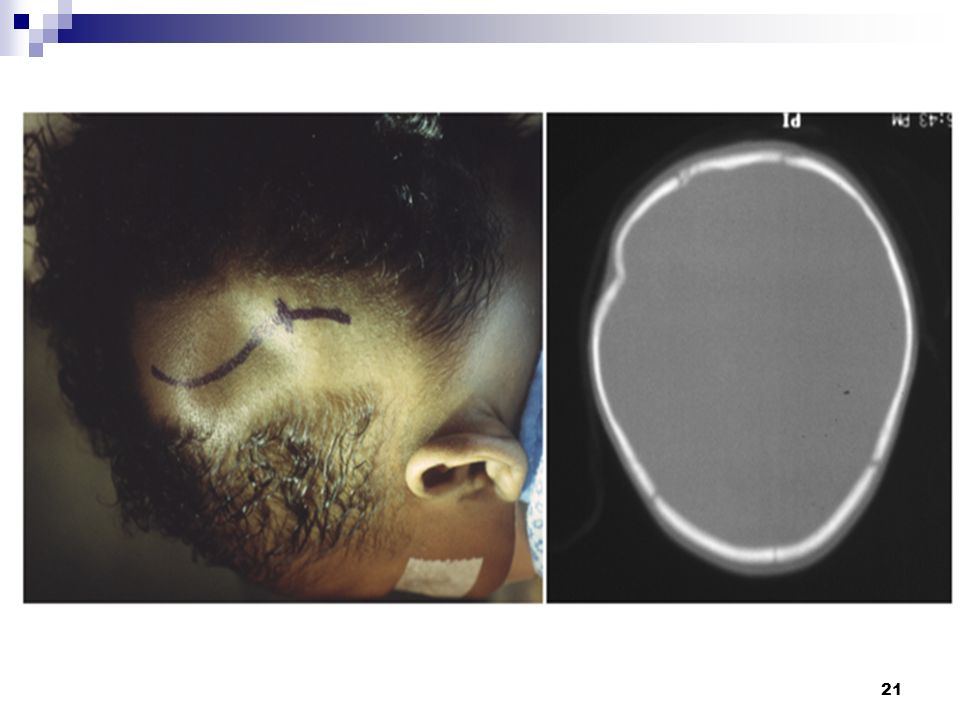
Injuries to the Head Skull Fractures
Uncommon because of compressible skull & open sutures Forceps/Prolonged labor Linear/Depressed Usually asymptomatic Associated intracranial hemorrhage may produce symptoms

22
Injuries to the Head Skull Fractures
Rx – conservative elevation of depressed fracture - Thumb pressure Hand pump - Vacuum extractor Surgical elevation Healing within a few months

23
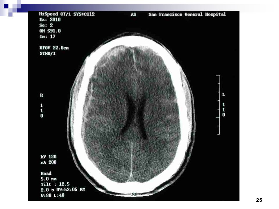
Injuries to the Head Intracranial hemorrhage
- Subdural/Subarachnoid/IVH Usually asymptomatic Forceps/Vacuum Prolonged labor Usually associated with fracture

24
Injuries to the Head Subarachnoid hemorrhage
-more frequent than realized -usually asymptomatic -may cause seizures (day 2-3) -bloody CSF - CT/MRI
 -bloody CSF. - CT/MRI.")
26
Injuries to the Head Subdural Hematoma
- may be silent for several days -head circumference poor feeding /vomiting /lethargy altered consciousness/seizures DX- CT/MRI RX- Subdural taps/surgical drainage

28
Injuries to the Head Fractures of Facial bones
-nasal fracture/dislocation -deviated nasal septum -maxillary fracture -mandibular fracture

29
EYE INJURIES Eye Lids edema/ecchymoses/laceration
Subconjuntival hemorrhage Orbital fracture/hemorrhage Extra Ocular Muscle injury Corneal Abrasion Intra Ocular hemorrhage

30
Injuries to the Ear Ecchymoses Abrasion Avulsion Hematoma

31
Neck and Shoulder injuries
Fractured Clavicle -most frequently fractured bone -difficult delivery -shoulder dystocia -breech -Crepitus or deformity at the site -movement/moro on affected side -associated brachial plexus palsy

32
Neck and Shoulder injuries
Fractured Clavicle DX- X-ray RX- conservative immobilization reduce pain pain subsides in 7-10 days good prognosis

33
Neck and Shoulder injuries
Fracture of the Humerus second most common fracture difficult delivery/traction shoulder dystocia breech deformity

34
Neck and Shoulder injuries
Fractured Humerus: Management Splinting/immobilization in adduction Closed reduction and casting when displaced Watch for evidence of radial nerve injury Callus formation occurs, and complete recovery expected in 2-4 weeks In 8-10 days, the callus formation is sufficient to discontinue immobilization

35
Intra-abdominal Organ Injury
Uncommon sometimes overlooked as a cause of death in the newborn Hemorrhage is the most serious acute complication liver is the most commonly damaged internal organ

36
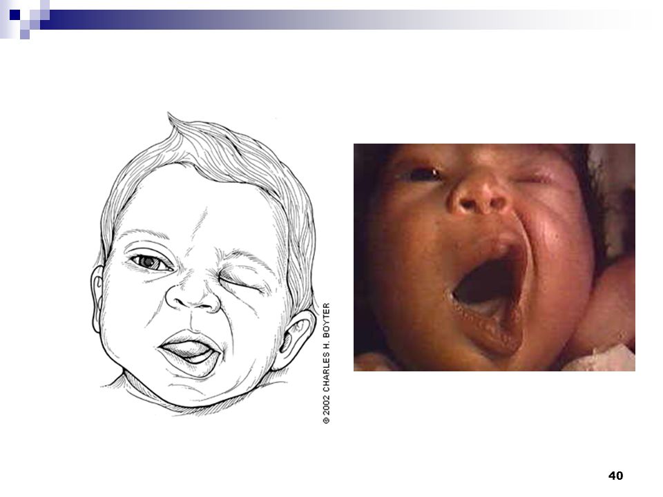
Nerve Palsies Facial Nerve Etiology Compression Of peripheral nerve
-forceps -prolonged labor -in-utero compression CNS Injury -temporal bone fracture -tissue destruction

37
Nerve Palsies Facial Nerve Clinical Manifestation
Paralysis apparent day 1-2 Unilateral/bilateral Affected side smooth/drooping Amplified by crying

38
Nerve Palsies Facial Nerve: central nerve injury
asymmetric facies with crying mouth is drawn towards the normal side wrinkles are deeper on the normal side movement of the forehead and eyelid is unaffected the paralyzed side is smooth with a swollen appearance absent nasolabial fold on affected side corner of the mouth droops on affected side no evidence of trauma is present on the face

39
Nerve Palsies Facial Nerve: peripheral nerve injury
asymmetric facies with crying Unable to close eye on affected side may be evidence of forceps mark

41
Nerve Palsies Facial Nerve Palsy: prognosis 85% recover in 1 week
90% recovery in 1 year Surgery if no resolution in 1 yr Palsy due to trauma usually resolves or improves palsy that persists is often due to absence of the nerve

42
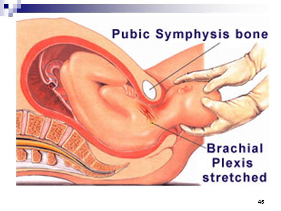
Nerve Palsies Brachial Plexus injury Types of Injury Stretch Rupture
Avulsion

43
Nerve Palsies Brachial Plexus injury Types of Injury
Stretch % recovery in 1 year Rupture-needs surgical repair Avulsion-needs surgical repair

44
Nerve Palsies Brachial Plexus injury
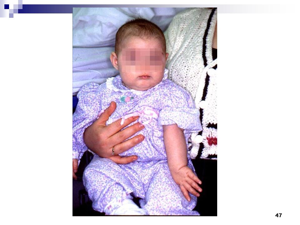
Weakness or total paralysis of muscles innervated by the brachial plexus C-5 to C-8 and T1 Erb's Palsy C5-C7- proximal muscle weakness Klumpke’s Palsy C8 and T1- weakness in the intrinsic muscles of the hand

46
Nerve Palsies Brachial Plexus injury Neurological Features
Erb's Palsy (C5-C6) The involved extremity lies: in adduction in pronation and internally rotated Moro, biceps and radial reflexes are absent Grasp reflex is usually present 2-5% ipsilateral phrenic nerve paresis The "waiter's tip" posture
 The involved extremity lies: in adduction. in pronation and internally rotated. Moro, biceps and radial reflexes are absent. Grasp reflex is usually present. 2-5% ipsilateral phrenic nerve paresis. The waiter s tip posture.")
48
Nerve Palsies Brachial Plexus Injury Neurological Features
Klumpke’s Palsy (C7-8, T1) weakness of the intrinsic muscles of the hand grasp reflex is absent
 weakness of the intrinsic muscles of the hand. grasp reflex is absent.")
49
Nerve Palsies Brachial Plexus Injury Neurological Features
Total Plexus Palsy Erb's Palsy + absent grasp reflex Sensory loss worse than Erb's

50
Nerve Palsies Brachial Plexus Prognosis
Depends on severity and extent of lesion 88% resolved by 4 months 92% by 12 months 93% by 48 months

51
Nerve Palsies Brachial Plexus Prognosis
Depends on severity and extent of lesion 88% resolved by 4 months 92% by 12 months 93% by 48 months

52
Nerve Palsies Brachial Plexus Management Prevention of contractures
immobilize limb gently across the abdomen for first week and then start passive range of motion exercises at all joints supportive wrist splints

53
Nerve Palsies Brachial Plexus Management Electrotherapy-controversial
Surgical exploration-if no significant functional recovery by 3 months Exploration after 6 months is of little benefit

54
Nerve Palsies Laryngeal nerve injury
The infant presents with a hoarse cry or respiratory stridor most often unilateral nerve paralysis Swallowing may be affected if the superior branch is involved Bilateral paralysis may be caused by trauma to both laryngeal nerves or, more commonly, by a CNS injury such as hypoxia or hemorrhage involving the brain stem Patients with bilateral paralysis often present with severe respiratory distress or asphyxia

55
Nerve Palsies Laryngeal nerve injury & Prognosis:
Paralysis often resolves in 4-6 wk, although full recovery may take 6-12 months Treatment symptomatic Small frequent feeds, once infant is stable Minimize the risk of aspiration Infants with bilateral involvement may require gavage feeding and tracheotomy

56
THANK YOU

Similar presentations


 Cyanotic heart diseases : lead to intrauterine growth retardation, due to.>")


 BY.DR.HINA ADNAN DNT 472.>")