Download presentation
Presentation is loading. Please wait.

2
Infrared (IR) Spectroscopy IR deals with the interaction of infrared radiation with matter. The IR spectrum of a compound can provide important information about its chemical nature and molecular structure. Most commonly, the spectrum is obtained by measuring the absorption of IR radiation, although infrared emission and reflection are also used. Widely applied in the analysis of organic materials, also useful for polyatomic inorganic molecules and for organometallic compounds.

3
Overview 1.Electromagnetic radiation 2.Vibrations 3.Principle of IR experiment 4.IR spectrum 5.Types of vibration 6.CGF/Fingerprint regions 7.IR activity of vibrations 8.Interpretation of IR spectra 9.Instrumentation 10.Sample preparation

4
Electromagnetic Radiation The propagation of electromagnetic radiation in a vacuum is constant for all regions of the spectrum (= velocity of light): c = × 1 Å = 10 –10 m 1 nm = 10 –9 m 1 m = 10 –6 m Another unit commonly used is the wavenumber, which is linear with energy: Work by Einstein, Planck and Bohr indicated that electromagnetic radiation can be regarded as a stream of particles or quanta, for which the energy is given by the Bohr equation:
: c = × 1 Å = 10 –10 m 1 nm = 10 –9 m 1 m = 10 –6 m Another unit commonly used is the wavenumber, which is linear with energy: Work by Einstein, Planck and Bohr indicated that electromagnetic radiation can be regarded as a stream of particles or quanta, for which the energy is given by the Bohr equation:")
5
The Electromagnetic Spectrum

6
LIMIT OF RED LIGHT: 800 nm, 0.8 m, 12500 cm -1 NEAR INFRARED: 0.8 -2.5 m, 12500 - 4000 cm -1 MID INFRARED: 2.5 - 50 m, 4000 - 200 cm -1 FAR INFRARED: 50 - 1000 m, 200 - 10 cm -1 Divisions arise because of different optical materials and instrumentation. Infrared region

7
Molecular spectra There are three basic types of optical spectra that we can observe for molecules: 1.Electronic or vibronic spectra (UV-visible-near IR) (transitions between a specific vibrational and rotational level of one electronic state and a vibrational and rotational level of another electronic state) 2.Vibrational or vibrational-rotational spectra (IR region) (transitions from the rotational levels of one vibrational level to the rotational levels of another vibrational level in the same electronic state) 3.Rotational spectra (microwave region) (transitions between rotational levels of the same vibrational level of the same electronic state)
 (transitions between a specific vibrational and rotational level of one electronic state and a vibrational and rotational level of another electronic state) 2.Vibrational or vibrational-rotational spectra (IR region) (transitions from the rotational levels of one vibrational level to the rotational levels of another vibrational level in the same electronic state) 3.Rotational spectra (microwave region) (transitions between rotational levels of the same vibrational level of the same electronic state)")
9
Infrared radiation in the range from 10,000 – 100 cm –1 is absorbed and converted by an organic molecule into energy of molecular vibration –> this absorption is quantized: Vibrational spectra (I): Harmonic oscillator model A simple harmonic oscillator is a mechanical system consisting of a point mass connected to a massless spring. The mass is under action of a restoring force proportional to the displacement of particle from its equilibrium position and the force constant f (also k in followings) of the spring.
 of the spring..")
10
The vibrational frequency is increasing with: increasing force constant f = increasing bond strength decreasing atomic mass Example: f c c > f c=c > f c-c The vibrational energy V(r) can be calculated using the (classical) model of the harmonic oscillator: Using this potential energy function in the Schrödinger equation, the vibrational frequency can be calculated:
 can be calculated using the (classical) model of the harmonic oscillator: Using this potential energy function in the Schrödinger equation, the vibrational frequency can be calculated:")
11
Vibrational spectra (II): Anharmonic oscillator model The actual potential energy of vibrations fits the parabolic function fairly well only near the equilibrium internuclear distance. The Morse potential function more closely resembles the potential energy of vibrations in a molecule for all internuclear distances- anharmonic oscillator model. Fig. 12-1

12
The energy difference between the transition from n to n+1 corresponds to the energy of the absorbed light quantum The difference between two adjacent energy levels gets smaller with increasing n until dissociation of the molecule occurs (Dissociation energy E D ) E VIB ( E n+1 – E n ) =h osc Note: Weaker transitions called “overtones” are sometimes observed. These correspond to =2 or 3, and their frequencies are less than two or three times the fundamental frequency ( =1) because of anharmonicity. Typical energy spacings for vibrational levels are on the order of 10 -20 J. from the Bolzmann distribution, it can be shown that at room temperature typically 1% or less of the molecules are in excited states in the absence of external radiation. Thus most absorption transitions observed at room temperature are from the =0 to the =1 level.
 because of anharmonicity. Typical energy spacings for vibrational levels are on the order of J. from the Bolzmann distribution, it can be shown that at room temperature typically 1% or less of the molecules are in excited states in the absence of external radiation. Thus most absorption transitions observed at room temperature are from the =0 to the =1 level..")
13
The IR absorption spectrum can be obtained with gas-phase or with condensed-phase molecules. For gas-phase molecules vibration-rotation spectra are observed, while in condensed phases, the rotational structure is lost. For most routine analytical applications of infrared spectrometry, spectra are obtained with condensed-phase samples. Hence, the discuss here centers around the vibrational transitions observed with molecules present as pure liquid, as solutions, or in the solid state.

14
Molecular vibrations How many vibrations are possible (=fundamental vibrations)? A molecule has as many degrees of freedom as the total degree of freedom of its individual atoms. Each atom has three degrees of freedom (corresponding to the Cartesian coordinates), thus in an N-atom molecule there will be 3N degree of freedom. In molecules, movements of the atoms are constrained by interactions through chemical bonds. Translation - the movement of the entire molecule while the positions of the atoms relative to each other remain fixed: 3 degrees of translational freedom. Rotational transitions – interatomic distances remain constant but the entire molecule rotates with respect to three mutually perpendicular axes: 3 rotational freedom (nonlinear), 2 rotational freedom (linear).
, thus in an N-atom molecule there will be 3N degree of freedom. In molecules, movements of the atoms are constrained by interactions through chemical bonds. Translation - the movement of the entire molecule while the positions of the atoms relative to each other remain fixed: 3 degrees of translational freedom. Rotational transitions – interatomic distances remain constant but the entire molecule rotates with respect to three mutually perpendicular axes: 3 rotational freedom (nonlinear), 2 rotational freedom (linear)..")
15
Fundamental Vibrations Vibrations – relative positions of the atoms change while the average position and orientation of the molecule remain fixed.

16
There are two different types of vibrational modes: Vibrations can either involve a change in bond length (stretching) or bond angle (bending) Vibration Types
 or bond angle (bending) Vibration Types")
17
The bending vibrations are often subdivided into scissoring, rocking, wagging, and twisting.

18
Principle of IR experiments E-vector in electromagnetic radiation has frequency Molecular vibrations involving change in dipole moment set up fluctuating electric field Vibrational energies: fundamental (= one quantum) Energy transferred to molecule by resonance when vibration frequency is the same as that of the electromagnetic radiation IR SAMPLE SAMPLE* (MOLECULE, GS) (VIB.)
 Energy transferred to molecule by resonance when vibration frequency is the same as that of the electromagnetic radiation IR SAMPLE SAMPLE* (MOLECULE, GS) (VIB.)")
19
Vibrations which do not change the dipole moment are Infrared Inactive (homonuclear diatomics). Selection Rules The energy associated with a quantum of light may be transferred to the molecule if work can be performed on the molecule in the form of displacement of charge. Selection rule: A molecule will absorb infrared radiation if the change in vibrational states is associated with a change in the dipole moment ( ) of the molecule. µ = qr q: electrical charge, r: directed distance of that charge from some defined origin of coordinates from the molecule. Dipole moment is greater when electronegativity difference between the atoms in a bond is greater. Some electronegativity values are: H 2.2; C 2.55; N 3.04; O 3.44; F 3.98; P 2.19; S 2.58; Cl 3.16
 of the molecule. µ = qr q: electrical charge, r: directed distance of that charge from some defined origin of coordinates from the molecule. Dipole moment is greater when electronegativity difference between the atoms in a bond is greater. Some electronegativity values are: H 2.2; C 2.55; N 3.04; O 3.44; F 3.98; P 2.19; S 2.58; Cl")
20
The theoretical number of fundamental vibrations (absorption frequencies) will seldom be observed –> overtones (multiples of a given frequency), combination (sum of two other vibrations) or difference (the difference of two other vibrations) tones increase the number of bands –> the following effects will reduce the number of theoretical bands: frequencies which fall outside the measured spectral region (400- 4000 cm –1 ) bands which are too weak bands are too close and coalesce occurrence of a degenerate band from several absorptions of the same frequency lack of change in molecular dipole Why not 3N-6/3N-5 bands in IR spectrum?
 will seldom be observed –> overtones (multiples of a given frequency), combination (sum of two other vibrations) or difference (the difference of two other vibrations) tones increase the number of bands –> the following effects will reduce the number of theoretical bands: frequencies which fall outside the measured spectral region ( cm –1 ) bands which are too weak bands are too close and coalesce occurrence of a degenerate band from several absorptions of the same frequency lack of change in molecular dipole Why not 3N-6/3N-5 bands in IR spectrum")
21
Infrared Spectrum of Carbon Dioxide

22
Vibrational Modes for a CH 2 Group

23
Absorption Regions

24
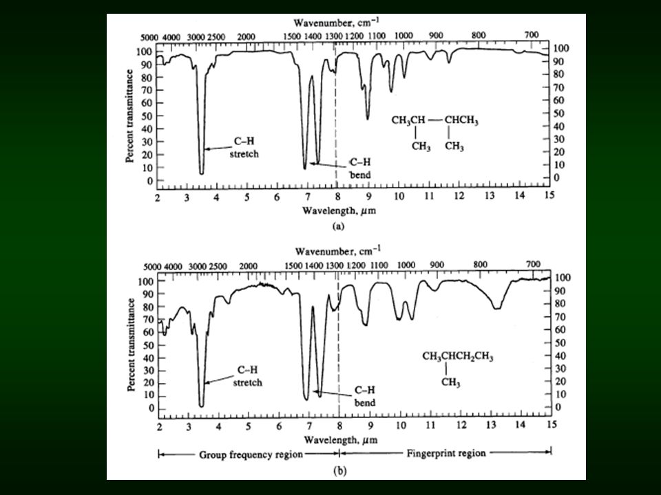
Group frequencies With certain functional or structural groups, it has been found that their vibrational frequencies are nearly independent of the rest of the molecule – group frequencies. Carbonyl group1650 to 1740 cm -1 various aldehydes and ketones For many groups involving only two atoms, the approximate frequency of the fundamental vibration can be calculated from a simple harmonic oscillator model. Calculations show that for most groups of interest, characteristic frequencies of stretching vibrations should lie in the region 4000 to 1000 cm -1. In practical, the region from 4000 to 1300 cm -1 is often called the group frequency region. The presence of various group vibrations in the IR spectrum is of great assistance in identifying the absorbing molecule.

26
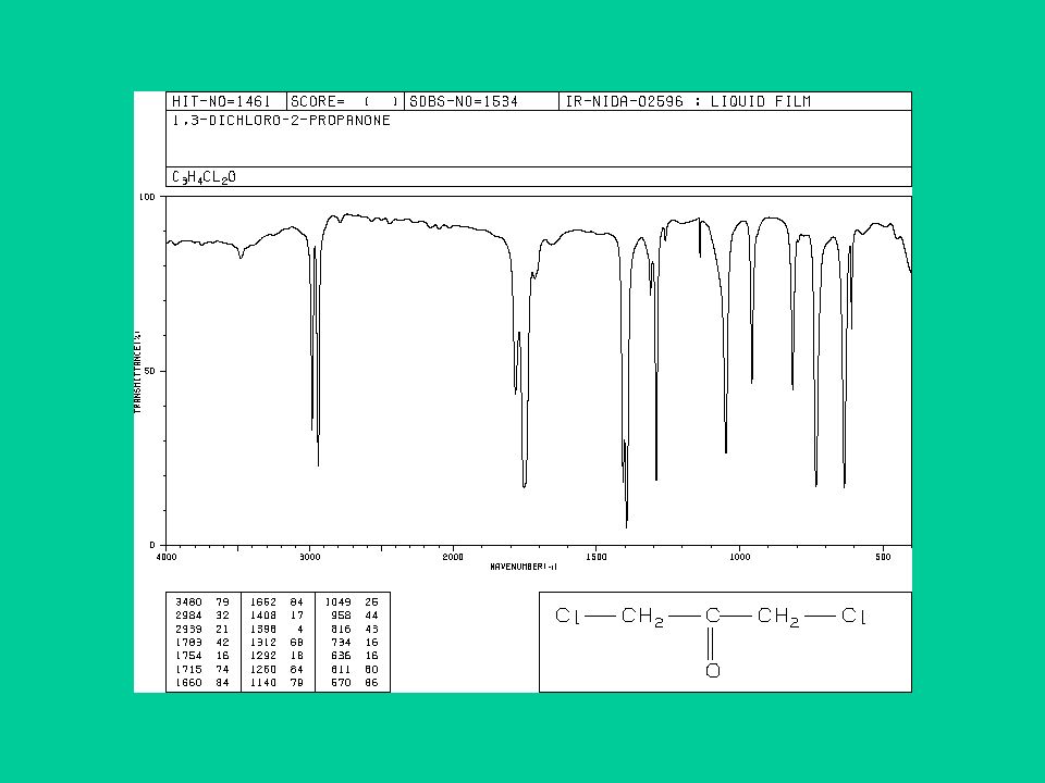
Fingerprint region In the region from 1300 to 400 cm -1, vibrational frequencies are affected by the entire molecule, as the broader ranges for group absorptions in the figure below – fingerprint region. Absorption in this fingerprint region is characteristic of the molecule as a whole. This region finds widespread use for identification purpose by comparison with library spectra.

29
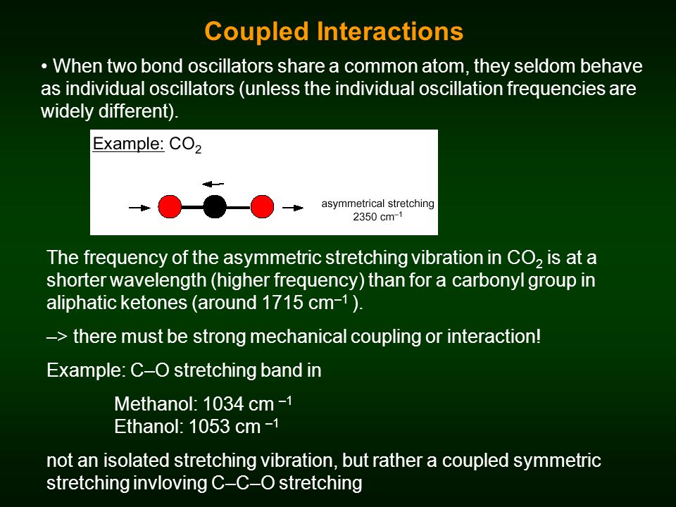
When two bond oscillators share a common atom, they seldom behave as individual oscillators (unless the individual oscillation frequencies are widely different). The frequency of the asymmetric stretching vibration in CO 2 is at a shorter wavelength (higher frequency) than for a carbonyl group in aliphatic ketones (around 1715 cm –1 ). –> there must be strong mechanical coupling or interaction! Example: C–O stretching band in Methanol: 1034 cm –1 Ethanol: 1053 cm –1 not an isolated stretching vibration, but rather a coupled symmetric stretching invloving C–C–O stretching Coupled Interactions
 than for a carbonyl group in aliphatic ketones (around 1715 cm –1 ). –> there must be strong mechanical coupling or interaction. Example: C–O stretching band in Methanol: 1034 cm –1 Ethanol: 1053 cm –1 not an isolated stretching vibration, but rather a coupled symmetric stretching invloving C–C–O stretching Coupled Interactions.")
30
Hydrogen bonding alters the force constant of both groups: – the X–H stretching bands move to lower frequency – the stretching frequency of the acceptor group (B) is also reduced, but to a lesser degree – The X–H bending vibration usually shifts to a shorter wavelength Effect of Hydrogen Bonding
 is also reduced, but to a lesser degree – The X–H bending vibration usually shifts to a shorter wavelength Effect of Hydrogen Bonding")
31
Carbon-Carbon Bond Stretching Stronger bonds absorb at higher frequencies: –C-C 1200 cm -1 –C=C 1660 cm -1 –C C 2200 cm -1 (weak or absent if internal) Conjugation lowers the frequency: –isolated C=C 1640-1680 cm -1 –conjugated C=C 1620-1640 cm -1 –aromatic C=C approx. 1600 cm -1 => Chapter 1230

32
Carbon-Hydrogen Stretching Bonds with more s character absorb at a higher frequency. –sp 3 C-H, just below 3000 cm -1 (to the right) –sp 2 C-H, just above 3000 cm -1 (to the left) –sp C-H, at 3300 cm -1 => Chapter 1231
 –sp 2 C-H, just above 3000 cm -1 (to the left) –sp C-H, at 3300 cm -1 => Chapter")
33
An Alkane IR Spectrum Chapter 1232 =>

34
An Alkene IR Spectrum Chapter 1233 =>

35
An Alkyne IR Spectrum Chapter 1234 =>

36
O-H and N-H Stretching Both of these occur around 3300 cm -1, but they look different. –Alcohol O-H, broad with rounded tip. –Secondary amine (R 2 NH), broad with one sharp spike. –Primary amine (RNH 2 ), broad with two sharp spikes. –No signal for a tertiary amine (R 3 N) => Chapter 1235
, broad with one sharp spike. –Primary amine (RNH 2 ), broad with two sharp spikes. –No signal for a tertiary amine (R 3 N) => Chapter")
37
An Alcohol IR Spectrum Chapter 1236 =>

38
Carbonyl Stretching The C=O bond of simple ketones, aldehydes, and carboxylic acids absorb around 1710 cm -1. Usually, it’s the strongest IR signal. Carboxylic acids will have O-H also. Aldehydes have two C-H signals around 2700 and 2800 cm -1. => Chapter 1237

39
A Ketone IR Spectrum Chapter 1238 =>

40
O-H Stretch of a Carboxylic Acid This O-H absorbs broadly, 2500-3500 cm -1, due to strong hydrogen bonding. Chapter 1239 =>

41
Carbon - Nitrogen Stretching C - N absorbs around 1200 cm -1. C = N absorbs around 1660 cm -1 and is much stronger than the C = C absorption in the same region. C N absorbs strongly just above 2200 cm - 1. The alkyne C C signal is much weaker and is just below 2200 cm -1. => Chapter 1240

42
A Nitrile IR Spectrum Chapter 1241 =>

43
Summary of IR Absorptions Chapter 1242 =>

44
Factors affecting the frequency of infrared peaks 1. Resonance and conjugation 2. Ring strain: A: on carbonyl frequencies B. on C-H stretching frequencies 3.Halogens A: on carbonyl frequencies B. on C-H stretching frequencies 4.Chirality 5.Phase: solid, liquid and gas (fundamentals in the gas phase are shifted to higher frequencies) ie. solvent or solute interactions lead to weakening of force constants; effects of H-bonding.
 ie. solvent or solute interactions lead to weakening of force constants; effects of H-bonding..")
45
Factors affecting the frequency of infrared peaks 1. Resonance, symmetry and conjugation

46
Effect of resonance, symmetry and conjugation on infrared frequencies What about the effect of conjugation? Do the facts support this interaction?

47
Carbonyl frequency: 1720-1680 (acid) 1700 cm -1 Ether frequency = 1000 to 1400 cm -1 1200 cm -1 Average of the two fundamentals = 1450 cm -1
 1700 cm -1 Ether frequency = 1000 to 1400 cm -1 1200 cm -1 Average of the two fundamentals = 1450 cm -1")
48
1410 cm -1 1560 cm -1 (1560+ 1410)/2 = 1485 cm -1
/2 = 1485 cm -1")
49
Figure IR-42. Sodium benzoate, KBr pellet: 15501400 Resonance

50
Sodium benzoate: (1550, 1400 cm -1 ); Average : 1475 cm -1 Sodium acetate: (1560, 1410 cm-1 ); Average : 1485 cm-1
; Average : 1475 cm -1 Sodium acetate: (1560, 1410 cm-1 ); Average : 1485 cm-1")
51
How important is resonance in amides?

52
In the barrier to rotation of the two CH 3 groups is approximately 18 kcal/mol (A C-C bond is worth about 60 kcal/mol)
")
54
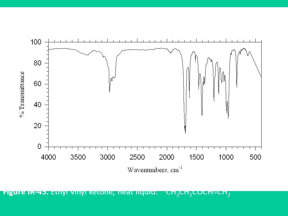
Other effects of conjugation on carbonyl frequencies Figure IR-45. Ethyl vinyl ketone, neat liquid: CH 3 CH 2 COCH=CH 2

55
Figure IR-44. 3-Nonen-2-one, 95%; neat liquid, thin film: Why the extra carbonyl peaks?

56
Factors affecting the intensities : Extent of interaction (dipole moment change) Concentration of each conformer
 Concentration of each conformer")
57
Effects of conjugation on double bonds

58
What’s this?

60
Factors affecting the frequency of infrared peaks 1. Resonance and conjugation 2. Ring strain: A: on carbonyl frequencies

61
1720 cm -1 1750 cm -1 1775 cm -1 cyclopropanone 1800 cm -1

62
Table. The Effect of Ring Strain on the Carbonyl Frequencies of Some Cyclic Molecules Ring Size ketone: cm -1 lactone: cm -1 lactam: cm -1 3 cyclopropanone: 1800 4 cyclobutanone: 1775 -propiolactone: 1840 5 cyclopentanone: 1751 -butyrolactone: 1750 -butyrolactam: 1690 6 cyclohexanone: 1715 -valerolactone: 1740 -valerolactam: 1668 7 cycloheptanone: 1702 caprolactone: 1730 caprolactam: 1658

63
Factors affecting the frequency of infrared peaks 1. Resonance and conjugation 2. Ring strain: A: on carbonyl frequencies B. on C-H stretching frequencies

65
What is the hybridization of a C-H bond in cyclopropane ?

66
Based on acidity, C-H bonds are somewhere between sp 2 and sp 3 This is also confirmed by 13 C-H coupling constant as we will see

69
Factors affecting the frequency of infrared peaks 1. Resonance and conjugation 2. Ring strain: A: on carbonyl frequencies B. on C-H stretching frequencies 3.Halogens A: on carbonyl frequencies B. on C-H stretching frequencies

70
1760 3011
72
Factors affecting the frequency of infrared peaks 1. Resonance and conjugation 2. Ring strain: A: on carbonyl frequencies B. on C-H stretching frequencies 3.Halogens A: on carbonyl frequencies B. on C-H stretching frequencies 4.Chirality

81
Capabilities of Infrared Analysis Identification and quantitation of organic solid, liquid or gas samples. Analysis of powders, solids, gels, emulsions, pastes, pure liquids and solutions, polymers, pure and mixed gases. Infrared used for research, methods development, quality control and quality assurance applications. Samples range in size from single fibers only 20 microns in length to atmospheric pollution studies involving large areas.

82
y axis is %T or A x axis is wavenumber (or wavelength) I o sample I T = I/I o %T = 100 I/I o T transmission / transmittance A = -log T A absorbance (no units) (Note A (but not T) concentration) IR spectrum
 I o sample I T = I/I o %T = 100 I/I o T transmission / transmittance A = -log T A absorbance (no units) (Note A (but not T) concentration) IR spectrum")
83
Absorbance spectrum of polystyrene –> generally used for quantitative work Ordinate Scaling Transmittance spectrum of polystyrene –> traditionally used for spectral interpretation

84
There is a substantial difference in “appearance”, whether the spectrum is linear in wavenumber (A, standard) or linear in wavelength (B): Abscissa Scaling (both spectra are recorded with the same sample) A B
 or linear in wavelength (B): Abscissa Scaling (both spectra are recorded with the same sample) A B")
85
BRUKE TENSOR TM Series Perkin Elmer TM Spectrum One Instrumentation

86
Dispersive instruments: with a monochromator to be used in the mid-IR region for spectral scanning and quantitative analysis. Fourier transform IR (FTIR) systems: widely applied and quite popular in the far-IR and mid-IR spectrometry. Nondispersive instruments: use filters for wavelength selection or an infrared-absorbing gas in the detection system for the analysis of gas at specific wavelength.
 systems: widely applied and quite popular in the far-IR and mid-IR spectrometry. Nondispersive instruments: use filters for wavelength selection or an infrared-absorbing gas in the detection system for the analysis of gas at specific wavelength..")
87
Dispersive IR spectrophotometers Simplified diagram of a double beam infrared spectrometer Modern dispersive IR spectrophotometers are invariably double-beam instruments, but many allow single-beam operation via a front-panel switch.

88
Double-beam operation compensates for atmospheric absorption, for the wavelength dependence of the source spectra radiance, the optical efficiency of the mirrors and grating, and the detector instability, which are serious in the IR region. single-beam instruments not practical. Double-beam operation allows a stable 100% T baseline in the spectra.

89
Dispersive spectrophotometers Designs Null type instrument

90
Instrumentation: IR Sources and Transducers Infrared sources consist of an inert solid that is electrically heated to a temperature between 1500 and 2200 K. The heated material will then emit infra red radiation. An ideal IR radiation source requirements: (1) be continuous over the wavelength range used, (2) cover a wide wavelength range, (3) be constant over long periods of time. The most common sources of IR radiation for the mid-IR region are: Nernst glowers, Globars, and heated wires. All of these heated sources emit continuous radiation, with a spectral output very similar to that of a blackbody radiation source. The range of light put out by mid-IR sources extends into both the NIR and far-IR regions, but intensity is at a maximum in the mid-IR region from 4000 to 400 cm -1. 64
 be continuous over the wavelength range used, (2) cover a wide wavelength range, (3) be constant over long periods of time. The most common sources of IR radiation for the mid-IR region are: Nernst glowers, Globars, and heated wires. All of these heated sources emit continuous radiation, with a spectral output very similar to that of a blackbody radiation source. The range of light put out by mid-IR sources extends into both the NIR and far-IR regions, but intensity is at a maximum in the mid-IR region from 4000 to 400 cm")
91
Components of dispersive spectrophotometers Nernst Glowerheated rare earth oxide rod (~1500 K) 1-50 µm (mid- to far-IR) Globarheated SiC rod (~1500 K)1-50 µm (mid- to far-IR) W filament lamp1100 K0.78-2.5 µm (Near-IR) Hg arc lampplasma50 - 300 µm (far-IR) CO2 laserstimulated emission lines9-11 µm 1. IR source

92
The Nernst glower Typically it was in the form of a cylindrical rod or tube having a diameter of 1-2 mm and length of 20 mm sealed by a platinum leads to the ends to permit electrical connection. It composed of a mixture of rare earth oxides such as zirconium oxide (ZrO 2 ), yttrium oxide (Y 2 O 3 ) and erbium oxide (Er 2 O 3 ) at a ratio of 90:7:3 by weight.zirconium oxideyttrium oxide They operated by being electrically heated to about 2000 °C. Initially they required external heating because the material is an insulator at room temperature. insulator Nernst glowers are fragile. They have a large negative temperature coefficient of electrical resistance and must be preheated to be conductive. Resistance decreases with increasing temperature the source circuit must be designed to limit the current to prevent rapid heating and destroyment 65
, yttrium oxide (Y 2 O 3 ) and erbium oxide (Er 2 O 3 ) at a ratio of 90:7:3 by weight.zirconium oxideyttrium oxide They operated by being electrically heated to about 2000 °C. Initially they required external heating because the material is an insulator at room temperature. insulator Nernst glowers are fragile. They have a large negative temperature coefficient of electrical resistance and must be preheated to be conductive. Resistance decreases with increasing temperature the source circuit must be designed to limit the current to prevent rapid heating and destroyment 65.")
93
The globar source A globar is a rod of silicon carbide (5 mm diameter, 50mm long) which is electrically heated to about 1500 K. Water cooling of the electrical contacts is needed to prevent arcing. no preheating necessary The spectral output is comparable with the Nernst glower, except at short wavelengths (less than 5 m) where it's output becomes larger. 67
 where it s output becomes larger. 67.")
94
The Tungsten Filament Lamp. Ordinary tungsten filament lamp (A quartz h alogen lamp contains a tungsten wire filament and iodine vapor sealed in a quartz envelope or bulb), used for near IR region of 4000-12,800 cm -1 (2.5-0.78 m) In a standard tungsten filament lamp, the tungsten evaporates from the filament and deposits on the lamp wall. This process reduces the light output as a result of the black deposit on the wall and the thinner filament. The halogen gas in a tungsten- halogen lamp removes the evaporated tungsten and redeposits it on the filament, increasing the light output and source stability 70
, used for near IR region of ,800 cm -1 ( m) In a standard tungsten filament lamp, the tungsten evaporates from the filament and deposits on the lamp wall. This process reduces the light output as a result of the black deposit on the wall and the thinner filament. The halogen gas in a tungsten- halogen lamp removes the evaporated tungsten and redeposits it on the filament, increasing the light output and source stability 70.")
95
IR Laser Sources A laser is a light source that emits very intense monochromatic radiation. Some lasers, called tunable lasers, emit more than one wavelength of light, but each wavelength emitted is monochromatic. The combination of high intensity and narrow line width makes lasers ideal light sources for some applications. Two types of IR lasers are available: gas phase and solid-state. Used in quantitative analysis 71

96
Diode Lasers Less expensive solid-state diode lasers with wavelengths in the NIR are available. Commercial instruments using multiple diode lasers are available for NIR analyses of food and fuels. Because of the narrow emission lines from a laser system, laser sources are often used in dedicated applications for specific analytes. They can be ideal for process analysis and product quality control, for example, but are not as flexible in their applications as a continuous source or a tunable laser. 73

97
Thermocouplethermoelectric effect - dissimilar metal junction cheap, slow, insensitive BolometerNi, Pt resistance thermometer (thermistor) Highly sensitive <400 cm -1 PyroelectricTri glycine sulfate piezoelectric material fast and sensitive (mid IR) PhotoconductingPbS, CdS, Pb Se light sensitive cells fast and sensitive (near IR) 2. Detector / transducer

98
First Type: Thermal Detectors 1- Thermocouple A thermocouple consists of a pair of junctions of different metals; for example, two pieces of bismuth fused to either end of a piece of antimony. The potential difference (voltage) between the junctions changes according to the difference in temperature between the junctions. Transducer junction: For IR radiation the junction is usually blackened to improve its heat absorbing capacity and sealed in an evacuated chamber with a window that is transparent to IR radiation. Reference Junction: Housed in the same chamber as the active junction but shielded from the incident radiation (have large heat capacity). Temperature response: In case of a well- designed thermocouple, temperature difference of 10 -6 K 79
 between the junctions changes according to the difference in temperature between the junctions. Transducer junction: For IR radiation the junction is usually blackened to improve its heat absorbing capacity and sealed in an evacuated chamber with a window that is transparent to IR radiation. Reference Junction: Housed in the same chamber as the active junction but shielded from the incident radiation (have large heat capacity). Temperature response: In case of a well- designed thermocouple, temperature difference of K 79.")
99
80

100
2- Thermopile To enhance sensitivity several thermocouples connected in series are called a thermopile. 81

101
3- Bolometer: Far-IR detector. A bolometer functions by changing resistance when heated. It is constructed of strips of metals such as platinum, germanium or nickel or from a semiconductor (thermistor). Relatively large change in resistance as a function of temperature is resulted (large response time). Rarely used in mid IR as other IR transducers. IR range of Ge bolometer 5- 400 cm -1 (2000-25 mm) 82
. Relatively large change in resistance as a function of temperature is resulted (large response time). Rarely used in mid IR as other IR transducers. IR range of Ge bolometer cm -1 ( mm) 82.")
102
Second Type: Pyroelectric detectors Pyroelectric detectors consists of a pyroelectric material which is an insulator (dielectric materials) with special thermal and electric properties. Triglycine sulphate (NH 2 CH 2 COOH) 3.H 2 SO 4 (usually deuterated or with a fraction of glycines replaced by alanine) is the most common material for pyroelectric infrared detectors. Unlike other thermal detectors the pyroelectric effect depends on the rate of change of the detector temperature rather than on the temperature itself. 83
 3.H 2 SO 4 (usually deuterated or with a fraction of glycines replaced by alanine) is the most common material for pyroelectric infrared detectors. Unlike other thermal detectors the pyroelectric effect depends on the rate of change of the detector temperature rather than on the temperature itself. 83.")
103
Detector Response Time The length of time that a detector takes to reach a steady signal when radiation falls on it. This varies greatly with the type of detector used and has a significant influence on the design of the IR instrument. Thermal detectors: such as thermocouples, thermistors, and bolometers have very slow response times, on the order of seconds. Consequently, when a spectrum is being scanned, it takes several seconds for the detector to reach an equilibrium point and thus give a true reading of the radiation intensity falling on it. 88

104
Infrared Instruments:Dispersive Instruments: Dispersive spectrometers, introduced in the mid- 1940s and widely used since, provided the robust instrumentation required for the extensive application of this technique. They are generally double beam, recording instruments, which use reflection gratings: Why double beam: Because of the low intensity of IR radiation. Low sensitivity of IR transducers. Large signal amplification needed 89

105
If the detector is not exposed to the light long enough, it will not reach equilibrium and an incorrect absorption trace will be obtained. Dispersive IR instruments with older-style thermal detectors to take on the order of 15 min to complete an IR scan. Attempts to decrease this time resulted in errors in the intensity of the absorption bands and recording of distorted shapes of the bands. It should also be remembered that when an absorption band is reached, the intensity falling on the detector decreases and the response depends on how fast the detector cools.

106
3. Optical system

107
Reflection gratings ( made from various plastics): the groove spacing is greater (e.g. 120 grooves mm -1 ). To reduce the effect of overlapping orders and stray radiation, filters or a preceding prism are usually employed. Two or more gratings are often used with several filters to scan a wide region. Mirrors but not lenses are used to focus and collimate the IR radiation. Generally made from Pyrex or another material with low coefficient of thermal expansion. Front surfaces coated with a vacuum-deposited thin metal film of Al, Ag, or Au.
. To reduce the effect of overlapping orders and stray radiation, filters or a preceding prism are usually employed. Two or more gratings are often used with several filters to scan a wide region. Mirrors but not lenses are used to focus and collimate the IR radiation. Generally made from Pyrex or another material with low coefficient of thermal expansion. Front surfaces coated with a vacuum-deposited thin metal film of Al, Ag, or Au..")
108
Windows are used for sample cells and to permit various compartment to be isolated from the environment. transparent to IR over the wavelength region inert to the various chemicals analyzed capable of being shaped, ground, and polished to the desired optical quality

109
The Fourier transform method provides an alternatives to the use of monochromators based on dispersion. In conversional dispersive spectroscopy, frequencies are separated and only a small portion is detected at any particular instant, while the remainder is discarded. The immediate result is a frequency- domain spectrum. Fourier transform infrared spectroscopy generates time-domain spectra as the immediately available data, in which the intensity is obtained as a function of time. Direct observation of a time-domain spectrum is not immediately useful because it is not possible to deduce, by inspection, frequency-domain spectra from the corresponding time-domain waveform (Fourier transform is thus introduced). Fourier Transform Infrared Spectrometer (FTIR)
. Fourier Transform Infrared Spectrometer (FTIR).")
110
In one arm of the interferometer, the IR source radiation travels through the beam splitter to the fixed mirror back to the beam splitter through the sample and to the detector. In the other arm, the IR source radiation travels to the beam splitter to the movable mirror, back through the beam splitter to the sample and to the detector. The difference in pathlengths of the two beams is the retardation . An He-NE laser is used as a monochromatic reference source. The laser beam is sent through the interferometer in the opposite direction to that of the IR beam. Single-beam FTIR Spectrometer

111
Double-beam FTIR Spectrometer

112
Interferometer Michelson interferometer If moving mirror moves 1/4 (1/2 round-trip) waves are out of phase at beam- splitting mirror - no signal If moving mirror moves 1/2 (1 round-trip) waves are in phase at beam-splitting mirror – signal...
 waves are out of phase at beam- splitting mirror - no signal If moving mirror moves 1/2 (1 round-trip) waves are in phase at beam-splitting mirror – signal...")
113
Principle of operation of FTIR spectrometer Determine s datapoint sampling interval Photodiode

114
very high resolution (< 0.1 cm –1 ) Two closely spaced lines only separated if one complete "beat" is recorded. As lines get closer together, must increase. (cm 1 ) 1/ Mirror motion is 1/2 Resolution governed by distance movable mirror travels very high sensitivity (nanogram quantity) can be coupled with GC analysis (–> measure IR spectra in gas-phase) High S/N ratios - high throughput Few optics, no slits mean high intensity of light Rapid (<10 s) Reproducible and Inexpensive Advantages of FTIR
 1/ Mirror motion is 1/2 Resolution governed by distance movable mirror travels very high sensitivity (nanogram quantity) can be coupled with GC analysis (–> measure IR spectra in gas-phase) High S/N ratios - high throughput Few optics, no slits mean high intensity of light Rapid (<10 s) Reproducible and Inexpensive Advantages of FTIR.")
115
Usually to improve resolution decrease slit width but less light makes spectrum "noisier" - signal to noise ratio (S/N) n # scans S/N improves with more scans (noise is random, signal is not!) To improve S/N ratio
 n # scans S/N improves with more scans (noise is random, signal is not!) To improve S/N ratio")
116
For routine instrument calibration, run the spectrum of polystyrene film (or indene) at resolution 2 cm -1. Band positions are available in the literature. Higher resolution calibrations may be made from gas- phase spectra (e.g. HCl gas). Spectrum calibration
. Spectrum calibration.")
117
Sample preparation techniques The preparation of samples for infrared spectrometry is often the most challenging task in obtaining an IR spectrum. Since almost all substances absorb IR radiation at some wave length, and solvents must be carefully chosen for the wavelength region and the sample of interest. Infrared spectra may be obtained for gases, liquids or solids (neat or in solution)
.")
118
A gas sample cell consists of a cylinder of glass or sometimes a metal. The cell is closed at both ends with an appropriate window materials (NaCl/KBr) and equipped with valves or stopcocks for introduction of the sample. Long pathlength ( 10 cm) cells – used to study dilute (few molecules) or weakly absorbing samples. Multipass cells – more compact and efficient instead of long-pathlength cells. Mirrors are used so that the beam makes several passes through the sample before exiting the cell. (Effective pathlength 10 m). To resolve the rotational structure of the sample, the cells must be capable of being evacuated to measure the spectrum at reduced pressure. For quantitative determinations with light molecules, the cell is sometimes pressurized in order to broaden the rotational structure and all simpler measurement. Gas samples
 and equipped with valves or stopcocks for introduction of the sample. Long pathlength ( 10 cm) cells – used to study dilute (few molecules) or weakly absorbing samples. Multipass cells – more compact and efficient instead of long-pathlength cells. Mirrors are used so that the beam makes several passes through the sample before exiting the cell. (Effective pathlength 10 m). To resolve the rotational structure of the sample, the cells must be capable of being evacuated to measure the spectrum at reduced pressure. For quantitative determinations with light molecules, the cell is sometimes pressurized in order to broaden the rotational structure and all simpler measurement. Gas samples.")
119
Pure or soluted in transparent solvent – not water (attacks windows) The sample is most often in the form of liquid films (“sandwiched” between two NaCl plates) Adjustable pathlength (0.015 to 1 mm) – by Teflon spacer Liquid samples
 The sample is most often in the form of liquid films ( sandwiched between two NaCl plates) Adjustable pathlength (0.015 to 1 mm) – by Teflon spacer Liquid samples")
120
Regions of transparency for common infrared solvents. The horizontal lines indicate regions where solvent transmits at least 25% of the incident radiation in a 1-mm cell.

121
Solid samples Spectra of solids are obtained as alkali halide discs (KBr), mulls (e.g. Nujol, a highly refined mixture of saturated hydrocarbons) and films (solvent or melt casting) Alkali halide discs: 1.A milligram or less of the fine ground sample mixed with about 100 mg of dry KBr powder in a mortar or ball mill. 2.The mixture compressed in a die to form transparent disc. Mulls 1.Grinding a few milligrams of the powdered sample with a mortar or with pulverizing equipment. A few drops of the mineral oil added (grinding continued to form a smooth paste). 2.The IR of the paste can be obtained as the liquid sample.
 and films (solvent or melt casting) Alkali halide discs: 1.A milligram or less of the fine ground sample mixed with about 100 mg of dry KBr powder in a mortar or ball mill. 2.The mixture compressed in a die to form transparent disc. Mulls 1.Grinding a few milligrams of the powdered sample with a mortar or with pulverizing equipment. A few drops of the mineral oil added (grinding continued to form a smooth paste). 2.The IR of the paste can be obtained as the liquid sample..")
122
1. Fundamental chemistry Determination of molecular structure/geometry. e.g. Determination of bond lengths, bond angles of gaseous molecules 2. Qualitative analysis – simple, fast, nondestructive Monitoring trace gases: NDIR.Rapid, simultaneous analysis of GC, moisture, N in soil. Analysis of fragments left at the scene of a crime Quantitative determination of hydrocarbons on filters, in air, or in water Main uses of IR spectroscopy:

123
Near-infrared and Far-infrared absorption The techniques and applications of near-infrared (NIR) and far-infrared (FIR) spectrometry are quite different from those discussed above for conventional, mid-IR spectrometry. Near-infrared: 0.8 -2.5 m, 12500 - 4000 cm -1 Mid-infrared: 2.5 - 50 m, 4000 - 200 cm -1 Far-infrared: 50 - 1000 m, 200 - 10 cm -1

124
Near-infrared spectrometry NIR shows some similarities to UV-visible spectrophotometry and some to mid-IR spectrometry. Indeed the spectrophotometers used in this region are often combined UV-visible-NIR ones. The majority of the absorption bands observed are due to overtones (or combination) of fundamental bands that occur in the region 3 to 6 m, usually hydrogen-stretching vibrations. NIR is most widely used for quantitative organic functional-group analysis. The NIR region has also been used for qualitative analyses and studies of hydrogen bonding, solute-solvent interactions, organometallic compounds, and inorganic compounds.
 of fundamental bands that occur in the region 3 to 6 m, usually hydrogen-stretching vibrations. NIR is most widely used for quantitative organic functional-group analysis. The NIR region has also been used for qualitative analyses and studies of hydrogen bonding, solute-solvent interactions, organometallic compounds, and inorganic compounds..")
125
Far-infrared spectrometry Almost all FIR studies are now carried out with FTIR spectrometers. The far-IR region can provide unique information. i)The fundamental vibrations of many organometallic and inorganic molecules fall in this region due to the heavy atoms and weak bonds in these molecules. ii)Lattice vibrations of crystalline materials occur in this region, iii)Electron valence/conduction band transition in semiconductors often correspond to far-IR wavelengths.
The fundamental vibrations of many organometallic and inorganic molecules fall in this region due to the heavy atoms and weak bonds in these molecules. ii)Lattice vibrations of crystalline materials occur in this region, iii)Electron valence/conduction band transition in semiconductors often correspond to far-IR wavelengths..")
126
Infrared Spectrum of CCl 4

129
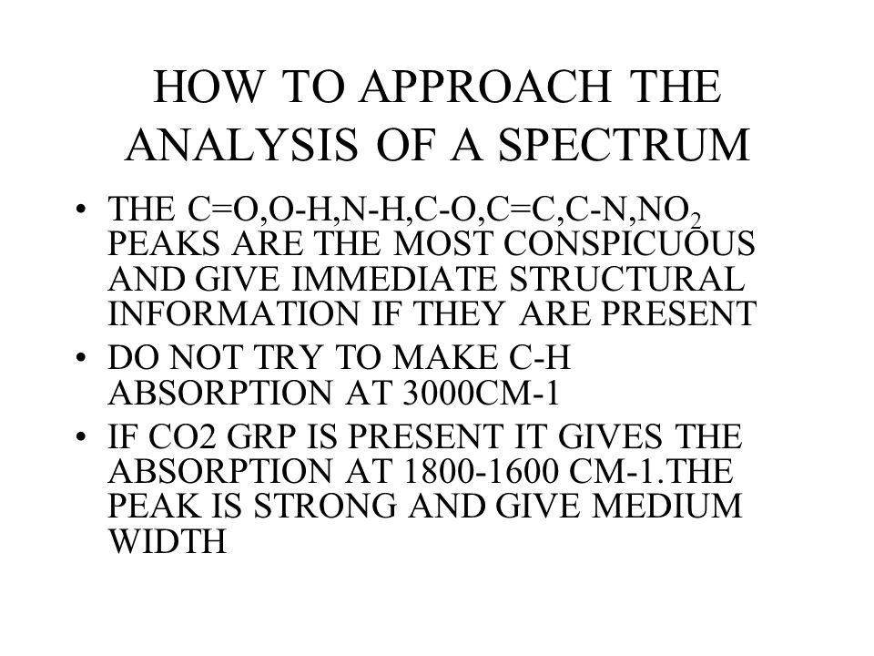
HOW TO APPROACH THE ANALYSIS OF A SPECTRUM THE C=O,O-H,N-H,C-O,C=C,C-N,NO 2 PEAKS ARE THE MOST CONSPICUOUS AND GIVE IMMEDIATE STRUCTURAL INFORMATION IF THEY ARE PRESENT DO NOT TRY TO MAKE C-H ABSORPTION AT 3000CM-1 IF CO2 GRP IS PRESENT IT GIVES THE ABSORPTION AT 1800-1600 CM-1.THE PEAK IS STRONG AND GIVE MEDIUM WIDTH

135
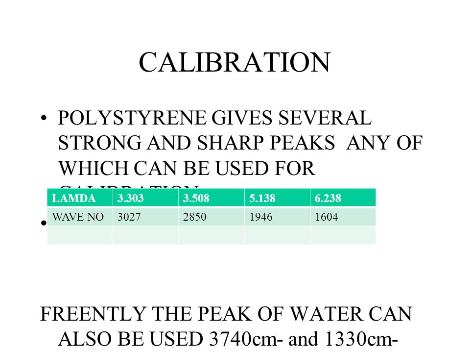
CALIBRATION POLYSTYRENE GIVES SEVERAL STRONG AND SHARP PEAKS ANY OF WHICH CAN BE USED FOR CALIBRATION FREENTLY THE PEAK OF WATER CAN ALSO BE USED 3740cm- and 1330cm- LAMDA3.3033.5085.1386.238 WAVE NO3027285019461604

136
Capabilities of Infrared Analysis Identification and quantitation of organic solid, liquid or gas samples. Analysis of powders, solids, gels, emulsions, pastes, pure liquids and solutions, polymers, pure and mixed gases. Infrared used for research, methods development, quality control and quality assurance applications. Samples range in size from single fibers only 20 microns in length to atmospheric pollution studies involving large areas.

137
ATTENUATED TOTAL REFLECTION SPECTROSCOPY

138
Outline Introduction to ATR technology Evanescent Waves Crystal Composition Data Collection Advantages & Disadvantages Sample Analysis Challenges Future Applications Conclusion

139
Attenuated total reflection (ATR) spectroscopy is a versatile and powerful technique for infrared sampling
 spectroscopy is a versatile and powerful technique for infrared sampling")
140
How an ATR accessory works Internal reflection spectroscopy passes infrared radiation through an infrared-transmitting crystal of high refractive index, allowing the radiation to reflect in the crystal one or more times An attenuated total reflection accessory measures the totally reflected infrared beam when the beam comes in contact with a sample In this way, an evanescent wave penetrates into the sample in contact with the crystal, producing a spectrum of the sample

141
Evanescent Waves The infrared radiation interacts with the sample through a series of standing waves, called evanescent waves An evanescent wave is a penetrating electromagnetic field whose intensity quickly decays as it moves away from its source

143
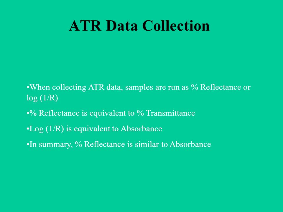
ATR Data Collection When collecting ATR data, samples are run as % Reflectance or log (1/R) % Reflectance is equivalent to % Transmittance Log (1/R) is equivalent to Absorbance In summary, % Reflectance is similar to Absorbance
 % Reflectance is equivalent to % Transmittance Log (1/R) is equivalent to Absorbance In summary, % Reflectance is similar to Absorbance")
144
Disadvantages ATR vs. Transmission The ATR crystal absorbs energy at lower energy levels If the sample does not have good contact with the crystal, the data will not be accurate Most ATR crystals have pH limitations

145
Crystal Composition Crystals are typically made of: Zinc Selenide (ZnSe) Germanium (Ge) Zinc Sulfide (ZnS) Silicon (Si) Diamond AMTIR: Germanium, Arsenic, Selenium (GeAsSe)
 Germanium (Ge) Zinc Sulfide (ZnS) Silicon (Si) Diamond AMTIR: Germanium, Arsenic, Selenium (GeAsSe)")
146
Advantages ATR vs. Transmission When an ATR accessory is used, most samples can be run “neat”, which means “in their natural state” ATR sampling is fast and easy because little or no sample preparation is required Other techniques, such as infrared transmission, often require the sample to be heated, pressed or ground in order to collect the spectrum

147
Solid analysis ATR is an excellent technique for measuring the composition of solids Some examples of solids are films, fabrics, paper, hard polymer sheets, glass, rubber ATR is an ideal technique for measuring dark colored materials which often absorb too much energy to be measured by IR transmission

148
Liquid Analysis ATR is an ideal technique for analyzing liquids Sample preparation is minimal Cleanup is easy and fast

149
Liquid Analysis ATR can be used to analyze non-aqueous solutions such a lubricants, oils, paints, glues, solvents, inks and dyes Gels and pastes can also be analyzed

150
Powder Analysis Powders are easier to run by ATR than by IR transmission, because little or no preparation is required This category includes pure samples and mixtures that are available in powdered from Some examples of pure samples and mixtures are pharmaceuticals and pigments

151
Challenges Alignment of the beamsplitter Technique will change with sample type

152
Future Applications Forensic Investigation Biomedical applications

153
Attenuated Total Reflectance is an easy-to-use, fast, and versatile technique for infrared sampling Solids, pastes, gels, liquids and powders can be analyzed with little or no preparation Conclusion

Similar presentations


 Spectroscopy>")


