Download presentation
Presentation is loading. Please wait.

1
Immunology Laboratory Equipment

2
1- Centrifuge Is equipment used to separate substances of greater and lesser density. Simple centrifuges are used in chemistry, biology, and biochemistry for isolating and separating suspensions. They vary widely in speed and capacity. They usually comprise a rotor containing two, four, six, or many more numbered wells within which the samples, contained in centrifuge tubes, may be placed.

3
Microcentrifuge tube Microcentrifuge tubes are small, cylindrical plastic containers with conical bottoms, typically with an integral snap cap. They are used in molecular biology and biochemistry to store and centrifuge small amounts of liquid.

4
2- Incubator In biology, an incubator is a device used to grow and maintain of course microbiological cultures or cell cultures. The incubator maintains optimal temperature, humidity and other conditions such as the carbon dioxide (CO2) and oxygen content of the atmosphere inside. Incubators are essential for a lot of experimental work in cell biology, microbiology and molecular biology and are used to culture both bacterial as well as eukaryotic cells. Most incubators include a timer; some can also be programmed to cycle through different temperatures, humidity levels, etc. Incubators can vary in size from tabletop to units the size of small rooms.
 and oxygen content of the atmosphere inside. Incubators are essential for a lot of experimental work in cell biology, microbiology and molecular biology and are used to culture both bacterial as well as eukaryotic cells. Most incubators include a timer; some can also be programmed to cycle through different temperatures, humidity levels, etc. Incubators can vary in size from tabletop to units the size of small rooms.")
5
3- Spectrophotometer Is a photometer (a devise for measuring light intensity) that can measure intensity as a function of the color, or more specifically, the wavelength of light.
 that can measure intensity as a function of the color, or more specifically, the wavelength of light.")
6
4- Micro plate reader (ELISA plate reader)
Microplate Readers (also known as plate readers) are laboratory instruments designed to detect biological, chemical or physical events of samples in microtiter plate. Sample reactions can be assayed in well format microtiter plates. The most common microplate format used in academic research laboratories or clinical diagnostic laboratories is 96-well. In most cases, a high-intensity lamp passes light to the microtiter well and detector quantifies the light emitted by the reaction happening in the microplate well.
 are laboratory instruments designed to detect biological, chemical or physical events of samples in microtiter plate. Sample reactions can be assayed in well format microtiter plates. The most common microplate format used in academic research laboratories or clinical diagnostic laboratories is 96-well. In most cases, a high-intensity lamp passes light to the microtiter well and detector quantifies the light emitted by the reaction happening in the microplate well.")
7
Micro plate detection uses:
ELISAs (Enzyme-linked immunosorbent assay, a biochemical technique used mainly in immunology to detect antibodies or antigens). Protein and cell growth assays Nucleic acid quantitation Molecular interactions Enzyme activity Cell toxicity, proliferation, and viability ATP quantification Immunoassays High throughput screening of compounds and targets in drug discovery such as FLIPR assays.
. Protein and cell growth assays. Nucleic acid quantitation. Molecular interactions. Enzyme activity. Cell toxicity, proliferation, and viability. ATP quantification. Immunoassays. High throughput screening of compounds and targets in drug discovery such as FLIPR assays.")
8
5- Microtiter plate (ELISA plate -8x12-) and ELISA pipette
Is a flat plate with multiple ‘’wells’’ used as small test tubes. The microplate has become a standard tool in analytical research and clinical diagnostic testing laboratories. A very common usage is in the enzyme-linked immunosorbent assay (ELISA).
.")
10
6- Autoclave An autoclave is a pressurized device designed to heat aqueous solutions above their boiling point to achieve sterilization.

11
7- pipette Also called a pipet, pipettor or medicine dropper.
Is a laboratory instrument used to transport a measured volume of liquid.

12
8- Homogenizer A homogenizer is a piece of laboratory equipment used for the homogenization of various types of material, such as tissue, plant, food, soil, and many others. Homogenization is a very common sample preparation step prior to the analysis of nucleic acids, proteins, cells, metabolism, pathogens, and many other targets.

13
9- Refrigerators For small cell culture laboratories, a domestic refrigerator (preferably one without an autodefrost freezer) is an adequate and inexpensive piece of equipment for storing reagents and media at 2–8°C. For larger laboratories, a cold room restricted to cell culture is more appropriate. Make sure that the refrigerator or the cold room is cleaned regularly to avoid contamination.
 is an adequate and inexpensive piece of equipment for storing reagents and media at 2–8°C. For larger laboratories, a cold room restricted to cell culture is more appropriate. Make sure that the refrigerator or the cold room is cleaned regularly to avoid contamination.")
14
10- Freezers Most cell culture reagents can be stored at –5°C to –20°C; therefore an ultradeep freezer (i.e., a –80°C freezer) is optional for storing most reagents. While most reagents can withstand temperature oscillations in an autodefrost (i.e., self-thawing) freezer, some reagents such as antibiotics and enzymes should be stored in a freezer that does not autodefrost.
 is optional for storing most reagents. While most reagents can withstand temperature oscillations in an autodefrost (i.e., self-thawing) freezer, some reagents such as antibiotics and enzymes should be stored in a freezer that does not autodefrost.")
15
11- PH meter is an electronic instrument used for measuring the pH (acidity or alkalinity) of a liquid . A typical pH meter consists of a special measuring probe (a glass electrode) connected to an electronic meter that measures and displays the pH reading.
 connected to an electronic meter that measures and displays the pH reading.")
16
Leukocyte Total and Differential Count
Lab 2

17
WBC TOTAL COUNT Can be determined:
manually by haemocytometer and microscope automated by haematological analysers by estimation from a blood smear

18
-Leukocyte estimation in a blood smear (400x):
Normal number Increased Decreased

19
DIFFERENTIAL LEUKOCYTE COUNT
Can be determined: from a stained blood smear automated by some haematological analysers, but these need prior validation

20
DIFFERENTIAL COUNT IN A STAINED BLOOD SMEAR: BATTLESHIP METHOD

21
How to perfom a differential count:
Count leukocytes, note each cellular type identified Avoid counting the same field twice Do not focus on one section of the smear

22
TYPES OF LEUKOCYTES GRANULOCYTES: Neutrophils, Eosinophils, Basophils
MONOCYTES - LYMPHOCYTES

23
Normal neutrophils and a lymphocyte
Neutrophils are similar in most domestic species, characterised by pale cytoplasm and dark nucleus segmented into 2-5 lobes, although granules are more readily seen in ruminants.

24
Small and medium lymphocyte, cow
Small lymphocytes, dog

25
Normal neutrophil and monocyte
Monocytes are similar in most species; large cells with basophilic cytoplasm (often vacuolated or with tiny pink granules), and a pale irregularly clefted or folded nucleus with clumped chromatin.
, and a pale irregularly clefted or folded nucleus with clumped chromatin.")
26
Normal eosinophil and basophil
Morphology of eosinophils varies widely with species; granules are large in horses, small & numerous in ruminants, accompanied by vacuoles in dogs, and rod-shaped in cats. Basophils have purple-black granules, are numerous in large animals and may be almost absent in healthy dogs and cats.

27
Normal WBCs in different species, some examples
(1) (2) Eosinophil Eosinophil and neutrophil (3) Basophil (4) Eosinophil and neutrophils
 (2) Eosinophil. Eosinophil and neutrophil. (3) Basophil. (4) Eosinophil and neutrophils.")
30
REPORTING A DIFFERENTIAL COUNT
By relative % of each WBC type (INCORRECT) Ways to report the values By absolute number of each WBC type (CORRECT) (%of each cell type x total WBC number)
 Ways to. report the. values. By absolute number of each WBC type. (CORRECT) (%of each cell type x total WBC number)")
31
Lab 3 Serological test

32
Serological test (Antigen antibody interactions)
Serology is a blood test to detect the presence of antibodies against a microorganism (antigen). Certain microorganisms stimulate the body to produce antibodies during an active infection.
. Certain microorganisms stimulate the body to produce antibodies during an active infection.")
33
Blood is drawn from a vein (venipuncture), usually from the inside of the elbow or the back of the hand. A needle is inserted into the vein, and the blood is collected in an air-tight vial or a syringe. Preparation may vary depending on the specific test.

34
There are several serology techniques that can be used depending on the suspected antibodies. Serology techniques include Primary serological tests: (Marker techniques) e.g. Enzyme linked immuono sorben assay (ELISA) Immuno flurescent antibody technique (IFAT) Radio immuno assay (RIA) Secondary serological tests: e.g. Agglutination tests Complement fixation tests (CFT) Precipitation tests Serum neutralization tests (SNT) Toxin-antitoxin test Tertiary serological test: e.g. Determination of the protective value of an anti serum in an animal.
 e.g. Enzyme linked immuono sorben assay (ELISA) Immuno flurescent antibody technique (IFAT) Radio immuno assay (RIA) Secondary serological tests: e.g. Agglutination tests. Complement fixation tests (CFT) Precipitation tests. Serum neutralization tests (SNT) Toxin-antitoxin test. Tertiary serological test: e.g. Determination of the protective value of an anti serum in an animal.")
35
Agglutination tests Agglutination is visible clumping of particles, cells and bacteria by an antigen combining with its specific antibody. Such antibodies called agglutinating antibodies and the reaction is called agglutination. The resulting clumps are referred to as agglutinates.

36
There are three types of Agglutination tests:
First: Direct whole pathogen Agglutination: When the antigen is particulate, the reaction of an antibody with the antigen can be detected by agglutination (clumping) of the antigen. The general term agglutinin is used to describe antibodies that agglutinate particulate antigens. When the antigen is an erythrocyte the term heamagglutination is used.
 of the antigen. The general term agglutinin is used to describe antibodies that agglutinate particulate antigens. When the antigen is an erythrocyte the term heamagglutination is used.")
37
To assess bacterial infections:
For antibody detect, bacterial agglutination tests can be performed on the surface of glass slides or in tubes. Tube agglutination tests are often more sensitive, since a long incubation period (allowing more antigen and antibodies to interact) can be used. The small volume of liquid used for slide tests requires rather rapid reading of the result before the liquid has evaporated. e.g. A rise in titer of an antibody to a particular bacterium indicates an infection with that bacterial type. Determination of blood types or antibodies to blood group antigens + Rh: A blood group is a classification of blood based on the presence or absence of inherited antigenic substances on the surface of red blood cells (RBCs). These antigens may be proteins, carbohydrates, glycoproteins, or glycolipids, depending on the blood group system. - ABO test
 can be used. The small volume of liquid used for slide tests requires rather rapid reading of the result before the liquid has evaporated. e.g. A rise in titer of an antibody to a particular bacterium indicates an infection with that bacterial type. Determination of blood types or antibodies to blood group antigens + Rh: A blood group is a classification of blood based on the presence or absence of inherited antigenic substances on the surface of red blood cells (RBCs). These antigens may be proteins, carbohydrates, glycoproteins, or glycolipids, depending on the blood group system. - ABO test.")
38
ABO test The ABO test shows that people have one of four blood types: A, B, AB, or O. If your red blood cells have: The A antigen, you have type A blood. The liquid portion of your blood (plasma) has antibodies that fight against type B blood. The B antigen, you have type B blood. Your plasma has antibodies that fight against type A blood. Neither the A nor B antigen, you have type O blood. Your plasma has antibodies that fight against both type A and type B blood. Both the A and B antigens, you have type AB blood. Your plasma does not have antibodies against type A or type B blood.
 has antibodies that fight against type B blood. The B antigen, you have type B blood. Your plasma has antibodies that fight against type A blood. Neither the A nor B antigen, you have type O blood. Your plasma has antibodies that fight against both type A and type B blood. Both the A and B antigens, you have type AB blood. Your plasma does not have antibodies against type A or type B blood.")
41
How to read your results
No Agglutination -ve result Agglutination +ve result

42
Second: Particle ( indirect ) Agglutination Tests:
These tests based on the detection of antibody via the agglutination of an artificial carrier particle with antigen bound to its surface. The carrier particle may be latex, treated erythrocytes and staphylococcal particle agglutination system (coagulation). When the carrier is erythrocyte the name of the test will be indirect or Passive Hemagglutination test: it is possible to coat erythrocytes with a soluble antigen (e.g. viral antigen, a polysaccharide or a hapten; A hapten is a small molecule that can elicit an immune response only when attached to a large carrier such as a protein) and use the coated red blood cells in an agglutination test for antibody to the soluble antigen. This is called passive hemagglutination. Antibody detection to Cytomegalovirus, Rubella virus, Varicella-zoster virus and others. The treponema pallidum particle agglutination assay (also called TPPA test) is an indirect agglutination assay used for detection and titration of antibodies against the causative agent of syphilis.
. When the carrier is erythrocyte the name of the test will be indirect or Passive Hemagglutination test: it is possible to coat erythrocytes with a soluble antigen (e.g. viral antigen, a polysaccharide or a hapten; A hapten is a small molecule that can elicit an immune response only when attached to a large carrier such as a protein) and use the coated red blood cells in an agglutination test for antibody to the soluble antigen. This is called passive hemagglutination. Antibody detection to Cytomegalovirus, Rubella virus, Varicella-zoster virus and others. The treponema pallidum particle agglutination assay (also called TPPA test) is an indirect agglutination assay used for detection and titration of antibodies against the causative agent of syphilis.")
43
Serological test (cont.)
Lab 4 Serological test (cont.)
")
44
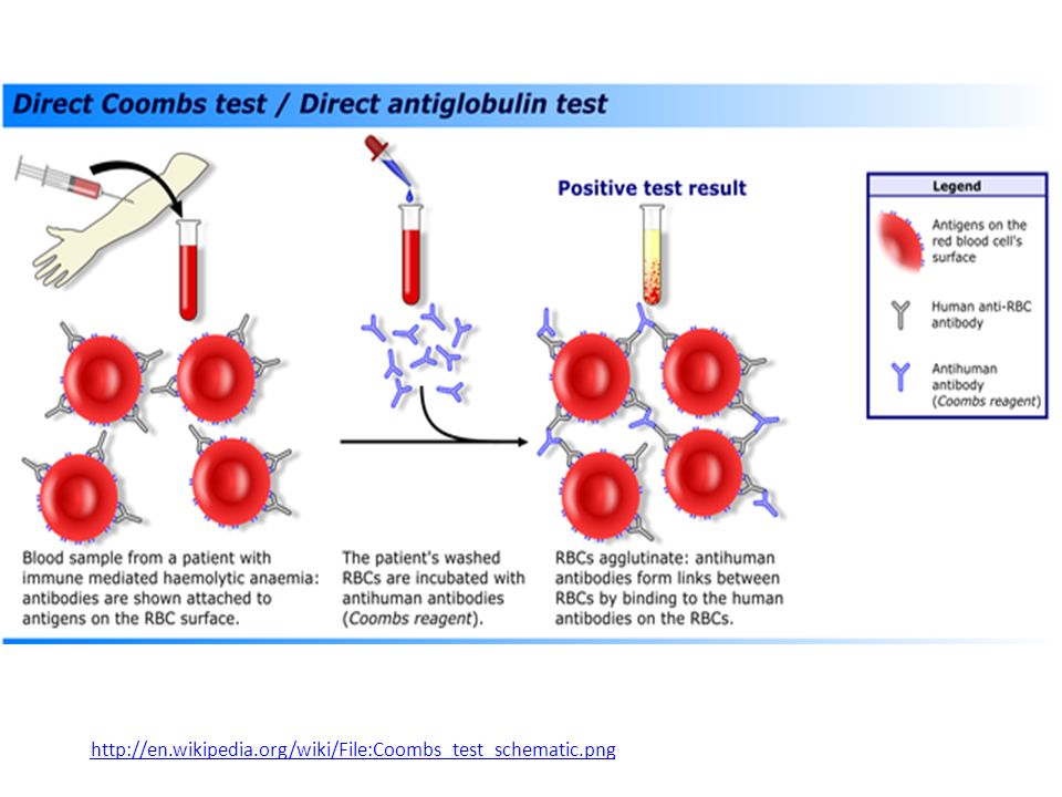
Third : Coombs Test (Antiglobulin Test)
This test used to detect antibodies which produced by the body itself and analyze the red blood cells. This condition happens in autoimmune hemolytic anemia or in hemolytic disease of new born (RH disease). There are two types of coombs test: The direct coombs test. The indirect coombs test.
. There are two types of coombs test: The direct coombs test. The indirect coombs test.")
45
The direct coombs test Also known as the direct antiglobulin test or DAT. The Direct Coombs test is used in vivo to test for autoimmune hemolytic anemia; ie, a condition of a low count of red blood cells (aka RBCs) caused by immune system lysis or breaking of RBC membranes causing RBC destruction. In certain diseases or conditions an individual’s blood may contain IgG antibodies that can specifically bind to antigens on the red blood cell surface membrane, and their circulating red blood cell can become coated with IgG autoantibodies. The direct Coombs test is used to detect these antibodies that are bound to the surface of red blood cells; a blood sample is taken and the RBCs are washed (removing the patient’s own plasma) and then incubated with antihuman globulin (also known as “Coombs reagent”. If this produces agglitination of RBCs, the direct Coombs test is positive, a visual indication that antibodies are bound to the surface of red blood cells.
 caused by immune system lysis or breaking of RBC membranes causing RBC destruction. In certain diseases or conditions an individual’s blood may contain IgG antibodies that can specifically bind to antigens on the red blood cell surface membrane, and their circulating red blood cell can become coated with IgG autoantibodies. The direct Coombs test is used to detect these antibodies that are bound to the surface of red blood cells; a blood sample is taken and the RBCs are washed (removing the patient’s own plasma) and then incubated with antihuman globulin (also known as Coombs reagent . If this produces agglitination of RBCs, the direct Coombs test is positive, a visual indication that antibodies are bound to the surface of red blood cells.")
47
The indirect coombs test.
Also known as the indirect antiglobulin test or IAT. Is used in prenatal testing of pregnant women and in testing blood prior to a blood transfusion (to detect in-vitro antibody-antigen reactions). It is used to detect very low concentrations of antibodies against RBCs that are present unbound in the patient's plasma/serum prior to a blood transfusion. In antenatal care, the IAT is used to screen pregnant women for antibodies that may cause hemolytic disease of the newborn. In this case, serum is extracted from the blood, and the serum is incubated with RBCs of known antigenicity. Then, the coombs reagent is added. If agglutination occurs, the indirect Coombs test is positive. The IAT can also be used for compatibility testing, antibody identification, RBC phenotyping, and titration studies.
. It is used to detect very low concentrations of antibodies against RBCs that are present unbound in the patient s plasma/serum prior to a blood transfusion. In antenatal care, the IAT is used to screen pregnant women for antibodies that may cause hemolytic disease of the newborn. In this case, serum is extracted from the blood, and the serum is incubated with RBCs of known antigenicity. Then, the coombs reagent is added. If agglutination occurs, the indirect Coombs test is positive. The IAT can also be used for compatibility testing, antibody identification, RBC phenotyping, and titration studies.")
49
Hemagglutination Inhibition test
The agglutination test can be modified to be used for the measurement of soluble antigens. This test is called hemagglutination inhibition. It is called hemagglutination inhibition because one measures the ability of soluble antigen to inhibit the agglutination of antigen-coated red blood cells by antibodies. In this test, a fixed amount of antibodies to the antigen in question is mixed with a fixed amount of red blood cells coated with the antigen. Also included in the mixture are different amounts of the sample to be analyzed for the presence of the antigen. If the sample contains the antigen, the soluble antigen will compete with the antigen coated on the red blood cells for binding to the antibodies, thereby inhibiting the agglutination of the red blood cells.

50
Hemagglutination inhibition test
Many human viruses have the ability to bind to surface structure on red blood cells from different species and cause agglutination. Patients sera are added to a system that contains the virus suspected of causing disease. If antibodies to the virus are present they will form complexes and block the binding sites on the viral surfaces. When the proper red blood cells are added to the solution, all of the virus particles which are bounded with antibodies will prevent the virus from agglutinate the red blood cells. Thus the patient serum is positive for hemagglutination inhibition test

51
How to Test for the Inhibition of Latex Agglutination From human chorionic gonadotrophin(HCG)=> HCG test (pregnancy test) : to detect human chorionic gonadotrophin hormone.
 : to detect human chorionic gonadotrophin hormone.")
52
Lab 5 Flow cytometry

53
Process for quantifying cells
What is flow Cytometry? Process for quantifying cells Measures different property of cells Able to categorize and quantify Even able to separate out subpopulations of cells

54
What is Flow Cytometry? Flow ~ cells in motion, Cyto ~ cell , Metry ~ measure, Measuring properties of cells while in a fluid stream. Flow cytometry (abbreviated: FCM) is a technique for counting and examining microscopic particles, such as cells and chromosomes, by suspending them in a stream of fluid and passing them by an electronic detection apparatus. It allows simultaneous multiparametric analysis of the physical and/or chemical characteristics of up to thousands of particles per second.
 is a technique for counting and examining microscopic particles, such as cells and chromosomes, by suspending them in a stream of fluid and passing them by an electronic detection apparatus. It allows simultaneous multiparametric analysis of the physical and/or chemical characteristics of up to thousands of particles per second.")
55
Flow Cytometry How it works
1. Draw cells, with excess fluid, from test tube into machine. 2. Cells pass in single file past laser. 3. Laser hits cell and light is scattered. 4. Photomultiplier multiplies light intensity and a light sensor measures the amount of light and scatter pattern. 5. Based on cell characteristics (size and shape), the computer categorizes and quantifies the cells.
, the computer categorizes and quantifies the cells.")
56
Flow-Cytometer

57
White Blood Cell Differentials
White Blood Cells, Platelets (stained purple), a T-Lymphocyte white cell (stained green), and a Monocyte white cell (stained gold) as seen through a scanning electron microscope. ©2000 Dennis Kunkel, Ph.D. Image Source: Neutrophils Lymphocytes Monocytes Eosinophils Basophils
, a T-Lymphocyte white cell (stained green), and a Monocyte white cell (stained gold) as seen through a scanning electron microscope. ©2000 Dennis Kunkel, Ph.D. Image Source: Neutrophils. Lymphocytes. Monocytes. Eosinophils. Basophils.")
58
Uses of Flow cytometry Flow cytometry is routinely used in the diagnosis of health disorders, especially blood cancers. The technology has applications in a number of fields, including molecular biology, pathology, immunology, plant biology and marine biology. It has broad application in medicine (especially in transplantation, hematology, tumor immunology and chemotherapy, genetics and sperm sorting for sex preselection). In protein engineering, flow cytometry is used in conjunction with yeast display and bacterial display to identify cell surface-displayed protein variants with desired properties.
. In protein engineering, flow cytometry is used in conjunction with yeast display and bacterial display to identify cell surface-displayed protein variants with desired properties.")
59
How Is It Done:

60
Flow Cytometry Data Analysis
The data generated by flow-cytometers can be plotted in a single dimension, to produce a histogram, or in two-dimensional dot plots or even in three dimensions. The regions on these plots can be sequentially separated, based on fluorescence intensity, by creating a series of subset extractions, termed "gates." Specific gating protocols exist for diagnostic and clinical purposes especially in relation to hematology. Data accumulated using the flow cytometer can be analyzed using software, e.g., WinMDI(depricated), Flowjo, or CellQuest Pro.
, Flowjo, or CellQuest Pro.")
61
Fluorescence And Antibodies To The Rescue

62
histogram gating plots Contour Plot Density Plot Dot Plot
Greyscale Density

63
Lab 6 Allergy tests

64
Allergy Allergy is an appropriate and harmful immune response to a normally harmless substance. It is usually caused by proteins called Allergen, so we can define allergen: the substance which causes allergy and it is usually proteins. Types of allergens: House dust mites, fungi, pollen, animal dander and saliva, food, insects, drugs, chemicals and latex. They may be indoors or outdoors. They enter the body through skin, digestive and respiratory systems.

65
Mechanism of allergic reaction
When an allergen gains entry the body, it is recognized as foreign by the immune system. The allergen stimulates B cells to proliferate and produce specific IgE antibodies. The antibodies bind to surface receptors of mast cells, which are found in most tissues. On subsequent exposure, the allergen react with antibodies leading to lysis of the mast cell. This causes the release of histamine and other substances which cause immediate symptoms of allergy.

67
Allergic diseases and symptoms
Respiratory diseases: Asthma: wheezing صعوبة التنفس, coughing and hard breathing. Allergic rhinitis التهاب مخاطية الأنف : sneezing عطس, rhinorrhoea إفرازات أنفية, nasal blockage and itching in nose, mouth, ears and eyes. Sinusitis التهاب الجيوب الانفية: face and teeth pain, headache, nasal discharge and blockage. Nasal polyps: nasal discharge and blockage, headache, hard breathing, snorting شخر and decrease the sense of smell. Dermatological diseases: Urticaria: redness and itching. Eczema: skin rash, redness and itching. Angioedema: swelling nose, eyes, tongue, neck, throat and lips. Allergic conjunctivitis: redness of eyes, itching, prickling and eye teary eyes. Gastroenteropathy: nausea, vomiting, diarrhea and abdomen or stomach pain. Anaphylactic shock: severe whole body allergic reaction can result in death (hypotension, trachea spasm, wheezing, hard breathing, fainting spell fast heart beating, sneezing, abdomen pain, urticaria, angioedema, diarrhea, nausea, vomiting and other symptoms). It can be treated with immediate adrenaline injection.
. It can be treated with immediate adrenaline injection.")
68
Allergy diagnosis Patient history. Physical examination.
Laboratory tests (in vivo – in vitro): involves having a skin (in vivo) or blood (in vitro) test to find out what substance, or allergen, may trigger an allergic response in a person. Skin tests are usually done because they are rapid, reliable, and generally less expensive than blood tests, but either type of test may be used.
: involves having a skin (in vivo) or blood (in vitro) test to find out what substance, or allergen, may trigger an allergic response in a person. Skin tests are usually done because they are rapid, reliable, and generally less expensive than blood tests, but either type of test may be used.")
69
Skin tests A small amount of a suspected allergen is placed on or below the skin to see if a reaction develops. There are three types of skin tests: Skin prick test. This test is done by placing a drop of a solution containing a possible allergen on the skin, and a series of scratches or needle pricks allows the solution to enter the skin. If the skin develops a red, raised itchy area (called a wheal), it usually means that the person is allergic to that allergen. This is called a positive reaction. Intradermal test. During this test, a small amount of the allergen solution is injected into the skin. An intradermal allergy test may be done when a substance does not cause a reaction in the skin prick test but is still suspected as an allergen for that person. The intradermal test is more sensitive than the skin prick test but is more often positive in people who do not have symptoms to that allergen (false-positive test results). Skin patch test. For a skin patch test, the allergen solution is placed on a pad that is taped to the skin for 24 to 72 hours. This test is used to detect a skin allergy called contact dermatitis.
, it usually means that the person is allergic to that allergen. This is called a positive reaction. Intradermal test. During this test, a small amount of the allergen solution is injected into the skin. An intradermal allergy test may be done when a substance does not cause a reaction in the skin prick test but is still suspected as an allergen for that person. The intradermal test is more sensitive than the skin prick test but is more often positive in people who do not have symptoms to that allergen (false-positive test results). Skin patch test. For a skin patch test, the allergen solution is placed on a pad that is taped to the skin for 24 to 72 hours. This test is used to detect a skin allergy called contact dermatitis.")
70
Skin prick test

71
Skin patch test

72
Blood test Allergy blood tests look for substances in the blood called antibodies. Blood tests are not as sensitive as skin tests but are often used for people who are not able to have skin tests. The most common type of blood test used is the enzyme-linked immunosorbent assay (ELISA, EIA). It measures the blood level of a type of antibody (called immunoglobulin E, or IgE) that the body may make in response to certain allergens. IgE levels are often higher in people who have allergies or asthma. Other lab testing methods, such as radioallergosorbent testing (RAST). In this test the serum to be tested for specific IgE is added to a solid phase allergen immunosorbent. After wash, this is incubated with radiolabelled purified or monoclonal anti IgE. The amount of radioactivity reflects the quantity of allergen specific antibodies in the patient serum.
. It measures the blood level of a type of antibody (called immunoglobulin E, or IgE) that the body may make in response to certain allergens. IgE levels are often higher in people who have allergies or asthma. Other lab testing methods, such as radioallergosorbent testing (RAST). In this test the serum to be tested for specific IgE is added to a solid phase allergen immunosorbent. After wash, this is incubated with radiolabelled purified or monoclonal anti IgE. The amount of radioactivity reflects the quantity of allergen specific antibodies in the patient serum.")
73
radioallergosorbent testing (RAST)
")
74
Advantages of allergy blood tests include:
Can be done at any time, regardless of any medications you are taking. Requires only one needle stick (unlike skin testing). This may be more attractive for people who are afraid of needles. Allergy blood testing is the preferred test for infants and very young children. Disadvantages of allergy blood tests include: More expensive than skin testing. Many health insurers do not cover allergy blood tests. May be less sensitive than skin tests. Takes days or weeks to get results because the blood sample must be sent to a laboratory for evaluation. Skin testing provides immediate results.
. This may be more attractive for people who are afraid of needles. Allergy blood testing is the preferred test for infants and very young children. Disadvantages of allergy blood tests include: More expensive than skin testing. Many health insurers do not cover allergy blood tests. May be less sensitive than skin tests. Takes days or weeks to get results because the blood sample must be sent to a laboratory for evaluation. Skin testing provides immediate results.")
75
Lab 7 ELISA

76
Enzyme Linked Immunosorbent Assay (ELISA)
")
77
ELISA Enzyme Linked Immunosorbent Assay (ELISA)
Term Was Coined By Engvall and Pearlmann in 1971 Different Types Sandwich Indirect Competitive Similar To RIA, Except No Radiolabel Can Be Used To Detect Both Antibody and Antigen Very Sensitive, pg/mL Relies on Monoclonal Abs

79
Sandwich ELISA 2 Antibodies Required Must Recognize Different Epitopes
1st Antibody Is Referred To As Capture Ab 2nd Antibody Detection Ab 2nd Antibody Is Biotinylated Enzymes Commonly Used: HRP (Horse Radish Peroxidase) And AKP (Alkaline Phosphatase) Substrate is TMB (Chromogen)
 And AKP (Alkaline Phosphatase) Substrate is TMB (Chromogen)")
80
ELISA Plate 96 well plate Made of plastic on which protein can be adsorbed (bind) easily Usually done 4C Special buffer used that will not denature Ab and maximize binding Blocking step ensures no empty spaces are left Blocking reagent is often 10% FBS

81
Sensitivity Of Elisa Typically the lowest cytokine concentration that can be detected above negative control 2-3 S.D Above Mean Background Signal Depending On Antibody Pair Used Sensitivity Varies Ex. 10 pg/mL

82
General Protocol Dilute capture Ab @ 1-4 g/mL In Binding Solution
Ex. Stock Solution Of Capture Ab: 0.5 mg/mL And Capture Ab Recommended Conc. 2 g/mL First Question To Ask Yourself ? How much volume would I use? Count 16 wells for S.C+ 3 wells for Negative Controls Your Samples (usually in triplicates) Add them up and multiply by 100 L (typical volume used per well) Let’s Say 4 mL Needed You will need 16 L of capture Ab Add capture Antibody, Seal plate (minimize evaporation) Incubate overnight at 4C
 Add them up and multiply by 100 L (typical volume used per well) Let’s Say 4 mL Needed. You will need 16 L of capture Ab. Add capture Antibody, Seal plate (minimize evaporation) Incubate overnight at 4C.")
83
Binding Solution Pharmingen Recommended Reagent
0.1 M Na HPO4, adjust to pH 9.0 or to pH 6.0 with 0.1 M NaH2PO4 pH Is Very Important, If Wrong No Binding Some Antibodies Require pH 6.0 Ex. Antibodies for mIL-10, mMCP-1, mTNF, rGM-CSF).
.")
84
Blocking Blocking Reagent 10% FBS in PBS
Alternatively 1% BSA (Immunoassay Grade) Filter To Remove Particulates Plate Is Brought To R.T Add 200 L per well Blocking Buffer Wait For 2 Hours At R.T Why Do We Block?
 Filter To Remove Particulates. Plate Is Brought To R.T. Add 200 L per well Blocking Buffer. Wait For 2 Hours At R.T. Why Do We Block")
85
After Blocking Wash x3 With PBS/Tween (detergent)
Add Standards + Samples Samples Are Typically Supernatants From Cultures Or Patient Serum/Plasma Use 100 L Often Dilution Is Required If Signal Is Too Strong Standards?

86
Standard Preparation Standards Are Diluted in Blocking Buffer/Tween
Start By Labeling eight, 1 mL Eppendorf Tubes Prepare Highest Conc. Tube (1 mL) Fill The Remaining Tubes with 0.5 mL Blocking Buffer Serially Dilute From Top To Lowest
 Fill The Remaining Tubes with 0.5 mL Blocking Buffer. Serially Dilute From Top To Lowest.")
87
Assume You Have A Stock Tube @ 2ng/L, Volume 5 L
Usually Remaining Standard Cytokine Is Thrown Away Thawing-Unthawing Affects Cytokine

88
After Standard Preparation
Add Samples, Standards, Negative Control Negative Control Should Be The Buffer You Use Dilute Standard or Culture Medium Incubate For 2 Hrs at R.T Aspirate And Wash 5x

90
Addition Of Detection Ab
Avidin is a Hen Oviduct Protein Avidin has very high affinity for biotin (B vitamin) B vitamin is conjugated on the detection Ab Add Working 100 L/well Ex. Stock Detection Antibody=0.5mg/mL You need to prepare 5 1 g/mL Use 10 L of Stock Antibody Add 5 L of Enzyme (Avidin-HRP) Dilution is 1:1000 Incubate for 60 R.T Wash 6x
 B vitamin is conjugated on the detection Ab. Add Working 100 L/well. Ex. Stock Detection Antibody=0.5mg/mL. You need to prepare 5 1 g/mL. Use 10 L of Stock Antibody. Add 5 L of Enzyme (Avidin-HRP) Dilution is 1:1000. Incubate for 60 R.T. Wash 6x.")
91
Addition Substrate Prepare Substrate by Mixing 1:1 volume
Add 100 L/well Incubate for 10 mins, Avoid Formation of Excessively Bright Color (Spec will not be able to read) Terminate Reaction by Adding 0.5 M H2SO4 (color changes from blue to yellow) Read Plate At Appropriate Wavelength (=450 nm)
 Terminate Reaction by Adding 0.5 M H2SO4 (color changes from blue to yellow) Read Plate At Appropriate Wavelength (=450 nm)")
92
Lab 8 Tissue Typing

93
Everyone has several antigens located on the surface of his/her leukocytes:
One particular group of these antigens is called the HLA (Human Leukocyte Antigens).
.")
94
The HLA Is responsible for stimulating the immune response to recognize tissue as self versus non-self. Is controlled by a set of genes located next to each other on chromosome 6 called the Major Histocompatibility Complex (MHC).
.")
96
The test that determines which HLA antigens are present is called tissue typing or HLA typing.
Tissue typing identifies the similarity of the antigens present in both the donor and the recipient.

97
The closer the HLA antigens on the transplanted organ match the recipient, the more likely that the recipient’s body will not reject the transplant. For this reason, tissue typing of the kidney donor and recipient is necessary before a kidney transplantation.

98
There are two main classes of HLA antigens:
Class I (HLA-A, HLA-B, and HLA-Cw) Class II (HLA-DR, HLA-DQ, and HLA-DP)
 Class II (HLA-DR, HLA-DQ, and HLA-DP)")
99
Every person inherits each of the following antigens from each parent:
HLA-A antigen HLA-B antigen HLA-Cw antigen HLA-DR antigen HLA-DQ antigen and HLA-DP antigen

101
The set of HLA antigens received from a parent is called a haplotype.
There are a variety of alleles for each of these HLA antigens.

102
The large number of possible variations and combinations of HLA antigens make finding a match in a family more likely than finding a match in the general public.

103
When performing an HLA typing test for a kidney transplant, the following HLA antigens are looked at: HLA-A HLA-B HLA-DR

104
The MHC genes are the most polymorphic known.
There are hundreds of known alleles for each HLA Antigen. Each allele is identified by a number (i.e. HLA-A1 or HLA-A2).
.")
105
Six HLA antigens are looked at for each person.
Remember each person has two of each of the antigens (one inherited from the mother and one inherited from the father).
.")
106
By analyzing which six of these HLA-antigens both the donor and recipient have, scientists are able to determine the closeness of tissue matching. A six-antigen match is the best compatibility between a donor and recipient. This match occurs 25% of the time between siblings الاشقاء who have the same mother and father.

107
HLA Typing Techniques Traditionally, HLA typing was done using serological techniques: Blood from the patient was mixed with serum containing known antibodies to determine which antigens were present. HLA typing now is predominantly done using molecular techniques: Patient’s DNA is isolated. PCR is used to amplify specific HLA genes. Genes are sequenced to determine which alleles are present.

108
Once the donor and recipient have been tested for tissue compatibility, the next step is an Antibody Screening (also called a Panel Reactive Antibody or PRA). A small amount of the organ recipient’s serum is mixed with cells from 60 different individuals (each test is done separately).
.")
109
Purpose of Antibody Screening
Scientists can determine how many different HLA antibodies a patient has in his/her blood. If a patient reacts with 30/60 cells, he/she is said to have 50 Percent Reactive Antibody (also known as PRA). The lower a person’s PRA, the less likely he/she is to reject a transplant.
. The lower a person’s PRA, the less likely he/she is to reject a transplant.")
110
Crossmatch Test After tissue typing and antibody screening are complete and a potential donor has been identified, the final test is called a crossmatch test. Crossmatch Test: A small amount of the potential donor’s white cells is mixed with a small amount of the recipient’s serum. By exposing the donor’s HLA to the recipient’s serum, scientists can determine if the recipient has antibodies to any of the donor’s HLA.

111
Positive Crossmatch: A reaction between the donor’s and recipient’s samples occurs.
Indicates that the recipient’s body will likely reject the implanted kidney. Indicates the transplant cannot be performed. Negative Crossmatch: No reaction between the donor’s and recipient’s samples occurs. Indicates that the recipient’s body will most likely not reject the implanted kidney. Indicates the transplant can be performed.

Similar presentations


 are gamma globulin proteins that are.>")


 Or to precipitate.>")


 response is to inoculate (immunize) an animal with an antigen (foreign substance) and then measure.>")


>")