Download presentation
Presentation is loading. Please wait.

1
2015-10-06 負責教師 : 唐世杰 海洋大學生命科學系、生物科技所

2
signaling cells target cells that have receptors for the signaling molecules. Extracellular signaling molecules small molecules (e.g., amino acid or lipid derivatives, acetylcholine), peptides proteins hydrophobic molecules: steroids, retinoids, and thyroxine, intracellular receptors; nuclear receptors
, peptides proteins hydrophobic molecules: steroids, retinoids, and thyroxine, intracellular receptors; nuclear receptors.")
3
Ligand extracellular or membrane-spanning domains of the receptor. conformational change Function or activity of ligand ?????? The overall process of converting signals into cellular responses, as well as the individual steps in this process, is termed signal transduction.

4
Figure 15.1 Overview of signaling by cell-surface receptors. exocytosis

5
In animals, signaling by extracellular, secreted molecules can be classified into three types endocrine, paracrine, or autocrine based on the distance over which the signal acts. In addition, certain membrane-bound proteins on one cell can directly signal an adjacent cell

6
Receptors Activate a Limited Number of Signaling Pathways external signals induce two major types of cellular responses: (1)changes in the activity or function of specific pre-existing proteins (2) changes in the amounts of specific proteins produced by a cell, most commonly as the result of modification of transcription factors leading to activation or repression of gene transcription. In general, the first type of response occurs more rapidly than the second type.

7
Second, some classes of receptors can initiate signaling via more than one intracellular signal-transduction pathway, leading to different cellular responses. This complication is typical of G protein–coupled receptors, receptor tyrosine kinases, and cytokine receptors.

8
The response of a cell or tissue to specific external signals is dictated by the particular receptors it possesses, by the signal-transduction pathways they activate, and by the intracellular processes ultimately affected. Each receptor generally binds only a single signaling molecule or a group of very closely related molecules. In contrast, many signaling molecules bind to multiple types of receptors, each of which can activate different intracellular signaling pathways and thus induce different cellular responses. Cell 10^10 molecules/cell 10^6 on the plasma membrane Receptors 0.1~5 % (1000~ 5*10^4 molecules) 15.2 Studying Cell-Surface Receptors Receptor Proteins Exhibit Ligand-Binding and Effector Specificity
 15.2 Studying Cell-Surface Receptors Receptor Proteins Exhibit Ligand-Binding and Effector Specificity.")
9
binding specificity for a particular ligand, and the resulting receptor-ligand complex exhibits effector specificity (i.e., mediates a specific cellular response). For instance, different types of acetylcholine ( 乙醯膽鹼, 見於中樞和周 邊神經系統 ) receptors are found on the surface of striated muscle cells: triggers contraction by activating a ligand-gated ion channel heart muscle cells : contraction via activation of a G protein–coupled receptor and pancreatic acinar cells: triggers exocytosis of secretory granules that contain digestive enzymes.
 receptors are found on the surface of striated muscle cells: triggers contraction by activating a ligand-gated ion channel heart muscle cells : contraction via activation of a G protein–coupled receptor and pancreatic acinar cells: triggers exocytosis of secretory granules that contain digestive enzymes..")
10
Experimental Figure 15.3 Growth hormone binds to its receptor through molecular complementary.

11
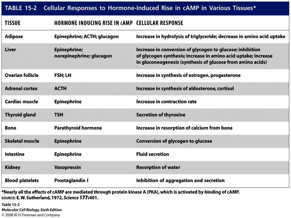
On the other hand, different receptors of the same class that bind different ligands often induce the same cellular responses in a cell. In liver cells, for instance, the hormones epinephrine, glucagon, and ACTH bind to different members of the G protein–coupled receptor family, but all these receptors activate the same signal-transduction pathway, one that promotes synthesis of cyclic AMP (cAMP). This small signaling molecule in turn regulates various metabolic functions, including glycogen breakdown. As a result, all three hormones have the same effect on liver cell metabolism.
. This small signaling molecule in turn regulates various metabolic functions, including glycogen breakdown. As a result, all three hormones have the same effect on liver cell metabolism..")
12
15.3 Highly Conserved Components of Intracellular Signal-Transduction Pathways The binding of ligands (“first messengers”) low-molecular-weight intracellular signaling molecules termed second messengers.
 low-molecular-weight intracellular signaling molecules termed second messengers.")
13
Second Messengers Carry Signals from Many Receptors 15-9 Phospholipase C: phosphatidylinositol derivatives Adenylyl cyclase Guanylyl cyclase

14
The elevated intracellular concentration of one or more second messengers following binding of an external signaling molecule triggers a rapid alteration in the activity of one or more enzymes or nonenzymatic proteins Ca+2 In muscle: contraction a similar endocrine cells and of neurotransmitter- containing vesicles in nerve cells: exocytosis of secretory vesicles Similarly, a rise in cAMP induces various changes in cell metabolism that differ in different types of human cells. Calcium ion (Ca+2) and several membrane-bound phosphoinositides also act as second messengers. Nitric Oxide, free radical (reactive oxygen species, ROS)
 and several membrane-bound phosphoinositides also act as second messengers. Nitric Oxide, free radical (reactive oxygen species, ROS).")
15
Many Conserved Intracellular Proteins Function in Signal Transduction

16
Figure 15.6 GTPase switch proteins cycle between active and inactive forms.

17
Figure 15.7 Switching mechanism of G proteins.

18
Protein Kinases and Phosphatases Activation of all cell surface receptors leads directly or indirectly to changes in protein phosphorylation through the activation of protein kinases or protein phosphatases. Localization Activity Stability Protein-protein interaction tyrosine kinases serine/threonine kinases

19
Figure 15.5 A simple signal transduction pathway involving one kinase and one target protein.

20
Animal cells contain two types of protein kinases : tyrosine residues serine or threonine Phosphatases At last count the human genome encodes 500 protein kinases and 100 different phosphatases. In some signaling pathways, the receptor itself possesses intrinsic kinase or phosphatase activity; in other pathways, the receptor interacts with cytosolic or membrane- associated kinases.

21
Figure 15.9 Amplification of an extracellular signal.

22
phosphorylates specific residues in a set of target proteins whose patterns of expression generally differ in different cell types. Many proteins are substrates for multiple kinases, and each phosphorylation event, on a different amino acid, modifies the activity of a particular target protein in different ways, activation inhibition Stability The catalytic activity of a protein kinase itself commonly is modulated by phosphorylation by other kinases, (MAP kinase) by direct binding to other proteins, ( receptor tyrosine kinase) by changes in the levels of various second messengers. (cAMP) protein kinases phosphatases
 by direct binding to other proteins, ( receptor tyrosine kinase) by changes in the levels of various second messengers. (cAMP) protein kinases phosphatases.")
23
The ability of cells to respond appropriately to extracellular signals also depends on regulation of signaling pathways themselves. Down-regulation For example, once the concentration of an external signal decreases, signaling via some intracellular pathways is terminated by degradation of a second messenger; in other pathways, signaling is terminated by deactivation of a signal transduction protein. Deactivation (activation): modification phosphorylation protein-protein interaction inhibitor Detection: anti-phospho-protein antibody, r-P32-ATP inhibitors : ATP analog or substrate analog
: modification phosphorylation protein-protein interaction inhibitor Detection: anti-phospho-protein antibody, r-P32-ATP inhibitors : ATP analog or substrate analog.")
24
Another important mechanism for assuring appropriate cellular responses is desensitization of receptors at high signal concentrations or after prolonged exposure to a signal. The sensitivity of a cell to a particular signaling molecule can be down- regulated by endocytosis of its receptors, thus decreasing the number on the cell surface, or by modifying their activity so that the receptors either cannot bind ligand or form a receptor-ligand complex that does not induce the normal cellular response. Such modulation of receptor activity often results from phosphorylation of the receptor, binding of other proteins to it, or both.

25
Synthetic analogs of natural hormones are widely used in research on cell-surface receptors and as drugs. These analogs fall into two classes: agonists, which mimic the function of a natural hormone by binding to its receptor and inducing the normal response, and antagonists, which bind to the receptor but induce no response. By occupying ligand-binding sites on a receptor, an antagonist can block binding of the natural hormone (or agonist) and thus reduce the usual physiological activity of the hormone.
 and thus reduce the usual physiological activity of the hormone..")
26
15.4 General Elements of G Protein–Coupled Receptor Systems All G protein–coupled receptors (GPCRs): seven membrane-spanning The GPCR: hormones and neurotransmitters, light-activated receptors (rhodopsins) in the eye, odorant receptors in the mammalian nose
: seven membrane-spanning The GPCR: hormones and neurotransmitters, light-activated receptors (rhodopsins) in the eye, odorant receptors in the mammalian nose")
27
FIGURE 1 Effect of GTP on glucagon-stimulated cAMP production from AMP-PNP by purified rat liver membranes. In the absence of GTP, glucagon stimulates cAMP formation about twofold over the basal level in the absence of added hormone. When GTP also is added, cAMP production increases another fivefold. [Adapted from M. Rodbell et al., 1971, J. Biol. Chem. 246:1877.]

28
Epinephrine Binds to Several Different G Protein–Coupled Receptors Epinephrine is particularly important in mediating the body’s response to stress, such as fright or heavy exercise, when all tissues have an increased need to catabolize glucose and fatty acids to produce ATP. Liver (hepatic cell): β- adrenergic receptors rapid breakdown of glycogen to glucose (glycogenolysis) adipose cells: β- adrenergic receptors triacylglycerols to fatty acids in (lipolysis). heart muscle cells smooth muscle cells intestinal tract, skin, and kidneys. Epinephrine and norepinephrine were originally recognized as products of the medulla, or core, of the adrenal gland and are also known as adrenaline and noradrenaline. Embryologically, nerve cells derive from the same tissue as adrenal medulla cells, and norepinephrine is also secreted by differentiated nerve cells. Both hormones are charged compounds that belong to the catecholamines, active amines containing a catechol moiety:
: β- adrenergic receptors rapid breakdown of glycogen to glucose (glycogenolysis) adipose cells: β- adrenergic receptors triacylglycerols to fatty acids in (lipolysis). heart muscle cells smooth muscle cells intestinal tract, skin, and kidneys. Epinephrine and norepinephrine were originally recognized as products of the medulla, or core, of the adrenal gland and are also known as adrenaline and noradrenaline. Embryologically, nerve cells derive from the same tissue as adrenal medulla cells, and norepinephrine is also secreted by differentiated nerve cells. Both hormones are charged compounds that belong to the catecholamines, active amines containing a catechol moiety:.")
29
Isoproterenol: an agonist that binds to epinephrine receptors on bronchial smooth muscle cells about tenfold more strongly than does epinephrine Because ligand binding to these receptors promotes relaxation of bronchial smooth muscle and thus opening of the air passages in the lungs, isoproterenol is used in treating bronchial asthma ( 支氣管的氣喘 ), chronic bronchitis ( 支氣管炎 ), and emphysema ( 肺氣腫 ). Activation of epinephrine receptors on cardiac muscle cells increases the contraction rate. The antagonist alprenolol and related compounds, referred to as beta-blockers, have a very high affinity for these epinephrine receptors. Such antagonists are used to slow heart contractions in the treatment of cardiac arrhythmias ( 心律不整 ) and angina ( 心絞痛 ).
 and angina ( 心絞痛 )..")
30
Figure 15-13 Ligand-Activated G Protein- Coupled Receptors Catalyze Exchange of GTP for GOP on the a Subunit of a Trimeric G Protein 瘦肉精 萊克多巴胺 (ractopamine) 、克倫特羅 (clenbuterol) 沙丁胺醇 (salbutamol) 和特布他林 (terbutaline) 。
 、克倫特羅 (clenbuterol) 沙丁胺醇 (salbutamol) 和特布他林 (terbutaline) 。")
31
Figure 15.17 General mechanism of the activation of effector proteins associated with G protein–coupled receptors.

32
Experimental Figure 15.18 Activation of G proteins occurs within seconds of ligand binding in amoeba cells. fluorescence energy transfer (FET)
.")
33
Fig 15-14 cyan fluorescent protein (CFP) 440nm 490 nm yellow fluorescent protein (YFP) 470 527 The GPCR-mediated dissociation of trimeric G proteins detected in living cells. fluorescence energy transfer (FET)
.")
35
rods cones Rhodopsin, a G protein–coupled receptor that is activated The trimeric G protein coupled to rhodopsin, often called transducin (Gt), is found only in rod cells. A human rod cell contains about 4 x10 7 molecules of rhodopsin, which consists of the seven-spanning protein opsin to which is covalently bound the light-absorbing pigment 11-cis-retinal. Light Activates G Protein-Coupled Rhodopsins in Rod Cells of the Eye

36
Figure 15.22 The light-triggered step in vision.

37
Figure 15.23 Light-activated rhodopsin pathway and the closing of cation channels in rod cells. cGMP phosphodiesterase (PDE) Ca2+ -Sensing Proteins That Activate Guanylate Cyclase
 Ca2+ -Sensing Proteins That Activate Guanylate Cyclase.")
38
Figure 15.24 Inhibition of rhodopsin signaling by rhodopsin kinase.

39
Figure 15.25 Schematic illustration of transducin and arrestin distribution in dark-adapted and light-adapted rod cells.

40
15.5 G Protein- Coupled Receptors That Activate or Inhibit Adenylyl Cyclase In multicellular animals virtually all the diverse effects of cAMP are mediated through protein kinase A (PKA), also called cAMP-dependent protein kinase. Figure 15.26 Synthesis and hydrolysis of cAMP by adenylyl cyclase and cAMP phosphodiesterase.

41
cAMP and Other Second Messengers Activate Specific Protein Kinases

42
Figure 15.27 Hormone-induced activation and inhibition of adenylyl cyclase in adipose cells. cAMP-Mediated Activation of Protein Kinase A Produces Diverse Responses in Different Cell Types Signal Amplification Occurs in the cAMP-Protein Kinase A Pathway CREB Links cAMP and Protein Kinase A to Activation of Gene Transcription Anchoring Proteins Localize Effects of cAMP to Specific Regions of the Cell

44
Glycogen Metabolism Is Regulated by Hormone- Induced Activation of Protein Kinase A The first cAMP-mediated cellular response to be discovered— the release of glucose from glycogen—occurs in muscle and liver cells stimulated by epinephrine or other hormones whose receptors are coupled to Gs protein. In both muscle and liver cells glucose 1-phosphate: convert to glucose 6-phosphate In muscle cells this metabolite enters the glycolytic pathway: generate ATP liver cells: phosphatase glucose glucose transporter (GLUT2)
.")
45
Several Mechanisms Regulate Signaling from G Protein–Coupled Receptors termination of the response of β-adrenergic receptors: First, the affinity of the receptor for hormone decreases : affinity & number hormone….receptor-Gs-GTP hormone----receptor-Gs-GDP Second, the GTP bound to Gs is quickly hydrolyzed, reversing the activation of adenylyl cyclase and production of cAMP. Third, cAMP phosphodiesterase acts to hydrolyze cAMP to 5- AMP, terminating the cellular response.

46
Prolonged treatment -adrenergic receptor kinase (BARK)
")
47
Inhibit the interaction with Gs Jun kinase (JNK) c-srcMAP kinase AP2 Clathrin B-adrenergic receptor kinase (BARK)
 c-srcMAP kinase AP2 Clathrin B-adrenergic receptor kinase (BARK)")
48
Heterologous desensitization

49
負責教師 : 唐世杰 海洋大學生命科學系、生物科技所

50
Some Bacterial Toxins Irreversibly Modify G Proteins Pertussis toxin is secreted by Bordetella pertussis, the bacterium causing whooping cough Gi. This irreversible modification prevents release of GDP, locking Gia in the GDP-bound state epithelial cells of the airways, promoting loss of fluids and electrolytes and mucus secretion Vibrio Cholera Cholera toxin intracellular cAMP the loss of electrolytes and water into the intestinal lumen, producing the watery diarrhea characteristic of infection by these bacteria

51
15.6 G Protein-Coupled Receptors That Trigger Elevations in Cytosolic Ca2+ Synthesis of DAG and IP3 from membrane-bound phosphatidylinositol (PI). Each membranebound PI kinase places a phosphate (yellow circles) on a specific hydroxyl group on the inositol ring, producing the phosphoinositides PIP and PIP2. Cleavage of PIP2 by phospholipase C (PLC) yields the two important second messengers DAG and IP3.
 on a specific hydroxyl group on the inositol ring, producing the phosphoinositides PIP and PIP2. Cleavage of PIP2 by phospholipase C (PLC) yields the two important second messengers DAG and IP3..")
52
Inositol 1,4,5-Trisphosphate (IP3) Triggers Release of Ca+2 from the Endoplasmic Reticulum Gq or Go - PLC β As endoplasmic reticulum Ca2 stores are depleted, the IP3-gated Ca2 channels bind to and open store-operated TRP Ca2 channels in the plasma membrane, allowing influx of extracellular Ca2 (step 7 ).
 Triggers Release of Ca+2 from the Endoplasmic Reticulum Gq or Go - PLC β As endoplasmic reticulum Ca2 stores are depleted, the IP3-gated Ca2 channels bind to and open store-operated TRP Ca2 channels in the plasma membrane, allowing influx of extracellular Ca2 (step 7 ).")
53
calmodulin A small cytosolic protein called calmodulin, which is ubiquitous in eukaryotic cells, functions as a multipurpose switch protein that mediates many cellular effects of Ca2 ions. Binding of Ca2 to four sites on calmodulin yields a complex that interacts with and modulates the activity of many enzymes and other proteins (see Figure 3-28). One well-studied enzyme activated by the Ca2/calmodulin complex is myosin light-chain kinase, which regulates the activity of myosin in muscle cells. Another is cAMP phosphodiesterase, the enzyme that degrades cAMP to 5-AMP and terminates its effects. This reaction thus links Ca+2 and cAMP, one of many examples in which two second messengers interact to fine-tune certain aspects of cell regulation.
. One well-studied enzyme activated by the Ca2/calmodulin complex is myosin light-chain kinase, which regulates the activity of myosin in muscle cells. Another is cAMP phosphodiesterase, the enzyme that degrades cAMP to 5-AMP and terminates its effects. This reaction thus links Ca+2 and cAMP, one of many examples in which two second messengers interact to fine-tune certain aspects of cell regulation..")
54
Diacylglycerol (DAG) Activates Protein Kinase C, Which Regulates Many Other Proteins protein kinase C (PKC) In the absence of hormone stimulation, protein kinase C is present as a soluble cytosolic protein that is catalytically inactive. A rise in the cytosolic Ca+2 level causes protein kinase C to bind to the cytosolic leaflet of the plasma membrane, where the membrane-associated DAG can activate it. Thus activation of protein kinase C depends on an increase of both Ca+2 ions and DAG, suggesting an interaction between the two branches of the IP3/DAG pathway. The activation of protein kinase C in different cells results in a varied array of cellular responses, indicating that it plays a key role in many aspects of cellular growth and metabolism. Phorbol ester (DAG analog): tumor promoter
: tumor promoter.")
55
Signal-Induced Relaxation of Vascular Smooth Muscle Is Mediated by cGMP-Activated Protein Kinase G Nitroglycerin has been used for over a century as a treatment for the intense chest pain of angina. It was known to slowly decompose in the body to nitric oxide (NO), which causes relaxation of the smooth muscle cells surrounding the blood vessels that “feed” the heart muscle itself, thereby increasing the diameter of the blood vessels and increasing the flow of oxygen-bearing blood to the heart muscle. One of the most intriguing discoveries in modern medicine is that NO, a toxic gas found in car exhaust, is in fact a natural signaling molecule. ❚
, which causes relaxation of the smooth muscle cells surrounding the blood vessels that feed the heart muscle itself, thereby increasing the diameter of the blood vessels and increasing the flow of oxygen-bearing blood to the heart muscle. One of the most intriguing discoveries in modern medicine is that NO, a toxic gas found in car exhaust, is in fact a natural signaling molecule. ❚.")
56
eNOS nNOS iNOS (inducible) cGMP phosphodiesterase guanylyl cyclase
 cGMP phosphodiesterase guanylyl cyclase")
57
Viagra acts by inhibiting cGMP-specific phosphodiesterase type 5, an enzyme that promotes degradation of cGMP, which regulates blood flow in the penis.cGMP-specific phosphodiesterase type 5enzyme

Similar presentations


>")


