Download presentation
Presentation is loading. Please wait.

1
DNA Packaging

2
Lecture Overview Structure of chromosomes and chromatin. DNA packing and histone modification.

3
Objectives: Understand the nature of chromatin - how DNA and histones interact to form nucleosomes. Know the basic structures of the 10 nm chromatin fiber and the 30 nm chromatin fiber. Realize that chromosomes are single pieces of DNA packaged into chromatin.

4
Genetic Material in the Living Cells Cells contain a nucleus surrounded by a nuclear membrane in eukaryotic cells, and a nuclear region in the prokaryotic cells. In a non-dividing cell the nucleus is filled with a thread-like material known as "chromatin". Chromatin is made up of DNA and proteins (mainly histones and some non-histone acidic proteins).
..")
5
The Normal Human Chromosomes Normal human cells contain 23 pairs of homologous chromosomes: i. 22 pairs of autosomes. ii. 1 pair of sex chromosomes. Autosomes are the same in males and females Sex chromosomes are: i. XX in females ii. XY in males. Both X are homologous. Y is much smaller than X and has only a few genes.

6
Chromosomes One member of each chromosome pair is derived from each parent. Somatic cells have diploid complement of chromosomes i.e. 46. Germ cells (Gametes: sperm and ova) have haploid complement i.e 23. Individual chromosomes are recognized by i. arm lengths ( p- short, q -long). ii. centromere position (metacentric, sub-metacentric, acrocentric, telocentric).
 have haploid complement i.e 23. Individual chromosomes are recognized by i. arm lengths ( p- short, q -long). ii. centromere position (metacentric, sub-metacentric, acrocentric, telocentric)..")
7
Composition of Chromosome DNA Histones (Major proteins) Non-histone (Small amounts) p q The chromosomes themselves are macromolecular entities that must be synthesized, packaged, protected, and properly distributed to daughter cells at cell division. Significant segments of every chromosome are dedicated to these functions.

8
DNA Condensation: Why?

9
A severe problem of packaging 1.Largest human chromosome: ~3 x 10 8 bp 2.A typical cell = 10 μm = 10 x 10 -6 m 3.Therefore the DNA must be compacted ~10 4 -fold. This is like fitting an 11-mile-long string into a 6-foot box How long is it?

10
Eukaryotes contain thousands of times more DNA than do bacteria, and as a result, the DNA– condensation problems of eukaryotes— compacting the DNA so that it fits in the cell nucleus—are more complex than those of bacteria. Bacteria do not contain nucleosomes, although they have small, basic (positively charged) proteins that are involved in condensing their DNA.
 proteins that are involved in condensing their DNA..")
11
Continue.. DNA compaction must be dynamic, because changes in the degree of condensation must occur quickly and when needed, as the cell passes through the stages of the cell cycle. Furthermore, when in its most highly compacted form, DNA is not accessible to transcription or replication enzymes, so it must be able to rapidly expose regions containing genes that are required at any given moment, and then condense again. Modification enzymes that alter the state of DNA condensation, and can target their activity to specific regions of the chromosome that must be transcribed or replicated.

12
Nucleosomes: The Basic Units of DNA Condensation The material of chromosomes, both protein and DNA, is often referred to as chromatin. The protein component is about equal in mass to the DNA component. Histones constitute the largest protein component of chromatin, are highly conserved, basic proteins that assemble into octameric complexes containing two each of four different histone subunits. DNA wraps around the histones to form condensed nucleosomes.

13
Nucleosome (10 nm diameter): 8 histones in bead & 1 outside. Each bead: is surrounded by 140 bpDNA and there are 60 bp in the linker region. Space between beads is about 14 nm.

14
Continue.. Histones are rich in the basic amino acids arginine and lysine, which together make up about 25% of the amino acid residues in any given histone protein. Histone proteins are highly conserved among eukaryotic cells. Histones H3 and H4 are nearly identical in all eukaryotes, suggesting strict conservation of their functions. Histones H1, H2A, and H2B show less sequence similarity, but on the whole, they are more conserved than other types of proteins. Salt bridges between positively charged histones and negatively charges DNA play a major role in stabilizing DNA-histone complex

15
Nucleosomes Kornberg suggested that most of the 200 bp of DNA in a protein-DNA unit is wrapped around a histone octamer composed of two copies each of histones H2A, H2B, H3, and H4. These four histones have come to be known as the core histones. The remainder of the DNA serves as a linker between nucleosomes, to which histone H1 binds. Under physiological conditions, formation of the histone octamer from individual histone proteins requires the presence of DNA. Nucleosomes as beads on a string. Regularly spaced nucleosomes consist of core histone proteins bound to DNA.

16
Chromosomes Condense into a Compact Chromatin Filament Nucleosomes condense into a compact filament with a width of about 30 nm, referred to as the 30 nm filament. H1 promotes condensation into the 30 nm filament, but it is not essential for forming the filament. In contrast, the N-terminal tails of the core histones are absolutely required, suggesting that the tails provide important nucleosome- nucleosome contacts needed for 30 nm filament formation.

17
Higher-Order Chromosome Structure Involves Loops and Coils Inside chromosomes, DNA is much more highly condensed than in the 30 nm filament. 30 nm filaments is appear to be organized in loops estimated at 40 to 100 kbp long. Regions of the DNA interact with chromosomal scaffold proteins to give a protein core with DNA loops sticking out of it. This protein core then coils up to further package the DNA into the chromatids that are visible by light microscopy in metaphase.

18
First then Overall, Higher-order DNA compaction in a eukaryotic chromosome. This model shows the levels of organization that could provide the observed degree of DNA compaction in the chromosomes of eukaryotes. First the DNA is wrapped around histone octamers, then H1 stimulates formation of the 30 nm filament. Further levels of organization are not well understood but seem to involve further coiling and loops in the form of rosettes, which also coil into thicker structures. Overall, progressive levels of organization take the form of coils upon coils upon coils. It should be noted that in cells, the higher-order structures (above the 30 nm filament) are unlikely to be as uniform as depicted here. DNA packing- Superstructure Heterochromatin- compacted DNA. Euchromatin-Less compacted.
 are unlikely to be as uniform as depicted here. DNA packing- Superstructure Heterochromatin- compacted DNA. Euchromatin-Less compacted..")
19
DNA Organization in Eukaryotic Chromosomes Animation 1 : http://www.biostudio.com/demo_freeman_dna_coiling.ht m http://www.biostudio.com/demo_freeman_dna_coiling.ht m Animation 2: http://www.pbs.org/wgbh/nova/genome/dna.html 19

20
Big Question 1: Why are there no male calico cats? Case Study - Calico Cats Chromosomes & Inheritance

21
Big Question 2: Why do CC and Rainbow not look exactly alike, even though they are clones? CC (Carbon Copy)Rainbow
Rainbow.")
22
Genes are found on chromosomes, in most cases, you receive 2 alleles. Shown is a human karyotype which contains 22 pairs of autosomes 1 pair of sex chromosomes 1. When would a person only have 1 allele for a trait?

23
Cat Karyotype 2. How is it similar to the human karyotype? How is it different?

24
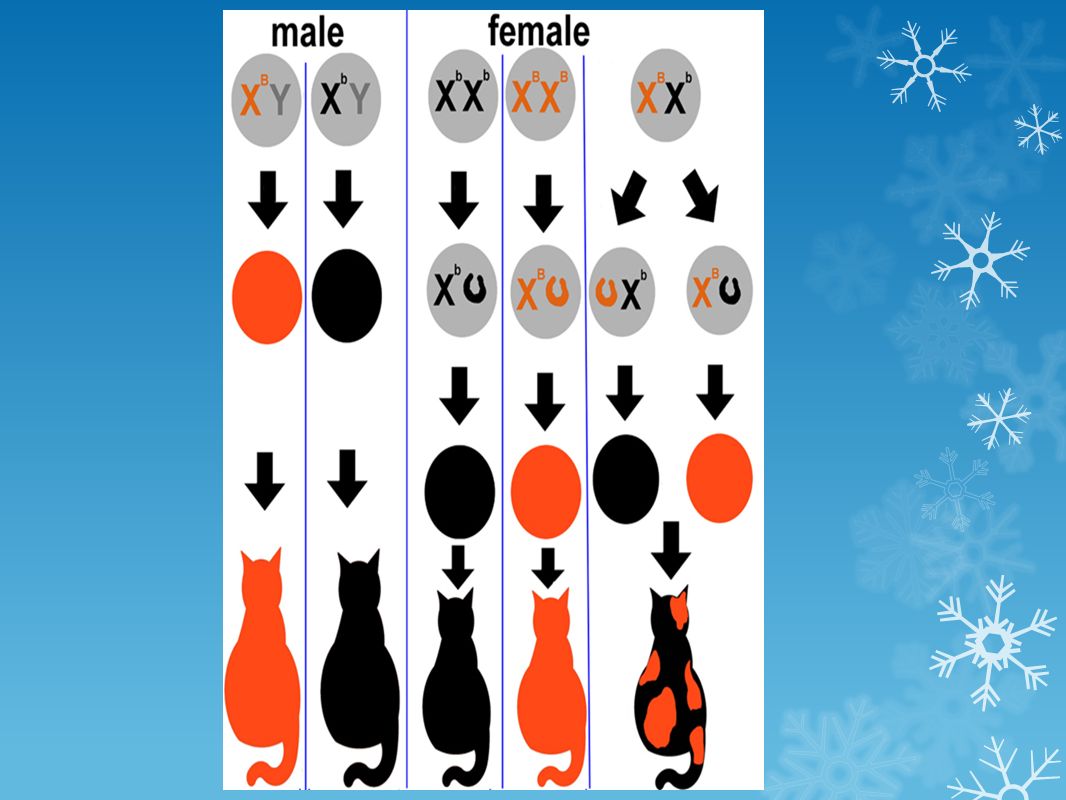
The genes for coat color in cats are located on the X chromosome. Alleles can be: ORange or Black Alleles are codominant What is Spaz’s genotype?

25
What are the genotypes of these 3 cats. The black and orange ones are both male.

26
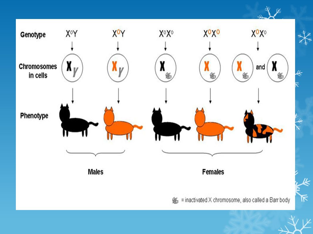
Question 2: Why do CC and Rainbow look different? Consider how color is expressed on a calico cat. Some areas express orange, some black…. Propose a reason for this.

27
A Barr body (named after discoverer Murray Barr) is the inactive X chromosome in a female somatic cell, rendered inactive in a process called lyonization, in those species in which sex is determined by the presence of the Y
 is the inactive X chromosome in a female somatic cell, rendered inactive in a process called lyonization, in those species in which sex is determined by the presence of the Y")
28
How do Barr Bodies explain CC and Rainbow? CC (Carbon Copy)Rainbow
Rainbow")
31
There are some rare cases of male calico cats. How does that happen?

32
During meiosis, each egg and sperm should receive only one copy of each chromosome (haploid) What is the diploid number for humans? And the haploid number?

33
The offspring ends up having an extra chromosome. Most of the time, this is FATAL, but not always.

34
Trisomy = extra chromosome Monosomy = one chromosome of the pair is missing Can you identify the abnormality in this karyotype of a girl with Turner Syndrome?

35
How would nondisjunction explain male calicos?

36
13. Consider a male calico cat. Do you think this calico would have cognitive disabilities similar to those found in children with Down Syndrome. Why or Why not?

37
Chapter 12: Chromosomal Abnormalities

38
Figure 12.10a NONDISJUNCTION CHANGES CHROMOSOME NUMBERS

39
Figure 12.10b NONDISJUNCTION CHANGES CHROMOSOME NUMBERS

40
Figure 12.12a

41
Figure 12.12b

42
Trisomy 18 - Edward's Syndrome ● Failure to grow and gain weight at the expected rate and severe feeding difficulties, diminished muscle tone and episodes in which there is temporary cessation of spontaneous breathing ● Developmental delays and intellectual disability ● A prominent back portion of the head, low-set, malformed ears, an abnormally small jaw, a small mouth an upturned nose, narrow eyelid folds, widely spaced eyes, and drooping of the upper eyelids

43
TRISOMY 13 (also known as Patau syndrome) - Of all babies born with the extra copy of chromosome 13 in all the cells of their body, around 50% die in the first month, and the rest within the first year Median survival age for children with Patau syndrome is 2.5 days
 - Of all babies born with the extra copy of chromosome 13 in all the cells of their body, around 50% die in the first month, and the rest within the first year Median survival age for children with Patau syndrome is 2.5 days")
44
CHROMOSOME MUTATIONS

45
Figure 12.13

46
Figure 12.13a

47
Figure 12.13b

48
Figure 12.13c

49
Figure 12.13d

50
Figure 12.14

51
DELETION MUTATION (WILLIAMS SYNDROME)
")
52
Williams Syndrome – Deletion of Chromosome 7

53
CRI DU CHAT Deletion of Chromosome #5

54
FRAGILE X - duplication mutation

Similar presentations