Download presentation
Presentation is loading. Please wait.

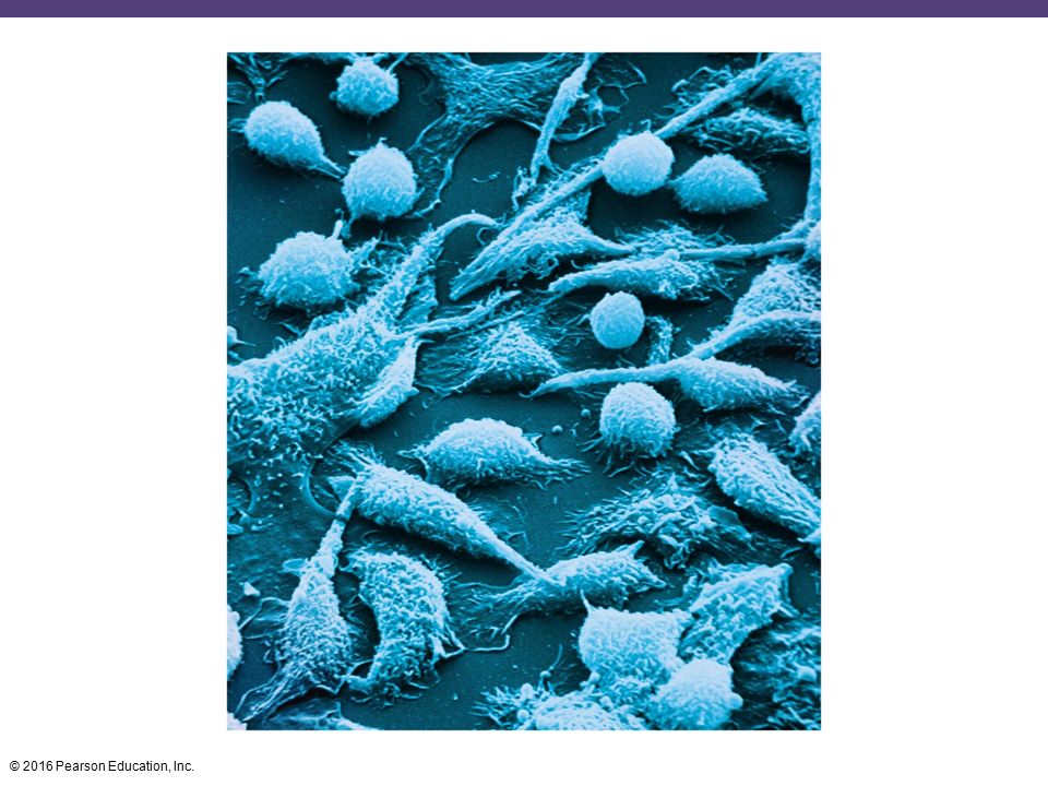
1
Adaptive Immunity: Specific Defenses of the Host
17 Adaptive Immunity: Specific Defenses of the Host

3
The Adaptive Immune System
Learning Objective 17-1 Compare and contrast adaptive and innate immunity.

4
The Adaptive Immune System
Adaptive immunity: defenses that target a specific pathogen Acquired through infection or vaccination Primary response: first time the immune system combats a particular foreign substance Secondary response: later interactions with the same foreign substance; faster and more effective due to "memory"

5
Host Defenses: The Big Picture
PLAY Animation: Host Defenses: The Big Picture

6
Check Your Understanding
Is vaccination an example of innate or adaptive immunity? 17-1

7
Dual Nature of the Adaptive Immune System
Learning Objective 17-2 Differentiate humoral from cellular immunity.

8
Dual Nature of the Adaptive Immune System
Humoral immunity Produces antibodies that combat foreign molecules known as antigens B cells are lymphocytes that are created and mature in red bone marrow Recognize antigens and make antibodies Named for bursa of Fabricius in birds

9
Humoral Immunity: Overview
PLAY Animation: Humoral Immunity: Overview

10
Dual Nature of the Adaptive Immune System
Cellular immunity (cell-mediated immunity) Produces T lymphocytes Recognize antigenic peptides processed by phagocytic cells Mature in the thymus T cell receptors (TCRs) on the T cell surface contact antigens, causing the T cells to secrete cytokines instead of antibodies
 Produces T lymphocytes. Recognize antigenic peptides processed by phagocytic cells. Mature in the thymus. T cell receptors (TCRs) on the T cell surface contact antigens, causing the T cells to secrete cytokines instead of antibodies.")
11
Figure 17.1 Differentiation of T cells and B cells.
Stem cells develop in bone marrow or in fetal liver Stem cell (diverges into two cell lines) Red bone marrow of adults Thymus Differentiate to B cells in adult red bone marrow Differentiate to T cells in thymus B cell T cell Migrate to lymphoid tissue such as spleen, but especially lymph nodes
 Red bone. marrow. of adults. Thymus. Differentiate to. B cells in adult. red bone marrow. Differentiate to. T cells in thymus. B cell. T cell. Migrate to lymphoid. tissue such as spleen, but especially lymph. nodes.")
12
Dual Nature of the Adaptive Immune System
Cellular immunity attacks antigens found inside cells Viruses; some fungi and parasites Humoral immunity fights invaders outside cells Bacteria and toxins

13
Check Your Understanding
What type of cell is most associated with humoral immunity, and what type of cell is the basis of cellular immunity? 17-2

14
Cytokines: Chemical Messengers of Immune Cells
Learning Objective 17-3 Identify at least one function of each of the following: cytokines, interleukins, chemokines, interferons, TNF, and hematopoietic cytokines.

15
Cytokines: Chemical Messengers of Immune Cells
Cytokines are chemical messengers produced in response to a stimulus Interleukins: cytokines between leukocytes Chemokines: induce migration of leukocytes Interferons (IFNs): interfere with viral infections of host cells Tumor necrosis factor (TNF): involved in the inflammation of autoimmune diseases Hematopoietic cytokines: control stem cells that develop into red and white blood cells Overproduction of cytokines leads to a cytokine storm
: interfere with viral infections of host cells. Tumor necrosis factor (TNF): involved in the inflammation of autoimmune diseases. Hematopoietic cytokines: control stem cells that develop into red and white blood cells. Overproduction of cytokines leads to a cytokine storm.")
16
Check Your Understanding
What is the function of cytokines? 17-3

17
Antigens and Antibodies
Learning Objectives 17-4 Define antigen, epitope, and hapten Explain antibody function, and describe the structural and chemical characteristics of antibodies Name one function for each of the five classes of antibodies.

18
Antigens Antigens: substances that cause the production of antibodies
Usually components of invading microbes or foreign substances Antibodies interact with epitopes, or antigenic determinants, on the antigen Haptens: antigens too small to provoke immune responses; attach to carrier molecules

19
Epitopes (antigenic determinants) on antigen
Figure Epitopes (antigenic determinants). Antibody A Epitopes (antigenic determinants) on antigen Antigens: components of cell wall Binding sites Bacterial cell Antibody B
. Antibody A. Epitopes (antigenic determinants) on antigen. Antigens: components. of cell wall. Binding sites. Bacterial cell. Antibody B.")
20
Figure Haptens.

21
Antibodies Globular proteins called immunoglobulins (Ig)
Valence is the number of antigen-binding sites on an antibody Bivalent antibodies have two binding sites

22
Antibodies Four protein chains form a Y-shape
Two identical light chains and two identical heavy chains joined by disulfide links Variable (v) regions are at the ends of the arms; bind epitopes Constant (Fc) region is the stem, which is identical for a particular Ig class Five classes of Ig (IgG, IgM, IgA, IgD, IgE)
 regions are at the ends of the arms; bind epitopes. Constant (Fc) region is the stem, which is identical for a particular Ig class. Five classes of Ig (IgG, IgM, IgA, IgD, IgE)")
23
Figure 17.4 The structure of a typical antibody molecule.
Antigen- binding site V Heavy chain V V V Antigen Light chain C C Epitope (antigenic determinant) Antibodies Fc (stem) region C C Hinge region Antigen-binding site Antibody molecule Enlarged antigen-binding site bound to an epitope Antibody molecules shown by atomic force microscopy
 Antibodies. Fc (stem) region. C. C. Hinge region. Antigen-binding. site. Antibody molecule. Enlarged antigen-binding site bound to an epitope. Antibody molecules. shown by atomic force microscopy.")
24
IgG Monomer 80% of serum antibodies In the blood, lymph, and intestine
Cross the placenta; trigger complement; enhance phagocytosis; neutralize toxins and viruses; protect fetus

25
IgM Pentamer made of five monomers held with a J chain
6% of serum antibodies Remain in blood vessels Cause clumping of cells and viruses First response to an infection; short-lived

26
IgA Monomer in serum; dimer in secretions 13% of serum antibodies
Common in mucous membranes, saliva, tears, and breast milk Prevent microbial attachment to mucous membranes

27
IgD Monomer 0.02% of serum antibodies Structure similar to IgG
In blood, in lymph, and on B cells No well-defined function; assists in the immune response on B cells

28
IgE Monomer 0.002% of serum antibodies
On mast cells, on basophils, and in blood Cause the release of histamines when bound to antigen; lysis of parasitic worms

29
Table 17.1 A Summary of Immunoglobulin Classes

30
Check Your Understanding
Does an antibody react with a bacterium as an antigen or as an epitope? 17-4 The original theoretical concepts of an antibody called for a rod with antigenic determinants at each end. What is the primary advantage of the Y-shaped structure that eventually emerged? 17-5 Which class of antibody is most likely to protect you from a common cold? 17-6

31
Humoral Immunity Response Process
Learning Objectives 17-7 Compare and contrast T-dependent and T-independent antigens Differentiate plasma cell from memory cell Describe clonal selection Describe how a human can produce different antibodies.

32
Clonal Selection of Antibody-Producing Cells
Major histocompatibility complex (MHC) genes encode molecules on the cell surface Class I MHC are on the membrane of nucleated animal cells Identify "self" Class II MHC are on the surface of antigen-presenting cells (APCs), including B cells
 genes encode molecules on the cell surface. Class I MHC are on the membrane of nucleated animal cells. Identify self Class II MHC are on the surface of antigen-presenting cells (APCs), including B cells.")
33
Clonal Selection of Antibody-Producing Cells
Inactive B cells contain surface Ig that bind to antigen B cell internalizes and processes antigen Antigen fragments are displayed on MHC class II molecules T helper cell (TH) contacts the displayed antigen fragment and releases cytokines that activate B cells B cell undergoes proliferation (clonal expansion)
 contacts the displayed antigen fragment and releases cytokines that activate B cells. B cell undergoes proliferation (clonal expansion)")
34
Figure 17.5 Activation of B cells to produce antibodies.
Extracellular antigens MHC class II with antigen displayed on surface B cell receptors Antigen fragments Cytokines Plasma cell Antibodies TH cell B cell B cell B cell receptors recognize and attach to antigen. Antigen is internalized into the B cell. Fragments of the antigen are presented on MHC proteins on the surface of the cell. A T helper cell that recognizes this antigen fragment is activated and releases cytokines, activating the B cell. The activated B cell begins clonal expansion, producing an army of antibody-producing plasma cells and memory cells.

35
Clonal Selection of Antibody-Producing Cells
Clonal selection differentiates activated B cells into: Antibody-producing plasma cells Memory cells Clonal deletion eliminates harmful B cells

36
Figure 17.6 Clonal selection and differentiation of B cells.
Stem cell Stem cells differentiate into mature B cells, each bearing surface immunoglobulins against a specific antigen. B cell Immunoglobin I II Antigens B cell II encounters its specific antigen and proliferates. Some B cells proliferate into long-lived memory cells, which at a later date can be stimulated to become antibody- producing plasma cells. Other B cells proliferate into antibody-producing plasma cells. Memory cell Plasma cell Plasma cells secrete antibodies into circulation. Blood vessel of cardiovascular system

37
Clonal Selection of Antibody-Producing Cells
T-dependent antigen Antigen that requires a TH cell to produce antibodies T-independent antigens Stimulate the B cell without the help of T cells Provoke a weak immune response, usually producing IgM No memory cells generated

38
(T-independent antigen)
Figure T-independent antigens. Polysaccharide (T-independent antigen) Epitopes B cell receptors
 Epitopes. B cell receptors.")
39
Antigen Processing and Presentation: Overview
PLAY Animation: Antigen Processing and Presentation: Overview

40
Humoral Immunity: Clonal Selection and Expansion
PLAY Animation: Humoral Immunity: Clonal Selection and Expansion

41
Check Your Understanding
Would pneumococcal pneumonia (see Figure 24.12, page 689) require a TH cell to stimulate a B cell to form antibodies? 17-7 Plasma cells produce antibodies; do they also produce memory cells? 17-8 In what way does a B cell that encounters an antigen function as an antigen-presenting cell? 17-9
 require a TH cell to stimulate a B cell to form antibodies Plasma cells produce antibodies; do they also produce memory cells In what way does a B cell that encounters an antigen function as an antigen-presenting cell")
42
Check Your Understanding
On what part of the antibody molecule do we find the amino acid sequence that makes the huge genetic diversity of antibody production possible? 17-10

43
Antigen–Antibody Binding and Its Results
Learning Objective Describe four outcomes of an antigen–antibody reaction.

44
Antigen–Antibody Binding and Its Results
An antigen–antibody complex forms when antibodies bind to antigens Strength of the bond is the affinity Protects the host by tagging foreign molecules or cells for destruction Agglutination Opsonization Antibody-dependent cell-mediated cytotoxicity Neutralization Activation of the complement system

45
Figure 17.8 The results of antigen–antibody binding.
PROTECTIVE MECHANISM OF BINDING ANTIBODIES TO ANTIGENS Agglutination Activation of complement Reduces number of infectious units to be dealt with Causes inflammation and cell lysis Complement Antibody Bacteria Bacterium Lysis Opsonization Antibody-dependent cell-mediated cytotoxicity Coating antigen with antibody enhances phagocytosis Antibodies attached to target cell cause destruction by macrophages, eosinophils, and NK cells Phagocyte Eosinophil Epitopes Perforin and lytic enzymes Large target cell (parasite) Neutralization Blocks adhesion of bacteria and viruses to mucosa Blocks attachment of toxin Virus Toxin Bacterium
 Neutralization. Blocks adhesion of bacteria. and viruses to mucosa. Blocks attachment. of toxin. Virus. Toxin. Bacterium.")
46
Humoral Immunity: Antibody Function
PLAY Animation: Humoral Immunity: Antibody Function

47
Check Your Understanding
Which antibodies may activate the complement system, and which antibodies are usually associated with agglutination? 17-11

48
Cellular Immunity Response Process
Learning Objectives Describe at least one function of each of the following: M cells, TH cells, TC cells, CTLs, Treg cells, NK cells Differentiate T helper, T cytotoxic, and T regulatory cells Differentiate TH1, TH2, and TH17 cells Define apoptosis Define antigen-presenting cell.

49
Cellular Immunity Response Process
T cells combat intracellular pathogens Mature in the thymus Thymic selection eliminates immature T cells Migrate from the thymus to lymphoid tissues Attach to antigens via T-cell receptors (TCRs)
")
50
Cellular Immunity Response Process
Pathogens entering the gastrointestinal tract pass through microfold cells (M cells) located over Peyer's patches Transfer antigens to lymphocytes and antigen-presenting cells (APCs)
 located over Peyer s patches. Transfer antigens to lymphocytes and antigen-presenting cells (APCs)")
51
Figure M cells. M cell on Peyer's patch. Note the tips of the closely packed microvilli on the surrounding epithelial cells. Microvilli on epithelial cell Antigen M cell TH cell Pocket B cells Macrophage Epithelial cell M cells facilitate contact between antigens passing through the intestinal tract and cells of the body's immune system.

52
Antigen-Presenting Cells (APCs)
Dendritic cells (DCs) Engulf and degrade microbes and display them to T cells Found in the skin, genital tract, lymph nodes, spleen, thymus, and blood Macrophages Activated by cytokines or the ingestion of antigenic material Migrate to the lymph tissue, presenting antigen to T cells
 Engulf and degrade microbes and display them to T cells. Found in the skin, genital tract, lymph nodes, spleen, thymus, and blood. Macrophages. Activated by cytokines or the ingestion of antigenic material. Migrate to the lymph tissue, presenting antigen to T cells.")
53
Figure 17.10 A dendritic cell.

54
Activated macrophages Resting (inactive) macrophage
Figure Activated macrophages. Activated macrophages Resting (inactive) macrophage
 macrophage.")
55
Antigen Processing and Presentation: MHC
PLAY Animation: Antigen Processing and Presentation: MHC

56
Classes of T Cells Clusters of differentiation (CD) CD4+ CD8+
T helper cells (TH) Cytokine signaling with B cells; interact directly with antigens Bind MHC class II molecules on B cells and APCs CD8+ Cytoxic T lymphocytes (CTL) Bind MHC class I molecules
 Cytokine signaling with B cells; interact directly with antigens. Bind MHC class II molecules on B cells and APCs. CD8+ Cytoxic T lymphocytes (CTL) Bind MHC class I molecules.")
57
T Helper Cells (CD4+ T Cells)
TCR on the TH cell recognize and bind to the antigen fragment and MHC class II on APC APC or TH secrete a costimulatory molecule, activating the TH cell TH cells produce cytokines and differentiate into: TH1cells TH2 cells TH17 cells Memory cells

58
Figure 17.12 Activation of CD4+ T helper cells.
Microorganism carrying antigens TH cell receptor (TCR) Cytokines Activated T cell Activated T cells proliferate T helper cell Costimulatory molecule, (required to activate T cells that have not previously encountered antigen) Complex of MHC class II molecule and antigen fragment MHC class II molecules Antigen fragments (short peptides) APC (dendritic cell) An APC encounters and ingests a microorganism. The antigen is enzymatically processed into short peptides, which combine with MHC class II molecules and are displayed on the surface of the APC. A receptor (TCR) on the surface of the CD4+ T helper cell (TH cell) binds to the MHC–antigen complex. This includes a Toll-like receptor. The TH cell or APC is stimulated to secrete a costimulatory molecule. These two signals activate the TH cell, which produces cytokines. The cytokines cause the TH cell (which recognizes a dendritic cell that is producing costimulatory molecules) to become activated.
 Cytokines. Activated. T cell. Activated T cells proliferate. T helper. cell. Costimulatory molecule, (required to activate T. cells that have not previously. encountered antigen) Complex of MHC. class II molecule and. antigen fragment. MHC class II. molecules. Antigen. fragments. (short peptides) APC (dendritic cell) An APC encounters and ingests a microorganism. The antigen is enzymatically processed into short peptides, which combine with MHC class II molecules and are displayed on the surface of the APC. A receptor (TCR) on the surface of the CD4+ T helper cell (TH cell) binds to the MHC–antigen complex. This includes a Toll-like receptor. The TH cell or APC is stimulated to secrete a costimulatory molecule. These two signals activate the TH cell, which produces cytokines. The cytokines cause the TH cell (which recognizes a dendritic cell. that is producing costimulatory molecules) to become activated.")
59
T Helper Cells (CD4+ T Cells)
TH17 cells produce IL-17 and contribute to inflammation TH1 cells produce IFN-g, which activates macrophages, enhances complement, and stimulates antibody production that promotes phagocytosis TH2 cells activate B cells to produce IgE; activate eosinophils

60
Figure 17.13 Lineage of effector T helper cell classes and pathogens targeted.
Antigen-presenting cell TH cells of various classes TH17 cells secrete cytokines that promote inflammatory responses; recruit neutrophils for protection against extracellular bacteria and fungi. TH1 cells are an important element of cellular immunity. Their cytokines (such as IFN-γ and IL-2) activate CD8+ T cells and NK cells, which control intracellular pathogens by killing infected host cells. They also enhance phagocytosis by antigen-presenting cells such as macrophages. IL-17 IFN-γ IL-4 TH2 cells Fungi Extracellular bacteria Neutrophil Intracellular bacteria and protozoa Macrophage Important in allergic responses, especially by production of IgE. Activate eosinophils to control extracellular parasites such as helminths (see ADCC discussion). Mast cell Basophil Eosinophil Helminth
 activate CD8+ T cells and NK cells, which control intracellular pathogens by killing infected host cells. They also enhance phagocytosis by antigen-presenting cells such as macrophages. IL-17. IFN-γ. IL-4. TH2 cells. Fungi. Extracellular bacteria. Neutrophil. Intracellular. bacteria and. protozoa. Macrophage. Important in allergic responses, especially by production of IgE. Activate eosinophils to control extracellular parasites such as helminths (see ADCC discussion). Mast cell. Basophil. Eosinophil. Helminth.")
61
Antigen Processing and Presentation: Steps
PLAY Animation: Antigen Processing and Presentation: Steps

62
T Regulatory Cells T regulatory cells (Treg)
Subset of CD4+ cells; carry an additional CD25 molecule Suppress T cells against self; protect intestinal bacteria required for digestion; protect fetus

63
T Cytotoxic Cells (CD8+ T Cells)
Activated into cytotoxic T lymphocyte (CTL) with the help of TH cell and costimulatory signals CTLs recognize and kill self-cells altered by infection Self-cells carry endogenous antigens on a surface presented with MHC class I molecules CTL releases perforin and granzymes that induce apoptosis in the infected cell
 with the help of TH cell and costimulatory signals. CTLs recognize and kill self-cells altered by infection. Self-cells carry endogenous antigens on a surface presented with MHC class I molecules. CTL releases perforin and granzymes that induce apoptosis in the infected cell.")
64
Figure 17.14 Killing of virus-infected target cell by cytotoxic T lymphocyte.
Processed antigen Processed antigen presented with MHC class I MHC class I T cell receptors Virus-infected cell (example of endogenous antigen) Virus-infected cell TH1 cell Cytokines CTLp CTL Infected target cell is lysed A normal cell will not trigger a response by a cytotoxic T lymphocyte (CTL), but a virus-infected cell (shown here) or a cancer cell produces abnormal endogenous antigens. The abnormal antigen is presented on the cell surface in association with MHC class I molecules. Binding of a TH1 cell promotes secretion of cytokines. The cytokines activate a precursor CTL, which produces a clone of CTLs. The CTL induces destruction of the virus-infected cell by apoptosis.
 Virus-infected cell. TH1 cell. Cytokines. CTLp. CTL. Infected target. cell is lysed. A normal cell will not trigger a response. by a cytotoxic T lymphocyte (CTL), but a. virus-infected cell (shown here) or a cancer cell produces abnormal endogenous antigens. The abnormal antigen is presented on the cell surface in association with MHC class I molecules. Binding of a TH1 cell promotes secretion of cytokines. The cytokines activate a precursor CTL, which produces a clone of CTLs. The CTL induces destruction of the virus-infected cell by apoptosis.")
65
T Cytotoxic Cells (CD8+ T Cells)
Apoptosis Programmed cell death Prevents the spread of infectious viruses into other cells Cells cut their genome into fragments, causing the membranes to bulge outward via blebbing

66
Figure Apoptosis.

67
Cell-Mediated Immunity: Cytotoxic T Cells
PLAY Animation: Cell-Mediated Immunity: Cytotoxic T Cells

68
Check Your Understanding
Which antibody is the primary one produced when an antigen is taken up by an M cell? 17-12 Which T cell type is generally involved when a B cell reacts with an antigen and produces antibodies against the antigen? 17-13 Which T cell type is generally involved in allergic reactions? 17-14

69
Check Your Understanding
What is another name for apoptosis, one that describes its function? 17-15 Are dendritic cells considered primarily part of the humoral or the cellular immune system? 17-16

70
Extracellular Killing by the Immune System
Learning Objective Describe the function of natural killer cells.

71
Extracellular Killing by the Immune System
Natural killer (NK) cells Granular leukocytes destroy cells that don't express MHC class I self-antigens Kill virus-infected and tumor cells and attack parasites Not always stimulated by an antigen Form pores in the target cell, leading to lysis or apoptosis
 cells. Granular leukocytes destroy cells that don t express MHC class I self-antigens. Kill virus-infected and tumor cells and attack parasites. Not always stimulated by an antigen. Form pores in the target cell, leading to lysis or apoptosis.")
72
Table 17.2 Principal Cells That Function in Cell-Mediated Immunity

73
Check Your Understanding
How does the natural killer cell respond if the target cell does not have MHC class I molecules on its surface? 17-17

74
Antibody-Dependent Cell-Mediated Cytotoxicity
Learning Objective Describe the role of antibodies and natural killer cells in antibody-dependent cell- mediated cytotoxicity.

75
Antibody-Dependent Cell-Mediated Cytotoxicity
Protozoans and helminths are too large to be phagocytized Protozoan or helminth target cell is coated with antibodies Immune system cells attach to the Fc regions of antibodies Target cell is lysed by chemicals secreted by the immune system cell

76
Figure 17.16 Antibody-dependent cell-mediated cytotoxicity (ADCC ).
KEY Macrophage Cytotoxic cytokines Lytic enzymes Perforin enzymes Eosinophil Extracellular damage Fc region Large parasite Epitope Antibody Organisms, such as many parasites, that are too large for ingestion by phagocytic cells must be attacked externally. Eosinophils Fluke Eosinophils adhering to the larval stage of a parasitic fluke

77
Check Your Understanding
What makes a natural killer cell, which is not immunologically specific, attack a particular target cell? 17-18

78
Immunological Memory Learning Objective Distinguish a primary from a secondary immune response.

79
Immunological Memory Secondary (memory or anamnestic) response occurs after the second exposure to an antigen More rapid, lasts many days, greater in magnitude Memory cells produced in response to the initial exposure are activated by the secondary exposure Antibody titer is the relative amount of antibody in the serum Reflects intensity of the humoral response IgM is produced first, followed later by IgG

80
Second exposure to antigen Initial exposure to antigen
Figure The primary and secondary immune responses to an antigen. IgG Second exposure to antigen IgM Initial exposure to antigen

81
Humoral Immunity: Primary Immune Response
PLAY Animation: Humoral Immunity: Primary Immune Response

82
Humoral Immunity: Secondary Immune Response
PLAY Animation: Humoral Immunity: Secondary Immune Response

83
Check Your Understanding
Is the anamnestic response primary or secondary? 17-19

84
Types of Adaptive Immunity
Learning Objective Contrast the four types of adaptive immunity.

85
Types of Adaptive Immunity
Naturally acquired active immunity Resulting from infection Naturally acquired passive immunity Transplacental or via colostrum Artificially acquired active immunity Injection of vaccination (immunization) Artificially acquired passive immunity Injection of antibodies
 Artificially acquired passive immunity. Injection of antibodies.")
86
Figure 17.18 Types of adaptive immunity.

87
Types of Adaptive Immunity
Antiserum: blood-derived fluids containing antibodies Serology: the study of reactions between antibodies and antigens Globulins: serum proteins Gamma () globulin: serum fraction containing antibodies
 globulin: serum fraction containing antibodies.")
88
Protein migration Cathode Anode γ β α Trough Globulins Albumin
Figure The separation of serum proteins by gel electrophoresis. Protein migration Cathode Anode γ β α Trough Globulins Albumin Globulins

89
Check Your Understanding
What type of adaptive immunity is involved when gamma globulin is injected into a person? 17-20

90
Figure 17.20 The Dual Nature of the Adaptive Immune System.

Similar presentations


>")


