Download presentation
Presentation is loading. Please wait.

1
T.A Nouf Alshareef KAU-Faculty of Science- Biochemistry department Analytical biochemistry lab (Bioc 343) 2012 nf.shareef@hotmil.com False colour scanning electron micrograph of Sephadex beads.
 2012 False colour scanning electron micrograph of Sephadex beads.")
2
Background Gel-filtration is liquid chromatography which separates molecules according to their size. Also known as: Size-Exclusion Chromatography (SEC) Gel - Permeation Chromatography Molecular sieve chromatography Used in separation of macromolecules such as proteins, peptides, nucleic acids and carbohydrates. Used in separation of macromolecules such as proteins, peptides, nucleic acids and carbohydrates.
 Gel - Permeation Chromatography Molecular sieve chromatography Used in separation of macromolecules such as proteins, peptides, nucleic acids and carbohydrates. Used in separation of macromolecules such as proteins, peptides, nucleic acids and carbohydrates..")
3
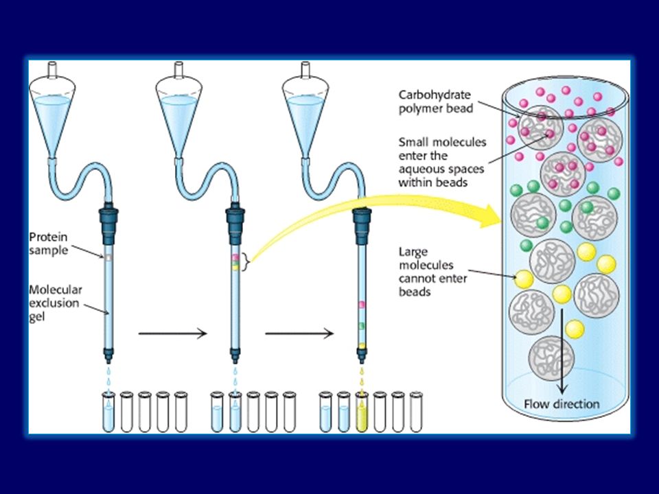
Stationary phase Is gel beads which contain pores of specific size. Usually gel is polysaccharides (dextran) or other polar polymers formulated into small beads Theses beads varying in degrees of cross-linking of the polysaccharide within the bead. Beads allowing smaller molecules to pass through their pores, while larger molecules are excluded.
 or other polar polymers formulated into small beads Theses beads varying in degrees of cross-linking of the polysaccharide within the bead. Beads allowing smaller molecules to pass through their pores, while larger molecules are excluded..")
4
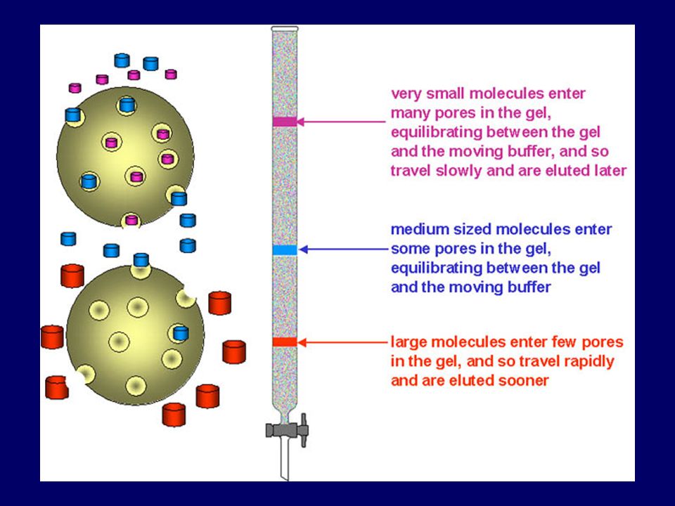
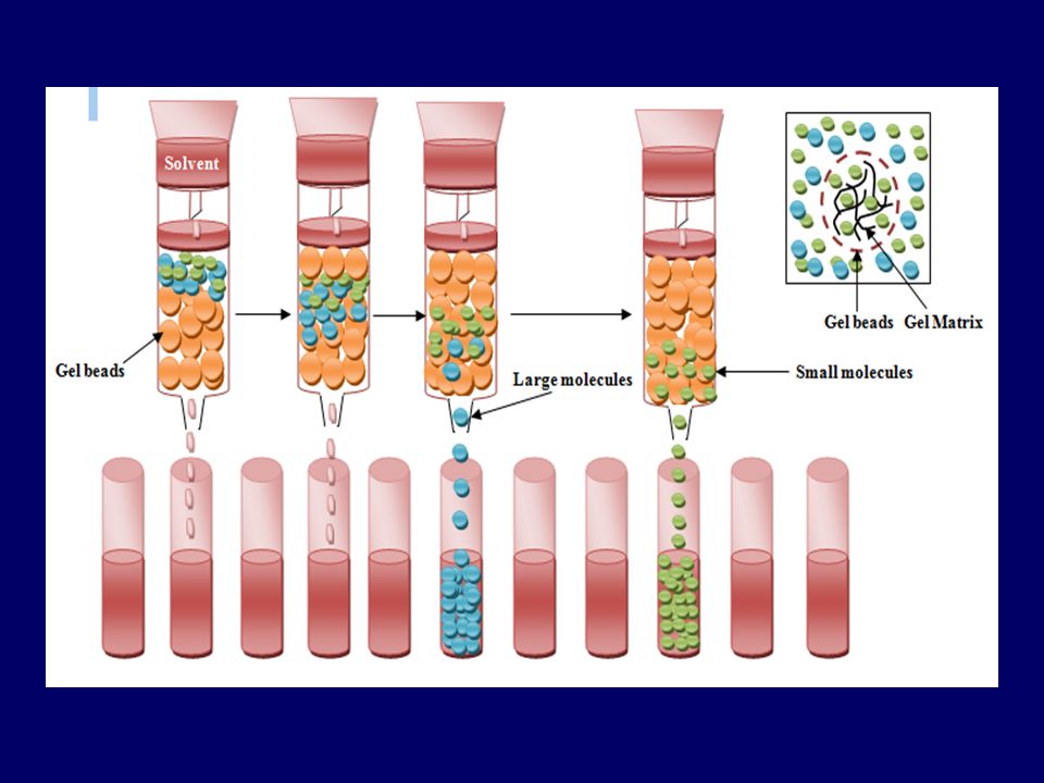
Principle: Sample pass through a column packed with a swollen gel. Separating of molecules occurs according to molecular weight: Separating of molecules occurs according to molecular weight: - Large molecules (that are larger than the largest pore): - Smaller molecules (the are retained because) Smaller molecules penetrate the pores to different degrees depends on their size molecules are eluted in order of decreasing size. So, Gel-filtration works opposite to sieve. can’t penetrate gel pores move around the beads excluded from gel pores pass through the column quickly elute first
: - Smaller molecules (the are retained because) Smaller molecules penetrate the pores to different degrees depends on their size molecules are eluted in order of decreasing size. So, Gel-filtration works opposite to sieve. can’t penetrate gel pores move around the beads excluded from gel pores pass through the column quickly elute first.")
7
Important parameters: Exclusion volume or void volume (Vo): volume between gel beads (Vo = elution volume of large molecules they do not enter the pores) Internal pore volume Vi: volume inside the beads. (Vi = elution volume of small molecules) Elution volume (Ve): volume required to elute a particular molecule Total volume (Vt ): is total volume of mobile phase in the column Vt = Vi+Vo
 Elution volume (Ve): volume required to elute a particular molecule Total volume (Vt ): is total volume of mobile phase in the column Vt = Vi+Vo.")
9
Partition coefficient (Kd) Kd: is the partition coefficient for solute, (the extent to which the molecules can penetrate the pores in stationary phase) its value range between 0 and 1 Kd = Ve – Vo Vi it is difficult to measure Vi precisely, the equation modified to determine available part of the resin (Kav). Kav = Ve – Vo Vt – Vo Sample components (solutes) are easily separated if their (Kav) value different from each other. (Vt = π r².h)
 are easily separated if their (Kav) value different from each other. (Vt = π r².h).")
10
Types of stationary phase The media used for gel exclusion chromatography are: – dextran (Sephadex™), – polyacrylamide (Bio-Gel P™) – dextran-polyacrylamide (Sephacryl™) – agarose (Sepharose™ and BioGel A™) Sephadex G-25 is most common gel used in gel-filtration chro. Each is available with a different ranges of pore size in the beads, permitting separation of macromolecules of different size.

11
Different types of matrix forming stationary phase: It is a strongly hydrophilic polymer, and swells in water before a column is prepared, the gel must be full hydrated. dextran Cross-linked dextran polymer (Sephadex G-10 to G-200) They are hydrophilic but are chemically more stable than dextran gels. polyacrylamide Cross-linked polyacrylamide (Biogel P-2 to G-300) They are also hydrophilic but are sold in the swollen form. Agarose Agarose-the largest pore size The polyacrylamide provide a three-dimensional structure which supports the interstitial agarose gel. polyacrylamide agarose Mixed gels of polyacrylamide and agarose (Ultragel) - Controlled-pore glass beads can use as porous gel
 They are hydrophilic but are chemically more stable than dextran gels. polyacrylamide Cross-linked polyacrylamide (Biogel P-2 to G-300) They are also hydrophilic but are sold in the swollen form. Agarose Agarose-the largest pore size The polyacrylamide provide a three-dimensional structure which supports the interstitial agarose gel. polyacrylamide agarose Mixed gels of polyacrylamide and agarose (Ultragel) - Controlled-pore glass beads can use as porous gel.")
13
Sephadex-25 It is an inert, bead-formed, cross-linked dexran (polymer of glucose). Sephadex beads are porous, Molecules larger than largest pores cannot enter the gel and are eluted first, Smaller molecules enter the beads and are retard. Sephadex G-25 exclude all molecules with a molecular weight greater than 5000, thereby eluting them first.

14
Advantages of gel filtration Reliable and simple Little equipment is required The procedures are straight forward Good separation and yields

15
Application of gel-filtration: Used in: Separate molecules of different sizes (biological molecules) Determination of the relative molecular mass: using a calibration curve prepared from the elution volumes of several reference substances of known relative molecular mass. Desalting or buffer exchange: The removal of solutes of low relative molecular mass from preparations of macromolecules.

16
Lab Practice In this lab, you will separate a mixture of blue dextran and cobalt chloride molecules through a gel-filtration column collecting elutes (Run-off), and calibrating a curve to show how the column separates molecules by molecular weight. Then, you will use the calibration curve to identify the molecular weights of the proteins.

17
Blue dextran: is a glucose polymer with high M.wt (M.wt =2000000 daltons) It is too large and can’t get into the beads and therefore excluded from the gel (Kd=0), and pass the column in the void volume (space between gel beads). Blue dextran is often used as a marker to measured void volume. Cobalt chloride: low molecular weight (Small molecule) is freely accessible to the gel particles (Kd=1), and elute at a volume equal to Vt
 is freely accessible to the gel particles (Kd=1), and elute at a volume equal to Vt.")
18
Both molecules (Blue dextran & CoCl 2.6H 2 O) are colored so the progress of the filtration can be followed by observing the separation of the colored bands. Fraction analysis: The completed fraction is then analyzed by measuring the extinction of each fraction at 625 nm and 510 nm Blue dextran ʎ max = 625 nm CoCl 2.6H 2 O ʎ max = 510 nm

20
Chemicals & other material: Sephadex G25 Blue dextran in saline Cobalt chloride in saline Sodium chloride (0.9 %), (Saline)
, (Saline)")
21
Procedure: Number 15 test tubes and arrange them in order on a rack. Prepare the gel bead column: Column (12 cm) is filled with semisolid (Swollen) gel beads of Sephadex G-25 [Sephadex gel soaked in the elution buffer 3-4 hrs]. Be gentle; do not allow gaps or bubbles to form. Allow small amount of saline to flow through the column between additions of beads (help the beads to settle) Equilibrate the column with saline by passing about 10 ml of saline through the column beads after it has completely settled.
 is filled with semisolid (Swollen) gel beads of Sephadex G-25 [Sephadex gel soaked in the elution buffer 3-4 hrs]. Be gentle; do not allow gaps or bubbles to form. Allow small amount of saline to flow through the column between additions of beads (help the beads to settle) Equilibrate the column with saline by passing about 10 ml of saline through the column beads after it has completely settled..")
22
It is important that the gel should be homogenous, free from bubbles, free from crack, and free from spaces between the walls. And it should be covered by the liquid "mobile phase" all the time. Avoid stirring up the top of the column bead when adding saline or samples, as this will give poor resolutions of the samples.

23
Drain saline solution down (keep1 mm of buffer above the gel) before adding the sample mixture Carefully add the sample mixture to the top of the column using a pasture pipette. (do not stir up the top of the gel) Turn off the stopcock to allow the mixture to enter the gel beads. Then add saline solution to the top, filling the space at the top of the column.
 Turn off the stopcock to allow the mixture to enter the gel beads. Then add saline solution to the top, filling the space at the top of the column..")
24
Collect the fractions (3 ml/tube) beginning with tube # 1 (Early fractions contain large molecules while later fractions contain smaller ones). Measure the absorbance spectrum of each fravtion in order to identify each molecule (Blue dextran at 625 nm, CoCl2.6H 2 O at 510 nm).
..")
25
Cobalt chloride Blue dextran Sephadex G25 Sodium chloride (0.9%), (Saline)
, (Saline)")
26
Large molecule Small molecule

28
Results: Tube No. Volume fraction (ml) Absorbance at 510 nm (Blue dextran) Absorbance at 625 nm (Cobalt chloride ) 1 3 26 39 412 515 618 721 824 927 1030 1133 1236 1339 1442 1545 1648 1751 1854
 Absorbance at 510 nm (Blue dextran) Absorbance at 625 nm (Cobalt chloride )")
Similar presentations


>")
 WWeb for lecture slides : E /QQ /MSN/gchat:>")


 Gel permeation chromatography (GPC) Gel Filtration Chromatography (GFC)>")
. Sample components enter pores.>")

 Normal Phase (very polar) Adsorption (very non-polar) Ion-Exchange (ionic)>")


>")


