Download presentation
Presentation is loading. Please wait.

1
Human Diseases A Systemic Approach Sixth Edition Chapter 12 Diseases of the Reproductive Systems Mary Lou Mulvihill Mark Zelman Paul Holdaway Elaine Tompary Jill Raymond

2
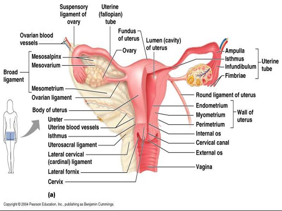
Female Reproductive System Organs of the Female Reproductive System Breasts Fallopian tubes Ovaries Vagina Vulva Uterus

3
Click on the screenshot to view an animation on the female pelvis.

4
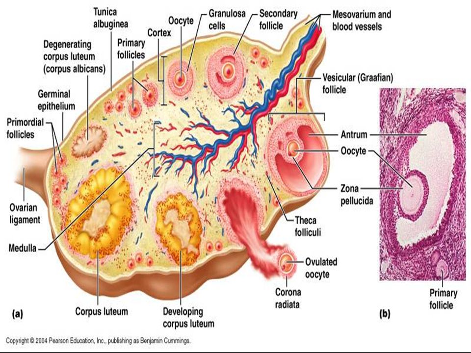
Sagittal section of the female pelvis, showing organs of the reproductive system.

5
The uterus, ovaries, and associated structures.

6
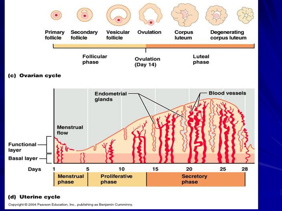
Uterus Pear shaped Hollow Contains thick muscular wall Lined with mucous membrane Rich blood supply Lies in the center of the pelvic cavity – Between bladder and rectum Bent forward - anteflexion Held in position by ligaments which anchor it to the perimetrium 3 sections – Fundus - upper – Corpus - body – Cervix - lower or neck

7
Endometrium Inner layer of uterus Contains rich blood supply Reacts to hormonal changes Prepares to receive ovum Fertilized egg implants here Provides nourishment and protection

8
Menstruation When pregnancy does not occur Endometrial lining is sloughed off Menstrual period Menarche – early teens Menopause – 40–55

9
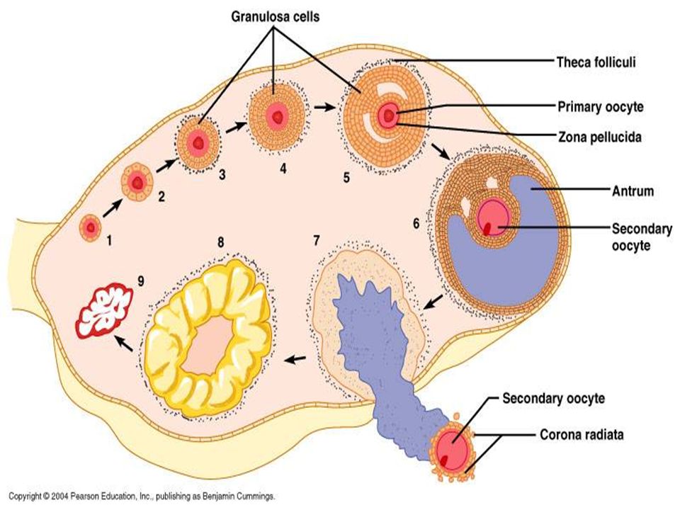
Ovaries 2, located on each side Within pelvic area Almond shaped glands Produce ovum and hormones – Follicle stimulating hormones and luteinizing hormones secreted by the anterior pituitary Stimulate ovulation – Estrogen and progesterone – Prepare endometrium to receive fertilized ovum

10
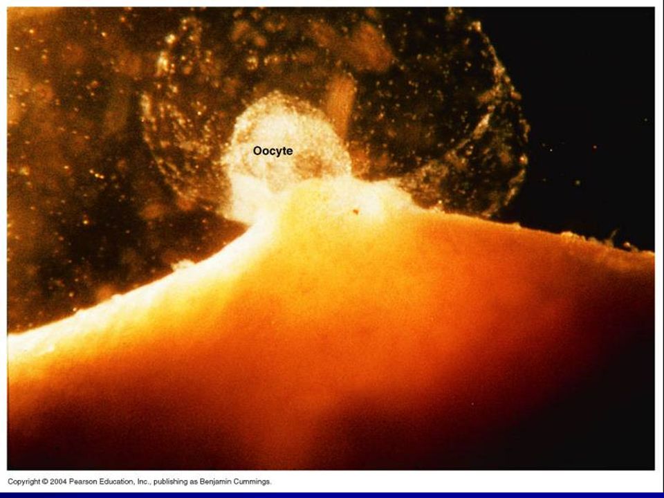
Click on the screenshot to view an animation showing oogenesis.

17
Fallopian Tubes Uterine tubes Oviduct 5½ inches long Project from either side of uterus End with finger-like projections – Fimbriae Purpose is to propel ovum from ovary to uterus Fertilization occurs within upper half of fallopian tubes.

18
Vagina Thin muscular tube Lined with mucous membranes Extends from cervix to outside of the body Allows for passage of menstrual flow During intercourse, receives the male penis and semen Serves as the birth canal Hymen is a thin membranous tissue – Covers external vaginal opening

19
Vulva General term meaning female external genitalia Bartholin’s glands – Secrete mucus for lubrication – Located on outer side of vaginal orifice Labia majora and minora are folds of skin – Serve as protection – Urinary meatus

20
Breasts Mammary glands – Produce milk – Lactation Milk-producing glands release milk at the nipple. Areola is the pigmented area around the nipple.

21
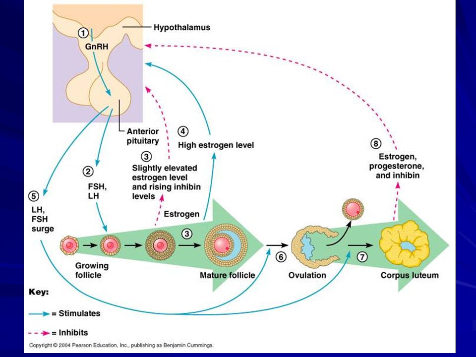
Physiology of the Female Reproductive System Reproductive cycle Regulation via secretion of female hormones – Estrogen, progesterone Governed by gonadotropic hormones of the anterior pituitary which is controlled by the hypothalamus

22
Female Reproductive Cycle Menarche: 10 to 15 years of aage Menopause: 40 to 50 years of age Gonadotropic hormones stimulate ovarian follicles to develop – Graffian follicles – Ovulation

23
First Half of the Cycle Estrogen is secreted – Endometrium becomes more vascular – Preparation for proliferative phase Corpus luteum follows release of the ovum Progesterone continues stimulation of endometrial growth and storage of nutrients for nourishing a fertilized ovum

25
Following Ovulation Corpus luteum ceases to secrete hormones approximately 8 to 12 days after ovulation. At the end of the monthly cycle, the level of estrogen and progesterone drops, and menstruation, the sloughing of the endometrial lining, occurs. If pregnancy occurs, the placenta gradually assumes the role of the corpus luteum in secreting these hormones.

28
Placenta and Umbilical Arteries The placenta is formed from both maternal and embryonic tissue. The endometrium thickens, becomes highly vascular, and develops large blood sinuses. An embryonic membrane, the chorion, develops fingerlike projections called villi, which dip into the maternal blood sinuses. This interdigitation of embryonic and maternal tissue constitutes the placenta. The umbilical arteries extend into the chorionic villi, where the exchange of carbon dioxide for oxygen and waste material for nutrients occurs. – Maternal and fetal bloods do not mix; the exchange of these substances is by diffusion across the blood vessel walls. Oxygen and nutrients return to the fetus through the umbilical vein.

29
Pregnancy Gestation period 40 weeks Birth before 37 weeks considered premature Embryo – from fertilization until 8 weeks Fetus – from 8 weeks to birth

30
Placenta Provides nourishment for fetus from mother Spongy structure that forms in the uterus next to the fetus Afterbirth Fetus attached to the placenta by the umbilical cord Surrounded by 2 membranous sacs – Amnion – holds amniotic fluid in which the fetus floats – Chorion – protective sac

31
Click on the screenshot to view a video on the topic of the placenta.

32
Labor Stage 1 Stage 2 Stage 3 Dilation stage Uterine muscles contract to expel fetus Thinning of the cervix – effacement Fetus presses on cervix, causing it to expand to 10 cm Expulsion Ends with birth of baby When the head appears, called crowning Placental stage Uterus again begins to contract Afterbirth delivered

33
Click on the screenshots to view videos showing labor.

36
Diseases of the Female Reproductive System Infections, tumors, and cysts develop in the reproductive organs and in the breasts. Abnormalities of the menstrual cycle and of pregnancy also occur.

37
Pelvic Inflammatory Disease Inflammation of the pelvic reproductive organs as a result of bacterial, viral, fungal, or parasitic invasion. Subsequent infection can ascend to the cervix (cervicitis) the endometrium (endometritis), fallopian tubes (salpingitis), and ovaries (oophoritis). The most common cause of PID is sexually transmitted disease; including gonorrhea and chlamydia. Streptococcal and staphylococcal organisms can enter the female reproductive tract after an abortion or delivery in which sterile procedures were not carefully followed.
 the endometrium (endometritis), fallopian tubes (salpingitis), and ovaries (oophoritis). The most common cause of PID is sexually transmitted disease; including gonorrhea and chlamydia. Streptococcal and staphylococcal organisms can enter the female reproductive tract after an abortion or delivery in which sterile procedures were not carefully followed..")
38
Pelvic Inflammatory Disease (continued) Symptoms: lower abdominal pain, fever resulting from the infection, chills, and leukorrhea, a white, foul-smelling vaginal discharge. Treatment: antibiotics, aspirin, bed rest, and fluids Untreated infections: risk of formation of abscesses, risk of ectopic pregnancy, and infertility from adhesions

39
Puerperal Sepsis An infection of the endometrium after childbirth or an abortion. Trauma and blood loss encountered during delivery provide a portal of entry for invading microorganisms through the birth canal. Lesions of the endometrium favor bacterial growth. Streptococci are the principal causative organisms, but staphylococci and E. coli enter the uterus through a lack of aseptic technique. Necrosis of the endometrium develops from the infection.

40
Puerperal Sepsis (continued) Infected blood clots can break loose and travel as septic emboli. Without proper treatment a systemic infection of the blood, or septicemia, or thrombophlebitis may result. The symptoms of puerperal sepsis are fever, chills, profuse bleeding, foul-smelling vaginal discharge, and pain in the lower abdomen and pelvis. Treatment: antimicrobials

41
Neoplasms of the Female Reproductive Organs Early detection, diagnosis, and treatment of any abnormal mass or lump are extremely important in preventing the growth and spread of cancer. Many tumors and cysts are harmless, but tests are required to differentiate between malignant and benign growths.

42
Click on the screenshot to view a video on the topic of cancer of the female reproductive organs.

43
Carcinoma of the Cervix Carcinoma of the cervix is one of the cancers most easily diagnosed in the early stages. Incidence of this malignancy has decreased significantly since the development of the Pap smear. Carcinoma in situ, a malignant lesion, is the earliest stage of cancer; the underlying tissue has not yet been invaded. Progression from carcinoma in situ to an invasive malignancy may be slow. Symptoms: ulceration, causing vaginal discharge and bleeding.

44
Carcinoma of the Cervix (continued) Cervical cancer may spread to surrounding organs: vagina, bladder, rectum, and pelvic wall. Widespread cancer becomes inoperable, and radiation therapy is the usual treatment. Carcinoma of the cervix is strongly associated with infection by human papilloma virus. Early sexual activity and promiscuity are also related to the incidence of this cancer.

45
Carcinoma of the Endometrium Carcinoma of the endometrium, the lining of the uterus, occurs most often in postmenopausal women who have had no children. The malignant tumor may grow into the cavity of the uterus or invade the wall itself. Ulcerations develop, and erosion of blood vessels causes vaginal bleeding. Surgery and radiation are the usual treatments.

46
Fibroid Tumors (Leiomyomas) Benign tumors of the smooth muscle of the uterus, or fibroid tumors The most common tumors of the female reproductive system and frequently cause no symptoms. Fibroids are often multiple and vary greatly in size. The cause of fibroid tumors is unknown although their growth is stimulated by estrogen. Symptoms include abnormal bleeding between periods or excessively heavy menstrual flow and pelvic pain.

47
Leiomyomas (continued) Fibroid tumors can also interfere with delivery of the newborn. Treatment for fibroid tumors depends on severity and childbearing plans. Myolyosis, a laparoscopic technique, may be used to knock out the blood vessels of the tumor, the tumor may be removed surgically or hysterectomy may be necessary.

48
Types of uterine fibroids.

49
Ovarian Neoplasm The ovaries are a common site for cancer to develop. The ovaries’ position deep in the pelvis makes early detection of the tumor difficult. Often extensive metastasis will occur before there are noticeable symptoms. Symptoms include abdominal and pelvic pain, weight loss, general malaise, and digestive disturbances. The cause of ovarian cancer is not known. Treatment may include surgical removal of the mass, hysterectomy, radiation, and chemotherapy.

50
Hydatidiform Mole A benign tumor of the placenta, it can develop after a pregnancy or be associated with an abnormal one. Hydatidiform mole is a developmental anomaly that occurs when the chorionic villi develop into a mass of grape-like vesicles. – The mass secretes chorionic gonadotropic hormone (CGH) – The uterus enlarges Bleeding usually occurs, and the mole is expelled Treatment: scraping of the uterus, the procedure of dilatation of the cervix and curettage (D&C), removes any fragments of the mass or placenta
 – The uterus enlarges Bleeding usually occurs, and the mole is expelled Treatment: scraping of the uterus, the procedure of dilatation of the cervix and curettage (D&C), removes any fragments of the mass or placenta.")
51
Choriocarcinoma A highly malignant tumor of the placenta A part of the placenta is formed by the embryonic membrane called the chorion. May develop after a hydatidiform mole, a normal delivery, or an abortion Tumor is highly invasive and metastasizes rapidly causing abdominal pelvic pain. A choriocarcinoma, like hydatidiform mole, secretes large amounts of CGH. Laparoscopy may be used to visualize the tumor. Chemotherapy rather than surgery is the usual treatment.

52
Adenocarcinoma of the Vagina Has been linked to the synthetic hormone diethylstilbestrol (DES) Rare cancer that has developed in some young girls whose mothers were given diethylstilbestrol during pregnancy. Diethylstilbestrol appears to have only slight effects in sons born to these women. – Testes have been found to be smaller than normal in some cases, and some cyst formation has been found in the epididymis. Symptoms include leukorrhea and a bloody vaginal discharge. Treatment may include surgical removal of the tumor, radiation, and chemotherapy.

53
Adenocarcinoma, Cancer of the Breast Ducts Most common breast malignancy, and leading cause of cancer death in women Sign: a hard fixed lump in the upper outer quadrant Benign tumors are encapsulated and not fixed to underlying structures Nipple retracts and skin dimples Axillary lymph nodes may be swollen

54
Adenocarcinoma, Cancer of the Breast Ducts The cause of adenocarcinoma is not known. Risk factors include family history, exposure to radiation or carcinogens, age, never being pregnant, having your first child after age thirty- five, early menarche, menopause after age fifty. Treatment: simple or radical mastectomy, lumpectomy, chemotherapy, radiation therapy.

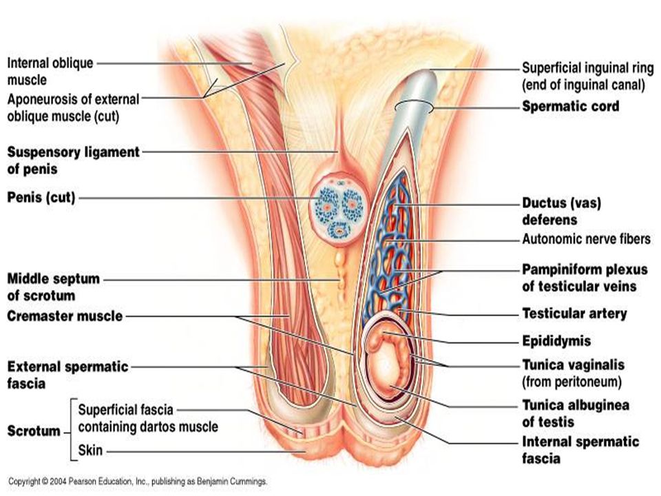
55
Figure 12-4: Cystic hyperplasia of the breast.

56
Paget’s Disease of the Nipple A rare cancer involving inflammatory changes that affect the nipple and the areola The nipple becomes granular and crusted with lesions resembling eczema. In advanced Paget’s disease, ulceration develops and there is a discharge from the nipple. The breast becomes edematous and is characterized as having a “pigskin” appearance. The cause of Paget’s disease is unknown. A significant feature in Paget’s disease is that an underlying infiltrating duct cancer accompanies it. Treatment depends on the extent of the disease and may include removal of the breast.

57
Benign Tumors of the Breast Fibroadenoma: a firm, movable mass easily removed by surgery. The fibroadenoma does not become malignant. Cystic hyperplasia or fibrocystic disease is very common and not serious. Develops at any age with the formation of numerous fluid filled lumps in the breast. Lumps tend to be painful at the time of the menstrual period as the breasts themselves respond to hormonal changes, enlarging and regressing. There may be a higher incidence of breast cancer development in women who have cystic hyperplasia. These women should be examined regularly to prevent mistaking a tumor for a cyst. The etiology of cystic hyperplasia is unknown. Treatment may include surgical removal of the tumor.

58
Menstrual Abnormalities Amenorrhea—absence of menstrual periods Dysmenorrhea—painful or difficult menses Menorrhagia—excessive or prolonged bleeding during menstruation Metrorrhagia—extreme irregularity of the menstrual cycle

59
Amenorrhea The absence of menstrual periods; known as primary amenorrhea if menstruation fails to begin Lack of gonadotropic hormones from the pituitary gland or a diseased ovary can cause the abnormality, and administration of hormones may be effective treatment. Secondary amenorrhea: The cessation of menstrual periods for more than one year – This can result from an ovarian or uterine disease, as well as hormonal imbalance; pituitary failure and thyroid disease can cause amenorrhea. Treatment may include hormone therapy.

60
Amenorrhea (continued) Certain psychological states such as extreme depression, eating disorders and/or excessive exercise, both of which deplete body fat, can cause amenorrhea. – The hypothalamus of the brain governs the release of pituitary hormones, including the gonadotropins. The condition of amenorrhea may correct itself if the stressful conditions can be eliminated.

61
Dysmenorrhea Painful or difficult menses; one of the most common gynecologic disorders Symptoms include dull to severe pelvic and lower back pain that may radiate to other areas. Causes include pelvic infections, endometriosis, and other unknown causes. Diagnosis is made based on pelvic examination; laparoscopy and D&C may be used to confirm the diagnosis. Treatment may include oral contraceptive therapy to regulate and decrease menstrual flow. Nonsteroidal anti-inflammatory medications may be given to reduce pain. Application of heat to the pelvic area may also be helpful.

62
Menorrhagia Excessive or prolonged bleeding during menstruation; it can result from tumors of the uterus, pelvic inflammatory disease, or endocrine imbalance. Failure to ovulate can also cause menorrhagia. If a corpus luteum is not formed, progesterone is not secreted and estrogen continues to stimulate endometrial thickening. Treatment varies according to the cause of the disease. Tumors should be removed surgically, pelvic inflammatory disease should be treated with antibiotics, and hormonal therapy should be administered for endocrine insufficiency.

63
Metrorrhagia Bleeding between menstrual periods or extreme irregularity of the cycle It results from an abnormal buildup and sloughing of endometrial tissue. Hormonal imbalance may be the cause of metrorrhagia, or the endometrial response to the hormones may be incorrect. A D&C is often performed and the endometrium returns to normal.

64
Toxic Shock Syndrome (TSS) Caused by an infection of Staphylococcus aureus Signs include high fever, rash, skin peeling, decreased blood pressure, gastrointestinal complaints, elevated liver enzymes, and neuromuscular disturbances. Treatment includes fluid replacement to counteract shock and administration of selected antibiotics. Etiology: tampon use associated with an increase in staphylococcal toxin from fibers used in “super” tampons to increase absorbency.

65
Toxic Shock Syndrome (TSS) (continued) The fibers apparently remove magnesium from the vagina, and this produces an environment that encourages growth of the bacteria that make toxins. – These fibers are no longer used. – It was found that some surgical dressings also contained the same fibers, a finding that may explain some cases of toxic shock syndrome in non-tampon users. Recommendations for women who use tampons include avoidance of the super-absorptive type, daytime use only, and frequent changes of tampons.

66
Premenstrual Syndrome Disabling symptoms prior to menstruation with disruption of family, business, and social relationships PMS consists of groups of severe symptoms, emotional, physical, and behavioral, which are associated with the menstrual cycle. They usually begin at the mid-point of the cycle and worsen until the onset of bleeding. Physical symptoms include lower abdominal bloating, breast swelling and soreness, headache, and constipation. Episodes of depression, anxiety, irritability, and hostility are characteristic of emotional changes. Typical behavioral symptoms include crying, binge eating, and clumsiness.

67
Premenstrual Syndrome (continued) Cause of PMS is unknown but researchers suspect that the production of cyclic ovarian hormones affect the production of other hormones and chemicals, specifically neurotransmitters. These chemicals may cause the symptoms, but it is not understood why some women are affected and others are not. Treatment has to be individually prescribed as women respond differently to various suggestions. For some women, dietary changes during the week before the onset of menstruation are helpful. These changes might include the avoidance of salt, sugar, caffeine, and alcohol. Aerobic exercise, brisk walking, or swimming is helpful for others. Antidepressant therapy or hormone therapy may also be used.

68
Click on the screenshot to view a video in the topic of premenstral syndrome. Return to Directory

69
Endometriosis A disease condition in which endometrial tissue from the uterus becomes embedded elsewhere The tissue may have been pushed backward through the fallopian tubes during menstruation or carried by blood or lymph. It then takes hold on some structure in the peritoneal cavity, such as the ovary. The endometrial tissue by nature responds to hormonal changes even when outside the uterus. Endometriosis causes pelvic pain, abnormal bleeding, and dysmenorrhea. Sterility and pain during sexual intercourse (dyspareunia) can result.
 can result..")
70
Endometriosis (continued) The etiology of endometriosis is unknown. The only certain means of diagnosing endometriosis is by seeing it. A tissue biopsy can be taken and examined. Treatment of endometriosis varies according to the extent of the abnormal growth and the age of the patient. Hormonal therapy is generally used for the young patient. Pregnancy, with the absence of menstruation, tends to hold the condition in check. Extensive proliferation of endometrial tissue requires surgery, and cysts filled with blood are usually found at this time.

71
Abnormalities of Pregnancy A most important factor during pregnancy is good prenatal health. The pregnant woman should be checked regularly for weight gain, blood pressure, and urine abnormalities. She should be instructed on the importance of proper diet and exercise. Most pregnancies progress normally, but occasionally some problems do arise.

72
Ectopic Pregnancy A pregnancy in which the fertilized ovum implants in a tissue other than the uterus The most common site of an ectopic pregnancy is in the fallopian tubes. The fertilized ovum becomes trapped because of a structure or obstruction such as a tumor. Salpingitis is a predisposing condition for a tubal pregnancy due to the inflammatory effect on the mucosal lining.

73
Ectopic Pregnancy (continued) Embryonic development proceeds for about 2 months, at which time the pregnancy terminates. The tube often ruptures, causing severe internal hemorrhage into the abdominal cavity. Intense pain and bleeding from the uterus result, and the embryo is usually destroyed by the trauma. Once the diagnosis has been made, the ruptured tube and embryo have to be removed surgically.

74
Spontaneous Abortion Commonly called a miscarriage Most likely due to genetic abnormality Usually occurs in the second or third month of pregnancy The first sign is vaginal bleeding with cramping. Prompt medical attention is needed to reduce the hazards of hemorrhage and infection. A D&C is usually performed to remove any tissue that remains in the uterus.

75
Morning Sickness Transient nausea or vomiting associated with the first trimester of pregnancy Cause of morning sickness is not known May be due to hormonal changes related to pregnancy Treatment is not necessary unless there is excessive vomiting that causes dehydration and weight loss.

76
Hyperemesis Gravidarum Excessive vomiting during pregnancy leading to dehydration, weight loss, and electrolyte and acid–base disturbances in the mother and baby. Cause is not known but is thought to be due to an increased production of chorionic gonadotropin by the fetus. This hypothesis is supported by hyperemesis gravidarum occurring more often in multiple fetus pregnancies. Diagnosis is made on the basis of symptoms, weight loss, and signs of dehydration. In severe cases the patient is treated with intravenous fluids and electrolyte replacement, all other fluids and food are withheld. Sedatives are given to control nausea and vomiting. Hyperemesis gravidarum usually subsides in the second trimester.

77
Click on the screenshot to view a video on breast cancer. Return to Directory

78
Multisystem effects of premenstrual syndrome.

79
Figure 12-6: Locations of endometriosis outside the uterus.

80
Table 12-1: Signs and Symptoms of Toxemia.

81
Click on the screenshot to view a video on the topic of preeclampsia.

82
Table 12-2 Risk factors for Gestational Diabetes Mellitus

83
Male Reproductive System Organs of the male reproductive system Bulbourethral gland Epididymis Penis Prostate gland Scrotum Seminal vesicle Testes Vas deferens

84
Click on the screenshot to view an animation comparing spermatogenesis and oogenesis.

85
Click on the screenshot to view an animation for spermatogenesis.

86
Anatomy and Physiology of the Male Reproductive System The male reproductive system is a combination of the reproduction and urinary systems. In the male, the major organs of reproduction are located outside the body The penis Contains the urethra, which carries both urine and semen to the outside of the body The scrotum – Contains the two testes, each with an epididymis

87
Click on the screenshot to view an animation on the male pelvis.

88
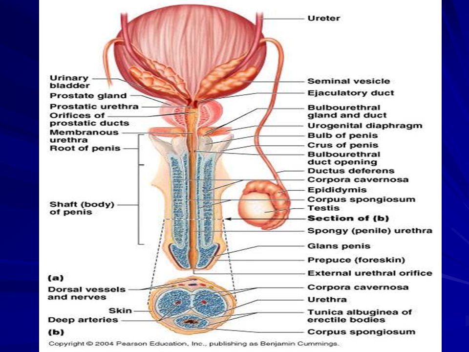
The male reproductive system.

89
Click on the screenshot to view animation showing sperm.

90
Internal Organs of Reproduction 2 seminal vesicles 2 vas deferens Prostate gland 2 bulbourethral glands

91
External Organs of Reproduction Scrotum is a sac that contains the testes or testicles Scrotum is divided by a septum; supports the testicles and lies between the legs and behind the penis During early childhood, the testes will frequently retract up into the pelvic cavity. However, as the young boy reaches one year in age, the testes will remain permanently in the scrotum. The perineum of the male is similar to that in the female. It is the area between the anus and the scrotum.

92
Testes The testes are oval in shape and are responsible for the development of sperm. This process, called spermatogenesis, takes place within the seminiferous tubules. The testes must be maintained at the proper temperature for sperm to survive. This lower temperature level is achieved by the placement of the testes suspended in the scrotum outside the body. The hormone testosterone, which is responsible for the growth and development of the male reproductive organs and sperm, is also produced by the testes. The singular for testes is testis.

94
Epididymis Each epididymis is a coiled tubule that lies on top of the testes within the scrotum. This elongated structure serves to store sperm as they are produced by the testes until they are ready to be released into the vas deferens.

95
Penis The penis is the male sex organ containing erectile tissue that is encased in skin. This organ delivers semen into the female vagina. The soft tip of the penis is referred to as the glans penis. It is protected by a covering called the prepuce or foreskin. It is this covering of skin that is removed during the procedure known as circumcision. The penis becomes erect during sexual stimulation, which allows it to be placed within the female for the ejaculation of semen.

96
Penis Consists of three cylindrical bodies of cavernous tissue also known as erectile tissue. – Tissue is filled with spaces, or sinuses, that become engorged with blood – The urethra passes through one of these cylindrical bodies as it extends to the outside and connective tissue supports the erectile structures. – The distal, expanded end of the penis is the glans penis. – A flap of loosely attached skin covering the glans, the prepuce or foreskin, is often removed shortly after birth; this is the procedure called circumcision.

97
Internal Organs of Reproduction Each vas deferens carries sperm from the epididymis up into the pelvic cavity. They travel up in front of the urinary bladder, over the top, and then back down the posterior side of the bladder to empty into the urethra. The vas deferens, along with nerves, arteries, veins, and lymphatic vessels running between the pelvic cavity and the testes, form the spermatic cord.

99
Seminal Vesicles The two seminal vesicles are small glands located at the base of the urinary bladder. These vesicles are connected to the vas deferens just before it empties into the urethra. The seminal vesicles secrete a fluid that nourishes the sperm. This liquid, along with the sperm, constitutes semen, the fluid that is eventually ejaculated during sexual intercourse.

100
Prostate Gland The single prostate gland is located just below the urinary bladder. It surrounds the urethra and when enlarged can cause difficulty in urination. The prostate is important for the reproductive process since it secretes an alkaline fluid that assists in keeping the sperm alive by neutralizing the pH of the urethra and vagina.

102
Bulbourethral Glands The bulbourethral glands are also known as Cowper’s glands. Two small glands located on either side of the urethra just below the prostate. They produce a mucus-like lubricating fluid that joins with semen to become a part of the ejaculate.

103
Urethra The male urethra extends from the urinary bladder to the external opening in the penis, the urinary meatus. Serves a dual function: the elimination of urine and the ejaculation of semen During the ejaculation process, a sphincter closes to keep urine from escaping.

104
Physiology of the Male Reproductive System Spermatogenesis, the formation of sperm, stimulated by gonadotropic hormones of the anterior pituitary Maturation of sperm continues in the epididymus Once ejaculated, sperm can live for 24 to 72 hours.

105
Accessory Glands They contribute to the nourishment and protection of sperm, and mucoid secretions from these glands form the semen. The seminal vesicles provide fructose, other nutrients, and prostaglandin, which increases uterine contractions. This helps to propel the sperm toward the fallopian tubes. The seminal vesicles release their secretions into the ejaculatory ducts at the same time the vas deferens empty the sperm. The muscular prostate gland, which surrounds the first part of the urethra, contracts during ejaculation, releasing its secretions. The secretion is alkaline, which buffers the highly acidic vaginal secretions that can inhibit sperm motility.

106
Accessory Glands (continued) Sexual stimulation of the male transmits impulses into the central nervous system, which initiates the male response. Sexual stimulation causes peristaltic contractions in the walls of the epididymis and vas deferens, propelling sperm into the urethra. The seminal vesicles and prostate gland simultaneously release their secretions, which mix with the mucous secretion of the bulbourethral glands forming the semen, the process of emission. Ejaculation of the semen, the culmination of the sexual act, occurs when contraction of this musculature increases pressure on the erectile tissue, and the semen is expressed.

107
Semen consists of sperm and the secretions of these glands. There are about 200–500 million sperm in one teaspoon of semen discharged during a typical ejaculation. A zygote is a product of one sperm and 1 egg. NOTE: A male can be considered infertile when his sperm count drops to 20 million per ml or less. During sexual arousal, erectile tissue in the penis swells with blood. Erection is essential for the insertion of the penis into the vagina. Like the clitoris, the penis consists of a glans that is richly supplied with nerve endings and a prepuce (foreskin) that covers the glans.
 that covers the glans..")
108
Diseases of the Prostate Inflammation from infections, sexually transmitted disease, benign hypertrophy Prostatitis Carcinoma of the prostate

109
Prostatitis The cause of prostatitis, inflammation of the prostate, is not always known. Infection frequently develops from gonococci in a male with gonorrhea or from E. coli that has caused a urinary tract infection. Symptoms: pain and a burning sensation during urination. The prostate may be tender, and pus from the tip of the penis is sometimes noted. Treatment: Penicillin is the usual treatment unless hypersensitivity to the drug necessitates the use of other antibiotics.

110
Figure 12-8: Enlarged prostate gland. Dashed line indicates the normal size.

111
Carcinoma of the Prostate Gland Carcinoma of the prostate is common in old age, but the tumor may be small and asymptomatic. Rectal examination may reveal an enlarged prostate that is very hard, harder than a benign enlargement. Prostatic carcinoma tends to metastasize before it is discovered. Symptoms may include weak urine flow, difficulty starting or stopping urine flow, pain and burning during urination, need to urinate at night, urinary incontinence, and urinary infection.

112
Carcinoma of the Prostate Gland (continued) Etiology of carcinoma of the prostate is unknown, although risk increases with age. Prognosis for this carcinoma is poor, as the malignancy spreads rapidly to nearby organs like the bladder and rectum. The cancer invades the lymph and blood vessels and metastasizes to the bone and other organs. Early diagnosis is key to a favorable outcome Treatment: surgery, removal of the testes, hormone therapy, chemotherapy, radiation therapy.

113
Some manifestations of prostate cancer.

114
Click on the screenshot to view a video on the topic of prostate cancer

115
Epididymitis Inflammation of the epididymis frequently caused by gonococci; a urinary tract infection or prostatitis can also be the source of the epididymitis. Abscesses sometimes form, and scar tissue develops that can cause sterility if both sides are affected. Symptoms include severe pain in the testes, swelling, and tenderness in the scrotum. Antibiotic treatment is effective when combined with rest and the avoidance of irritants such as alcohol and spicy foods.

116
Orchitis Inflammation of the testes, can follow an injury or viral infection such as mumps, with the development of inflammatory edema and pain. The most common cause of orchitis is mumps in an adult man. Swelling of the testes and severe pain usually develops about a week after mumps, (an inflammation of the parotid salivary glands). In severe cases, atrophy of the testes can occur, and if both sides are affected, sterility results. Treatment: rest and supportive measures
. In severe cases, atrophy of the testes can occur, and if both sides are affected, sterility results. Treatment: rest and supportive measures.")
117
Cryptorchidism Cryptorchidism is not a disease but a failure of the testes to descend from the abdominal cavity, where they develop during fetal life, to the scrotum. This condition should be corrected through surgery or hormonal therapy. Sterility results if this condition is not rectified. Maturation of the sperm cannot occur in the abdominal cavity, where the temperature is slightly higher than that of the scrotum. If the testes are not brought down into the scrotum, they should be removed. Undescended testes atrophy and may become the potential site of cancer.

118
Testicular Tumors Tumors of the testes are rare, but when they occur it is usually in young men, and these tumors are highly malignant. A painless lump develops in the testicle. Etiology is unknown, however predisposing factors include cryptorchidism, inguinal hernia during childhood, and history of mumps. Monthly testicular self-examinations are key to early detection. Treatment may include surgical removal of the testes, radiation, and chemotherapy.

119
Sexually Transmitted Infections (STIs) The incidence has increased in recent years. If untreated, serious conditions may develop that can gravely affect a person’s life. An estimated 1 million women contract pelvic infections each year as a result of undetected STIs. Infected individuals are often asymptomatic and spread the diseases to other sexual partners. Infected women may spread STIs to their offspring during pregnancy and childbirth. Sterility and life- threatening ectopic pregnancies are common complications of sexually transmitted infections.

120
Gonorrhea Gonorrhea, also known as “clap,” is one of the most common and widespread of sexually transmitted infections. Caused by the bacterium Neisseria gonorrhoeae Transmitted through sexual contact and during childbirth Treatment: good response with antibiotics

121
Chronic Gonorrhea Early detection and treatment is necessary Complications from untreated infections – Inflammation with fibrosis in the urethra and vas deferens – Fallopian tubes: salpingitis with pus in the peritoneal cavity – Pelvic inflammatory disease with abscesses, fibrosis – If untreated, can lead to life-threatening meningitis, endocarditis

122
Newborn Complications of Gonorrhea The baby of an infected mother can be born with acute purulent conjunctivitis, inflammation of the conjunctiva. The gonococcal organisms enter the eye during delivery, and if the cornea becomes ulcerated, blindness results. To prevent this infection from developing, a drop of erythromycin is routinely placed in the eyes of newborn babies.

123
Syphilis – Primary Phase The causative bacterium is a spirochete, Treponema pallidum, transmitted by sexual intercourse or intimate contact with an infectious lesion Primary State chancre, or ulceration, develops on the genitals in the primary stage of infection; appears within a few days to a few weeks after sexual contact. The chancre usually develops on the vulva of the female and on the penis of the male. The lesion, which sometimes goes unnoticed, heals after a few weeks. If untreated with penicillin, the secondary phase of the disease occurs in a matter of weeks.

124
Figure 12-9: Chancre of primary syphilis on the penis.

125
Syphilis – Secondary Phase The principal sign of the secondary phase is a non- itching rash that affects any part of the body: the trunk, soles of the feet, palms, mouth, vulva, or rectum. The individual is highly contagious during this stage, but he or she can be treated with penicillin. An untreated case of syphilis may be dormant for many years, but the organisms remain in the bloodstream and cause a systemic infection known as tertiary syphilis. The appearance of symptoms, years after the primary infection, marks the tertiary and most serious phase of syphilis.

126
Syphilis – Tertiary Phase The cardiovascular system is severely damaged at this stage of infection. The inflammatory response to the spirochetes in the blood causes fibrosis, scarring, and obstruction of blood vessels, particularly of the aorta. Lesions develop on the cerebral cortex, causing mental disorders, deafness, and blindness. Loss of sensation in the legs and feet due to spinal cord damage cause a characteristic gait to develop. Paresis, a general paralysis associated with organic loss of brain function, results in death if untreated. The tertiary lesions of the syphilitic infection are irreversible.

127
Syphillis Congenital Defects Congenital defects are numerous in an infant born to an infectious mother – Mental retardation – Physical deformities – Deafness – Blindness – The syphilitic infection can cause death of the fetus and spontaneous abortion. The severe consequences of syphilis point out the urgent need for early detection and treatment. Treatment with penicillin is successful except in reversing tertiary lesions. Development of resistant strains is a serious threat.

128
Genital Herpes Painful, viral disease that tends to recur periodically and for which there is no cure. Herpes virus is transmitted by intimate contact between mucous membrane surfaces. There are two types of herpes simplex virus-type I, causing “fever blisters” or “cold sores,” and type II, involving the mucous membranes of the genital tracts. Symptoms generally appear within 3 weeks after exposure to the virus. The symptoms intensify from a burning, itching sensation to severe pain. Multiple blisters appear on the genitalia and at times on the buttocks or thigh. As the blisters rupture, they become secondarily infected and ulcerate. Painful urination and vaginal discharge are common.

129
Transmission, Risks, and Treatment Active phase subsides as the lesions heal, but the virus remains dormant in ganglia until reactivated. The disease is transmitted by contact with an active sore that is releasing (shedding) the infectious virus. The virus can be spread from a cold sore on the lips to the genitals; the reverse is also true. There is no cure for a herpes infection, but secondary infections can be prevented and healing promoted. The lesions must be kept clean and dry, and ice-cold compresses may be used to relieve the pain.
 the infectious virus. The virus can be spread from a cold sore on the lips to the genitals; the reverse is also true. There is no cure for a herpes infection, but secondary infections can be prevented and healing promoted. The lesions must be kept clean and dry, and ice-cold compresses may be used to relieve the pain..")
130
Transmission, Risks, and Treatment (continued) Prescription medications can control the activation of dormant infections. Examples include acyclovir or AZT. Active herpes genitalis has very serious consequences during pregnancy, not only causing spontaneous abortion or premature delivery, but also increasing the risk of transmitting the infection to the newborn.

131
Figure 12-10: Genital herpes blisters as they appear on the labia.

132
Click on the screenshot to view a video on the topic of genital herpes.

133
Genital Warts Can develop in both men and women and are caused by a virus in the group called HPV (human papillomavirus) The warts may appear within weeks after sexual relations; vaginal, anal, or oral with an infected partner, or they might not develop for several months. In men, the warts occur on the penis or scrotum. In women, the most common site is the peritoneum, but they may occur on the vulva, vaginal opening, or skin of the thighs. The warts may even develop within the vagina and on the cervix.

134
Genital Warts (continued) Symptoms: itching or bleeding, although often they are first detected during a physical exam. An abnormal Pap smear might be an indication of human papilloma virus infection. The types of human papilloma virus that cause genital warts are considered as risk factors for cervical cancer. Treatment of genital warts depends on their size and number. Some are treated with medication applied by a healthcare provider, but the procedure is very painful. Electrocautery (burning), cryosurgery (freezing), and laser surgery are alternative treatments. Surgical removal does not mean a cure, as recurrence of genital warts is common.
, cryosurgery (freezing), and laser surgery are alternative treatments. Surgical removal does not mean a cure, as recurrence of genital warts is common..")
135
Figure 12-11: Genital warts. (Courtesy of the CDC / Dr. Wiesner, 1972.)
")
136
Chlamydial Infections The most prevalent STI in the United States Chlamydia trachomatis Causative Organism: Chlamydia trachomatis A leading cause of pelvic inflammatory disease in women, with resultant infertility, and severe urethritis in both sexes. – Women are often asymptomatic carriers of the infection and continue to infect partners and offspring. Symptomatic females experience vaginal discharge with burning and itching of the genital area. Males with chlamydial infection are usually symptomatic with penile discharge, burning and itching with urination, and epididymitis. The disease responds to certain antibiotics but not to penicillin. The infection often coexists with gonorrhea.

137
Trichomoniasis Caused by the protozoan Trichomonas vaginalis Symptomatic males may experience urethritis, epididymitis, and prostatitis. Symptomatic females may experience itching and burning in the genital area with a green frothy vaginal discharge with a fishy odor. Treatment with anti-parasitic medication such as metronidazole is effective.

138
Infertility Failure to conceive a child after one year of regular, unprotected intercourse. Approximately 10% of couples are infertile and 50% of couples that are treated for infertility become pregnant. The inability of a couple to conceive can originate in the male, female, or both. In males, causes of infertility include low sperm count or decreased sperm mobility, the presence of an STI or other infections of the reproductive system, blockage in the reproductive tract, structural anomalies, and endocrine disorders.

139
Infertility (continued) In females, causes of infertility include STIs or other infections of the reproductive system, hormonal problems, structural anomalies, blockage of the reproductive tract, and tumors. Treatment of infertility may include STI treatment if applicable, surgery to remove reproductive tract blockages, surgical correction of any anomalies, hormone therapy, artificial insemination and in vitro fertilization.

140
Sexual Dysfunction Any disorder that interrupts cycle from arousal to orgasm to resolution May be a physical or psychological condition Occurs in males and females

141
Impotence The inability of the male to achieve and maintain an erection sufficient for sexual intercourse Etiology: emotional disturbances or physiological diseases Emotional: Stress decreases the output of gonadotropic hormones, and, consequently, testosterone production and spermatogenesis are diminished. The dilation of penile arteries that leads to engorgement of the erectile tissue of the penis and then erection is under the control of the autonomic nervous system. Anxiety, fear, and worry are emotions that affect the nervous system.

142
Impotence (continued) Physiological: fatigue, arteriosclerosis, inadequate blood flow, diabetes mellitus, surgical complications, urologic disorders, and premature ejaculation are all possible causes. The onset of impotence may be caused by certain medications, drug abuse, or alcoholism; changes in these areas may correct the impotence.

143
Click on the screenshot to view a video on the topic of erectile dysfunction.

144
Treatment of Impotence Whether the cause is psychological or physiological in nature, treatment should be directed toward the source of the problem – Psychological therapy, sex therapy – Medications: Viagra – Evaluation of medications that may be the source of impotence such as certain heart medications or antidepressants – Control of atherosclerosis, diabetes mellitus With correct treatment, impotence can usually be overcome.

145
Dyspareunia Experienced by men and women, though more common in women Dyspareunia, painful sexual intercourse In women, physical causes may include an intact hymen, insufficient lubrication, STI, endometriosis, PID, and cysts or tumors. In men, physiological causes may include anatomic abnormalities, prostatitis, or STI. Psychological causes may include guilt, trauma, sexual abuse, and anxiety. Treatment begins with causal evaluation. Treatments may include the use of lubricants during intercourse or a gentle stretching of the vaginal opening, treatment of underlying infections, surgery, and counseling.

146
Female Arousal-Orgasmic Dysfunction Also known as frigidity, the lack of sexual desire or response in a woman Seldom caused by physical conditions, although medical problems that cause nerve damage can result in frigidity. Frigidity is usually due to a psychological condition such as stress, fatigue, depression, sexual abuse, guilt, and anxiety. Treatment may include therapy and arousal devices.

147
Premature Ejaculation Regularly ejaculating during foreplay, or immediately after beginning sexual intercourse This disorder can prevent the male from satisfying his partner or impregnating a woman. Premature ejaculation may have a psychological cause such as guilt or anxiety. Physical causes include degenerative neurological conditions. Any underlying physical causes are treated. Therapy may be necessary to help with psychological causes. Techniques that delay ejaculation including altering sexual positions and the squeeze technique may also be used.

148
Age-Related Diseases of the Reproductive System In both older females and males cancer of the reproductive organs is more common and is frequently related to hormone levels. Female changes and resultant disorders – Menopause – Uterine prolapse – Cystocele – Rectocele Males – Benign prostatic hyperplasia

149
Female – Age Related Physical Changes Reproductive organs – shrink in size Decrease in vaginal secretions Decrease in breast tissue volume Vaginal pH becomes more alkaline Menopause – Decrease estrogen and progesterone – Cessation of menstruation

150
Menopause and Health Risks Cardiac disease Osteoporosis Psychological symptoms: depression, sleep disorders, mood disorders, and decreased sex drive

151
Uterine Prolapse Uterus dropping or protruding downward into the vagina Condition results from trauma to the fascia, muscle, and pelvic ligaments during pregnancy and delivery, or atrophy of the pelvic floor muscles with age. The ligaments and muscles become so overstretched they can no longer hold the uterus in place so the uterus falls or sags downward. Symptoms include feelings of heaviness in the pelvic area, incontinence, and lower back pain. Treatment consists of strengthening the pelvic floor muscles (Kegel exercises), inserting a pessary into the vagina to support the uterus, or a hysterectomy
, inserting a pessary into the vagina to support the uterus, or a hysterectomy.")
152
Cystocele A downward displacement of the urinary bladder into the vagina This condition results from trauma to the fascia, muscle, and pelvic ligaments during pregnancy and delivery, or atrophy of the pelvic floor muscles with age. Symptoms include pelvic pressure, urinary urgency, frequency, and incontinence. Treatment consists of Kegel exercises. If cystocele is severe or exercise is ineffective, surgery may be necessary.

153
Rectocele The protrusion of the rectum into the posterior aspect of the vagina. This condition results from trauma to the fascia, muscle, and pelvic ligaments during pregnancy and delivery, or atrophy of the pelvic floor muscles with age. Symptoms include discomfort, constipation, and fecal incontinence. Treatment consists of surgical repair of the posterior wall of the vagina.

154
Benign Prostatic Hyperplasia Enlargement of the prostate gland Symptoms are a result of the enlarged prostate partially blocking the flow of urine from the bladder. – If the bladder cannot be fully emptied, residual urine provides a medium for bacterial infection and cystitis develops. Other symptoms include difficulty starting urination, weak urine stream. The blockage of urine outflow places backpressure on the ureters, which causes them to become congested with urine, a condition called hydroureters.

155
Benign Prostatic Hyperplasia (continued) This backpressure can extend to the kidneys; they swell with fluid, and hydronephrosis results. An imbalance of sex hormones frequently causes prostatic enlargement. The level of testosterone generally decreases with age, but estrogen from the adrenal cortex continues to be secreted, changing the ratio of the two. Treatment for benign prostatic hyperplasia, which is highly symptomatic, is surgical removal.

156
Diagnostic Procedures for Reproductive Diseases Sexual history, physical exam Pelvic examination PAP smear Laparoscopy Mammography Ultrasound Rectal Exam Cystoscopy

157
Click on the screenshot to view a video on the topic of vasectomy. Return to Directory

Similar presentations


- Organs mature, pubic and armpit hair, regulates release of.>")


