Download presentation
Presentation is loading. Please wait.

1
SKELETAL SYSTEM

2
SKELETAL SYSTEM FUNCTIONS Support (Primary function) Movement (Passive) Protection of Vital Organs Mineral Storage Blood Cell Formation (Hematopoiesis or Hemopoiesis)
 Movement (Passive) Protection of Vital Organs Mineral Storage Blood Cell Formation (Hematopoiesis or Hemopoiesis)")
3
OSSEOUS C.T. Compact (dense) Bone -Hard & heavy -Forms surface & diaphysis -Osteons = building blocks Cancellous (spongy) Bone -Lightweight -Fills epiphyses, Contains red marrow -Trabeculae = building blocks Matrix -Mineral Salts (hardness) -Collagen (strong & flexible)
 Bone -Hard & heavy -Forms surface & diaphysis -Osteons = building blocks Cancellous (spongy) Bone -Lightweight -Fills epiphyses, Contains red marrow -Trabeculae = building blocks Matrix -Mineral Salts (hardness) -Collagen (strong & flexible).")
4
Two Types of Bone

5
Compact Bone

6
Spongy Bone

7
Bone Cells Osteoblasts – Secrete to form bone Osteocytes *Mature bone cells *Maintain matrix *“Trapped” osteoblasts Osteoclasts – destroy bone *Enzymes digest protein *Acids dissolve minerals *Forms Marrow Cavity; Involved in Remodeling

8
Osteoblasts & Osteocytes

9
SEM Osteocyte

10
Osteoclast

11
SEM Osteoclast http://www.ectsoc.org/gallery/images/5lg.jpg http://www.ectsoc.org/gallery/images/5lg.jpg

12
SKELETAL DIVISIONS Axial Appendicular

13
Classification: Shape & Location Long Short Flat Irregular Sesamoid (develop in tendons; patella) Sutural (between cranial bones)
 Sutural (between cranial bones)")
15
Torus Bones

16
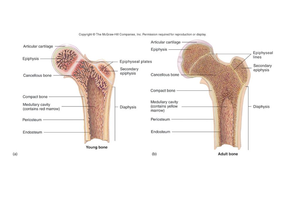
LONG BONE ANATOMY Diaphysis = shaft made of compact bone Epiphyses = ends filled with spongy bone containing red marrow Articular cartilage covers epiphyses Epiphyseal line indicates earlier location of epiphyseal (growth) plate
 plate")
18
LONG BONE ANATOMY Periosteum is C.T. covering bone Endosteum is C.T. lining medullary cavity Medullary cavity contains yellow marrow Nutrient Foramina – holes allowing for penetration of arteries

19
BONE DEVELOPMENT Ossification = replacement of other connective tissue with bone Begins during the 2 nd month of gestation Size increases until late teens (females) to mid-twenties (males) Two different ossification processes: -Intramembranous bone formation -Endochondral bone formation
 to mid-twenties (males) Two different ossification processes: -Intramembranous bone formation -Endochondral bone formation")
20
INTRAMEMBRANOUS OSSIFICATION Occurs in flat bones of skull, clavicles, mandible Begins with fibrous C.T. membrane Membrane calcifies & ossifies into bone Fontanels -“Soft spot”, not yet ossified -Allows for birth & brain growth

22
Process of Intramembranous Ossification Embryonic cells form osteoblasts Osteoblasts produce matrix = ossification center Matrix calcifies Matrix traps osteoblasts, Osteocytes form

23
Process of Intramembranous Ossification Trabeculae form Surface trabeculae form compact bone

24
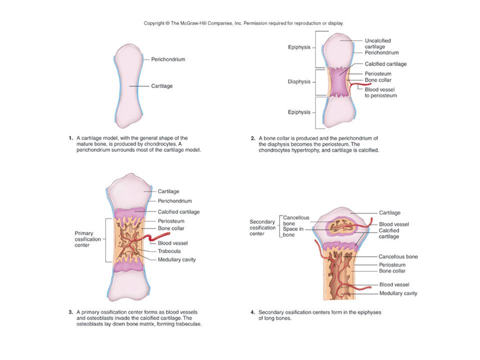
ENDOCHONDRAL OSSIFICATION Occurs in remainder of skeleton Begins with hyaline cartilage model Cartilage is replaced by bony tissue Steps include: -Bone collar forms -Cartilage in shaft calcifies -Primary Ossification center forms in shaft -Secondary Ossification centers in epiphyses

26
Formation of Bone Collar & Primary Ossification Center

27
Formation of Secondary Ossification Centers

28
Epiphyseal Plates & Articular Cartilage

30
APPOSITIONAL GROWTH Bone Widens Osteoclasts enlarge medullary cavity Osteoblasts add bone to surface of diaphysis

31
BONE REMODELING Replacement of old bone with new bone Involves resorption (osteoclasts) & deposition (osteoblasts) Alters bone shape in response to stress Benefits: -Allows for growth -Removes injured bone -New bone is more resistant to fracture
 & deposition (osteoblasts) Alters bone shape in response to stress Benefits: -Allows for growth -Removes injured bone -New bone is more resistant to fracture")
32
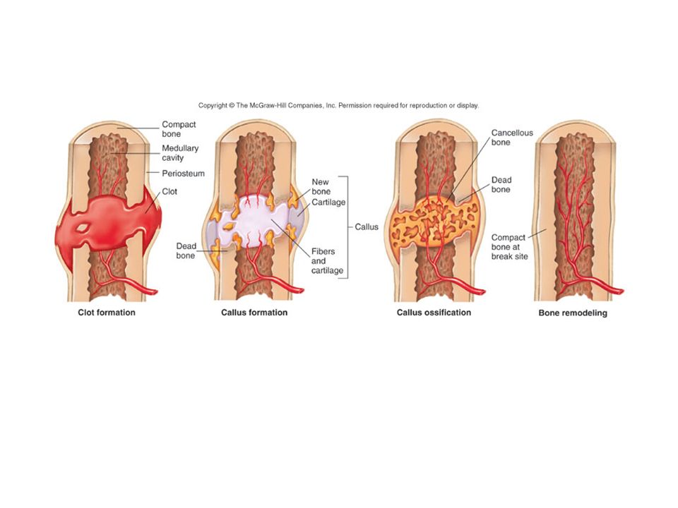
FRACTURES AND THEIR REPAIR Definition: Any break in a bone Repair may take months Requires: -Adequate minerals -Vitamins -Hormones -Weight-bearing exercise

33
STEPS IN FRACTURE REPAIR Broken blood vessels form a hematoma (blood clot) C.T. and Capillaries invade site; C.T. cells form fibrocartilage callus Bony callus of spongy bone replaces fibrocartilage callus Bony callus is remodeled

35
Types of Fractures Open (Compound) – Broken bone ends protrude through the skin Closed (Simple) – Bone does not penetrate the skin
 – Broken bone ends protrude through the skin Closed (Simple) – Bone does not penetrate the skin")
36
Types of Fractures Comminuted – splintered, crushed; small pieces between broken ends. Elderly. Most difficult to treat. Greenstick – Partial fracture; one side breaks, other side bends. Children.

37
Types of Fractures Impacted – One end of fractured bone forcefully driven into other end Spiral – fracture spirals around long bone axis from twisting force

38
Types of Fractures Pott’s – distal end of fibula, tibia or both Colles’ – distal end of radius

39
Types of Fractures Stress Fracture -Fracture without visible break -Microscopic fissures -No apparent damage to surrounding tissues -Results from repeated, strenuous activities -Can result from reduced calcium deposition in disease -25% involve tibia

40
Osteoarthritis of the Knee

41
BONES AS LEVERS Lever (bone): A rigid rod that moves about a fixed point Fulcrum (joint): The fixed point around which a lever moves Forces: Applied to levers at two points -Resistance: Force to be overcome -Effort or Work: Force required to overcome resistance; supplied by skeletal muscles
: A rigid rod that moves about a fixed point Fulcrum (joint): The fixed point around which a lever moves Forces: Applied to levers at two points -Resistance: Force to be overcome -Effort or Work: Force required to overcome resistance; supplied by skeletal muscles")
42
CLASSES OF LEVERS First Class: The fulcrum is between the effort/force and the resistance -Seesaw -Tilting head backward

43
FIRST CLASS LEVER F R E R E R E R E R E R E R E

44
CLASSES OF LEVERS CONTINUED Second Class: Resistance is between the fulcrum and the effort/force -Wheelbarrow -Rising up on one’s toes

45
SECOND CLASS LEVER F R E R E R E R E R E R E R E R E

46
Third Class: The effort/force is between the fulcrum and the resistance -Most common type in the human body -Flexing the elbow CLASSES OF LEVERS CONTINUED

47
THIRD CLASS LEVER F R E R E R E R E R E E R R E R E

48
CLASSIFICATION OF JOINTS Structural -Based on what tissues or structures are found between the bones -Fibrous, Cartilagenous, Synovial Functional -Based on amount of movement (& amount of movement is determined by structures found between bones) -Freely movable, Slightly movable, Immovable
 -Freely movable, Slightly movable, Immovable")
49
ARTICULATIONS: CLASSIFICATION BY STRUCTURE

50
ARTICULATIONS: CLASSIFICATION BY FUNCTION

51
ARTICULATIONS: EXAMPLES Sutures Functional: Synarthrosis Structural: Fibrous Knee Functional: Diarthrosis Structural: Synovial Pubic symphysis Functional: Amphiarthrosis Structural: Cartilagenous

52
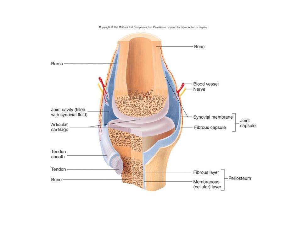
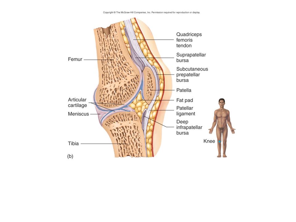
STRUCTURE OF A SYNOVIAL JOINT Articular cartilage – covers bone ends Synovial membrane – lines joint capsule Synovial fluid – lubricates & nourishes cartilage Synovial cavity – space between the bones Joint capsule – fibrous C.T. Ligaments – reinforce joint Bursae – synovial sacs at other sites of friction

55
TYPES OF SYNOVIAL JOINTS Classified based on shape of articular surfaces Gliding (plane) Hinge Pivot Ellipsoidal (condyloid) Saddle Ball-and-socket
 Hinge Pivot Ellipsoidal (condyloid) Saddle Ball-and-socket")
Similar presentations


 Movement (Passive) Protection of Vital Organs Mineral Storage Blood Cell Formation.>")

 or an organ –Bone referred to as a connective tissue consists of: cells.>")

 or an organ –Bone referred to as a connective tissue consists of: cells extracellular.>")


 Movement (Passive) Protection of Vital Organs Mineral Storage Blood Cell Formation.>")


. BONE FUNCTION: Support and Protection bones shape and form body structures bones support and protect softer,>")
