Download presentation
Presentation is loading. Please wait.

1
Fasciola hepatica sheep liver fluke

2
Fasciola hepatica Common name: The sheep liver fluke
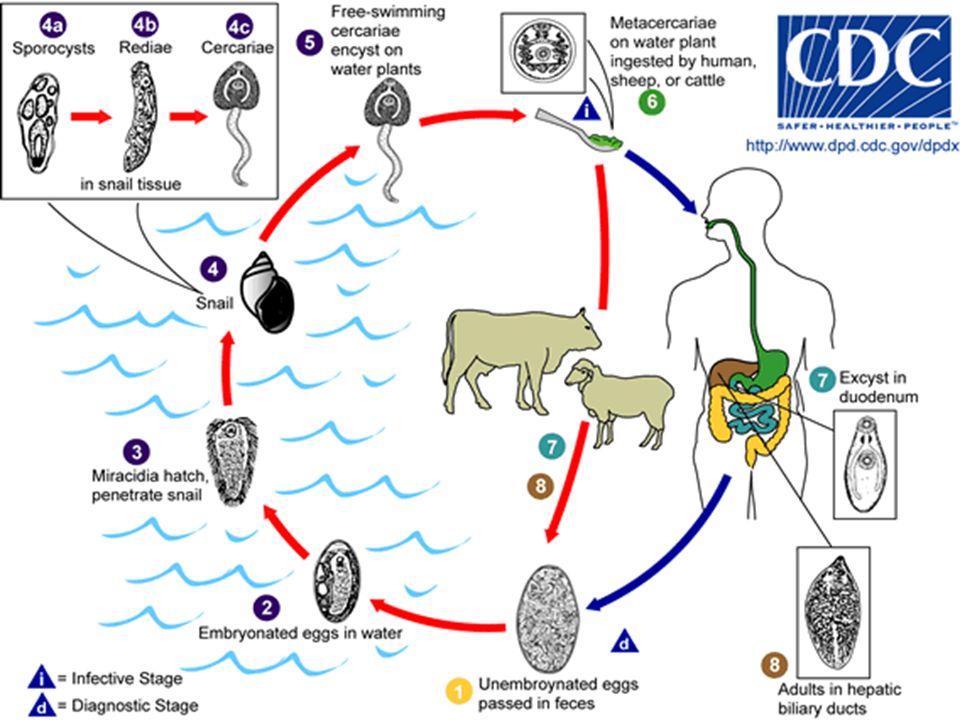
Habitat: Bile duct of liver. Route of infection: Man eat aquatic plants with encysted metacercariae. Definitive host: Usual host sheep, infects liver of various mammals, including humans. Intermediate host: Fresh water snails. Infective stage: Encysted metacercariae on vegetations. Diagnostic stage: Eggs in stool specimen. Disease: Fascioliasis.

3
Fasciola hepatica adult
Morphological characteristics 2-3 cm. Has conical projection Oral and ventral sucker. Pharynx. Branched caecum. Coiled uterus Genital formula : O ( ovary) T ( Testis)
 T ( Testis)")
4
Fasciola hepatica Eggs
Unembyonated. Thin egg shell. operculated. X um. Diagnostic stage

5
egg capsule with emerging miracidium of Fasciola hepatica

6
Life cycle The parasite browses on liver tissue for a period of up to 5-6 weeks and eventually finds its way to the bile duct where it matures into an adult and begins to produce eggs. Up to 25,000 eggs per day per fluke can be produced, and in a light infection, up to 500,000 eggs per day can be deposited onto pasture by a single sheep.

8
Pathology and clinical symptoms.
Most of the damage results from worms are migrating through the liver parenchyma feeding on liver cells and blood Worms in the bile ducts cause inflammation and edem The triad of fever, hepatomegaly, and eosinophilia. Symptoms and signs are associated with biliary obstruction Acute epigastric pain, and jaundice are common.

9
diagnosis Laboratory diagnosis: finding large operculated eggs in the feces.

10
Intestinal fluke Fasciolopsis buski

11
Fasciolopsis buski Common name: The large intestinal fluke
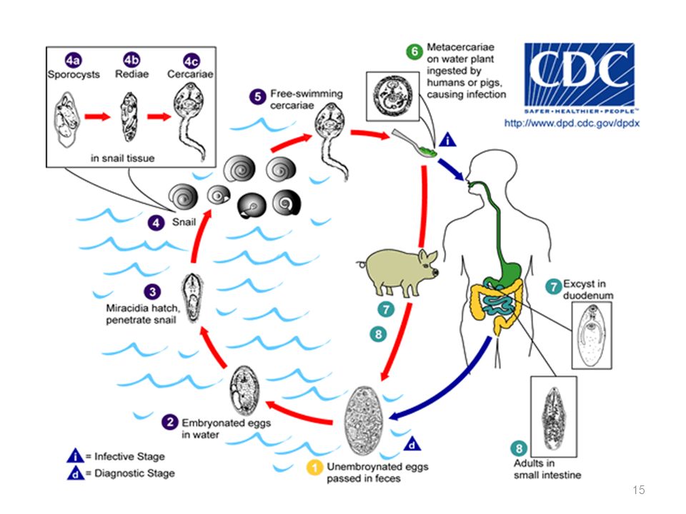
Habitat: Lumen of small intestine. Route of infection: Man eat uncooked plants with encysted metacercariae. Definitive host: Mainly human, other hosts; pigs and dogs. Intermediate host: Fresh water snails. Infective stage: Encysted metacercariae on vegetations. Diagnostic stage: Eggs in stool specimen. Disease: Fasciolopiasis.

12
Fasciolopsis buski adult
Morphological characteristics 2-7x cm. Oral and ventral sucker. Esophagus . Unbranched caecum. Coiled uterus. Branched ovary. Branched Testes. Genital formula : O ( ovary) T ( Testis)
 T ( Testis)")
13
This photo is to compare the sizes of Fasciolopsis buski (left) and Fasciola hepatica (right)
 and Fasciola hepatica (right)")
14
Fasciolopsis buski Eggs
Unembyonated. Thin egg shell. Inconspicuous. operculum. X um

16
Clinical features Laboratory diagnosis:
Most infections are light and asymptomatic. In heavier infections, symptoms include diarrhea, abdominal pain, fever, ascites, and intestinal obstruction. Laboratory diagnosis: operculated eggs and some times the adults are found in the feces.

17
Lung Fluke Paragonimus westermani

18
Paragonimus westermani
Common name: The Lung Fluke. Habitat: Encapsulated in Lungs. Definitive host: Human, also other mammals. First intermediate host: Water snail. Second intermediate host: Crustaceans,Crabs. Infective stage: Encysted metacercariae. Diagnostic stage: Eggs in sputum or feces. Disease: Paragonimiasis.

19
morphology 7 – 12 x 4 – 6 mm. Oral & Ventral Suckers
Unbranched caecum. Pharynx. Coiled Uterus ( black color) Genital formula: O T T
 Genital formula: O. T T.")
20
Paragonimus westermani

21
Paragonimus westermani Eggs
x um Large, thick, dark shell. Prominent operculum at the broad end. Thick posterior end. Unembryonated.

22
Life Stages Egg Miracidio RediaI Redia II
Cercarias Metacercarias

23
Paragonimus westwermani

24
Pathology: Adults in lungs stimulate inflammatory response resulting in granulomas. Movement of worms to heart or brain causes death. Symptoms: Chronic cough , difficulties breathing , sputum with blood. When moves to brain, can cause blindness, paralysis , disequilibrium , epilepsy.

25
DIAGNOSIS based on detection of characteristic eggs in sputum, or stool, serology helpful; standard test is complement fixation (CF) – has advantage to detect rapid decline in antibody levels
 – has advantage to detect rapid decline in antibody levels.")
26
Fasciola hepatica Miracidium
Raed Z. Ahmed, Medical Parasitology Lab.,2012

27
Fasciola hepatica cercariae
Raed Z. Ahmed, Medical Parasitology Lab.,2012

28
Fasciola hepatica rediae larvae
Raed Z. Ahmed, Medical Parasitology Lab.,2012

29
Schistosoma spp.

30
Raed Z. Ahmed, Medical Parasitology Lab.,2012
Schistosoma spp. Also known as bilharzia, cause schistosomiasis or bilhariziasis. Schistosoma spp. have 4 stages: Eggs, miracidia, cercaria, and adult stage. Eggs are passed through urine or feces to fresh water, where larvae stage can infect a new host by penetrating the skin. Schistosoma eggs are non- operculated but spined and have miracidum. Eggs hatch and release miracidia in water. Miracidia move in water looking for a special snail, and penetrate a snail tissue and developed to sporocyst. Cercaria releasaed by snail into water and free swimming, cercaria has a bifid tail and penetrate intact skin Cercaria lose tail during penetration and become schitosomulae, that circulate in the blood and migrate to portal blood of liver and mature into adult. Raed Z. Ahmed, Medical Parasitology Lab.,2012

31
Schistosoma spp. (cont….)
There are three medically important species: Schistosoma mansoni, lives in the mesenteric venules of large intestine, and cause intestinal bilharziasis. Schistosoma japonicum, lives in the mesenteric venules of small intestine. Schistosoma haematobium, lives in the venous plexus of the urinary bladder and cause schistosomal hematuria or urinary bilhariziasis. S. mansoni and S. japonicum are produce their eggs in stool, but S. haematobium produce eggs in urine. Raed Z. Ahmed, Medical Parasitology Lab.,2012

32
Schistosoma spp. (cont….)
Intermediate host: snail. Definitive host: human. Cercaria is the infective stage but eggs are the diagnostic stage. Diagnosis: Depends on finding the characteristic ova in feces or urine. Three species can be distinguished by the appearance of their eggs under microscope: S. mansoni eggs have prominent lateral spine. S. japonicum eggs have a very small round lateral spine. S. Haematobium eggs have a terminal spine. Raed Z. Ahmed, Medical Parasitology Lab.,2012 32

33
Snail for Schistosoma spp.
Snails Schistosome species Bulinus spp. S.haematobium Biomphalaria spp. S.mansoni Oncomelania spp. S.japonicum Raed Z. Ahmed, Medical Parasitology Lab.,2012

34
Raed Z. Ahmed, Medical Parasitology Lab.,2012
Snail Oncomelania spp. Biomphalaria spp. Bulinus spp. Raed Z. Ahmed, Medical Parasitology Lab.,2012

35
Raed Z. Ahmed, Medical Parasitology Lab.,2012
Schistosoma spp. Eggs S. japonicum S. haematobium S. mansoni Lateral spine Terminal spine Rounded spine Raed Z. Ahmed, Medical Parasitology Lab.,2012

36
Schistosoma miracidium
Raed Z. Ahmed, Medical Parasitology Lab.,2012

37
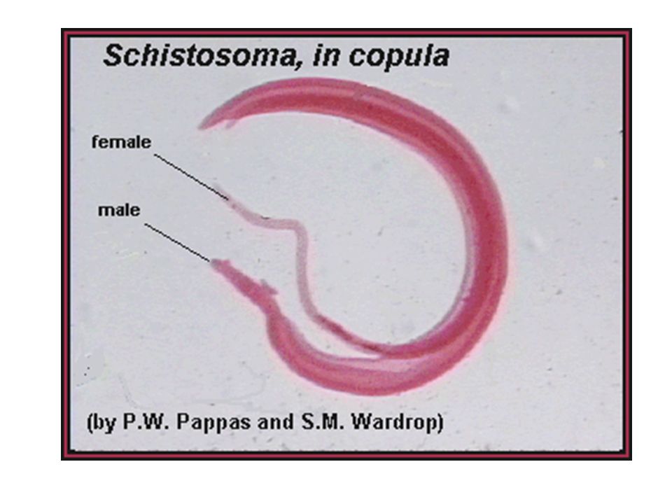
Schistosoma adult male
Adult male short, thick and have gyanchophoric canal Raed Z. Ahmed, Medical Parasitology Lab.,2012

38
Raed Z. Ahmed, Medical Parasitology Lab.,2012

39
Raed Z. Ahmed, Medical Parasitology Lab.,2012
Schistosoma cercaria Bifid tail Oval head Raed Z. Ahmed, Medical Parasitology Lab.,2012

Similar presentations


>")


>")


