Download presentation
Presentation is loading. Please wait.

1
The Heart Section 10.2

2
The Heart Pump to circulate blood. Right side receives deoxygenated blood from the body and pumps it to the lungs. Left side receives oxygenated blood from the lungs and pumps it to the body.

3
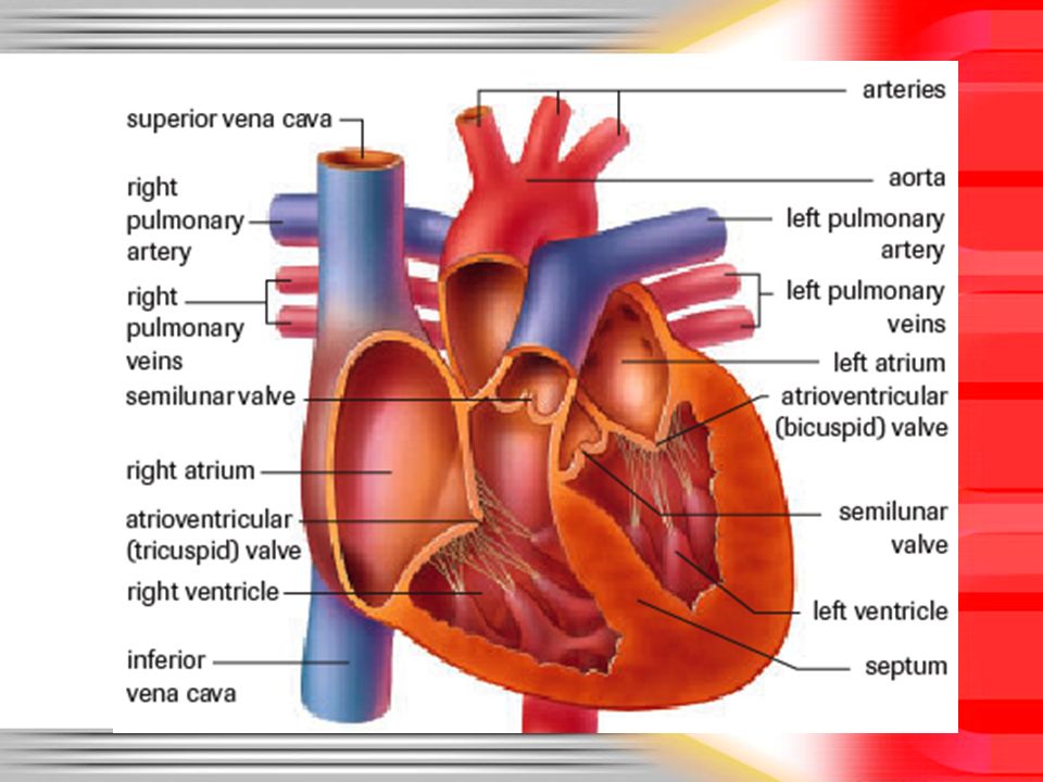
But before we can really talk about how the heart works….We need to know what all the pieces are called. Please label the diagram using your textbook, the internet, the-person-beside- you, me (etc.) if you need help.
 if you need help..")
4
Key Structures of the Heart Atria, Ventricles, Septum, Aorta, Atrioventricular (AV) valves, Semilunar Valves The heart itself is surrounded by the pericardium –A fluid filled membrane surrounding the heart that reduces friction
 valves, Semilunar Valves The heart itself is surrounded by the pericardium –A fluid filled membrane surrounding the heart that reduces friction")
6
Now that we know what it’s called, what does it do? The heart basically works like 2 pumps that run at the same time. Each “pump” pushes blood through a separate circuit: – the Pulmonary circuit, or – the Systemic circuit

7
Pulmonary Circulatory System Vessels that carry blood to and from the lungs

8
Systemic Circulatory System Carries blood to and from the body

9
One-Way Blood Flow Deoxygenated blood is carried from the body into the right atrium by large veins –Inferior vena cava: lower body –Superior vena cava: upper body Blood is pushed into the right ventricle, then into the pulmonary artery and lungs to become oxygenated

10
One Way Blood Flow Once oxygenated, blood returns to the left atrium via the pulmonary vein. It is pushed into the left ventricle, then exits the heart through the aorta, and eventually to the rest of the body

11
One way blood flow Atrioventricular (AV) valves: separate atria and ventricles Semilunar valves: separate ventricles from arteries Both types of valves prevent blood from flowing backward https://www.youtube.com/watch?v=H04d3rJCLCE
 valves: separate atria and ventricles Semilunar valves: separate ventricles from arteries Both types of valves prevent blood from flowing backward v=H04d3rJCLCE")
12
Summary Body (superior and inferior vena cava) Right Atrium Through AV Valve Right Ventricle Through Semilunar Valve Lungs (pulmonary arteries, then veins) Left Atrium Through AV Valve Left Ventricle Through Semilunar Valve Aorta and arteries
 Right Atrium Through AV Valve Right Ventricle Through Semilunar Valve Lungs (pulmonary arteries, then veins) Left Atrium Through AV Valve Left Ventricle Through Semilunar Valve Aorta and arteries")
13
Back to your Diagram… Draw in arrows that show the direction of blood flow for each section of the heart Use one colour to show when blood is oxygenated, and another to show when it is deoxygenated. For veins and arteries sticking out of the heart, label where the blood in it is headed

14
So, the heart pumps oxygenated blood to everything in the body… but how does the heart get oxygen?

15
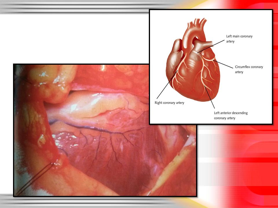
Coronary Arteries Like any other muscle cells, the heart cells needs blood (carrying oxygen) to survive It is supplied by the coronary arteries, which branch off the aorta
 to survive It is supplied by the coronary arteries, which branch off the aorta")
17
Coronary Blockage Unlike other muscles, the heart can’t slow down if it doesn’t get enough oxygen When a coronary artery gets blocked, chest pain (angina) occurs Like other arteries, coronary arteries can develop plaques…
 occurs Like other arteries, coronary arteries can develop plaques…")
18
Bypass Surgery In some cases, medications can be used to increase blood flow In other severe cases, the blood must be rerouted The heart must be stopped to graft the new artery in place!

19
In the past… Doctors had to rely on external symptoms or surgery to detect coronary artery blockages Buuut external symptoms can be for other problems, and surgery is dangerous Cardiac Catheterization is a more accurate, less dangerous tool for detecting blockages

20
Cardiac Catheterization Hollow tube inserted in a small incision in the groin, and passed up through an artery into the heart. A dye is injected into the heart through the catheter. Dye will travel through body and accumulate at a point of blockage in the circulatory system. Helps doctors locate blocks easier.

22
Angioplasty Catheter contains a tiny balloon that can be inflated to open up blocked blood vessels. https://www.youtube.com/watch?v=e13TGGccvT4 https://www.youtube.com/watch?v=e13TGGccvT4

23
Review Questions How many chambers does the heart have? What are they called? What is the difference between the pulmonary and systemic circuits? Why are coronary arteries so important?

24
Summary The Heart: Double pump with 4 chambers Pericardium, atria, ventricles, septum, AV valves, semilunar valves, coronary arteries Blood flows in 2 circuits: Pulmonary Systemic Coronary Grafts, Cardiac Catheterization, Angioplasty Catheter inserted in major artery, brought to heart Used to detect or repair blocked coronary arteries

25
Summary Body (superior and inferior vena cava) Right Atrium Through AV Valve Right Ventricle Through Semilunar Valve Lungs (pulmonary arteries, then veins) Left Atrium Through AV Valve Left Ventricle Through Semilunar Valve Aorta and arteries
 Right Atrium Through AV Valve Right Ventricle Through Semilunar Valve Lungs (pulmonary arteries, then veins) Left Atrium Through AV Valve Left Ventricle Through Semilunar Valve Aorta and arteries")
26
The Heart- part 2 So, we know the heart is a pump, and we know where it pumps to, but how does it actually work?

27
The Heart as a Muscle Cardiac muscle is myogenic, which means that it can contract without nerve stimulus –This is why a heart will beat for a few minutes after being removed from the body https://www.youtube.com/watch?v=uZVHroDfDv4

28
Setting the Heart’s Tempo The sinoatrial node (SA node) is a bundle of nerves that controls the beating of the heart –This is the “pacemaker” –Located in the upper right atrium –Avg. = about 70 beats per minute.

30
Setting the Hearts Tempo These contractions travel through atria to the atrioventricular (AV) node AV node acts as a conductor that passes the impulse to the Purkinje fibres –Two large nerves –Located mostly in the septum but branch
 node AV node acts as a conductor that passes the impulse to the Purkinje fibres –Two large nerves –Located mostly in the septum but branch")
31
Setting the Hearts Tempo Purkinji Fibers carry impulse to the base of the heart, then up the sides of the ventricles A wave of contraction is stimulated: both atria and both ventricles contract Congratulations, you have a beating heart

32
https://www.youtube. com/watch?v=HC_ds qELgDQ

33
SA node sends message to beatPasses through atria to the AV node AV node acts as a conductor and passes impulse to Purkinji fibers Purkinji fibers carry impulse to the base, then up the sides of the ventricles = A beating heart

34
Heart Sounds http://depts.washington.edu/physdx/heart/ demo.htmlhttp://depts.washington.edu/physdx/heart/ demo.html

35
Heart Sounds Sounds are created by the closing of heart valves There are two phases: –Diastole: period of relaxation –Systole: period of contraction

36
Heart Sounds Diastole: period of relaxation Both Atria and Ventricles are at rest As Atria relax, they fill with blood.

37
“Lubb” Systole: period of contraction When full, atria contract, causing AV valves to open –Blood flows into ventricles When full, ventricles also contract – this forces semilunar valves open – Also makes AV valves shut= the louder “lubb” sound

38
“Dubb” The semilunar valves keep blood from re-entering ventricles. Closing of semilunar valves makes the softer “dubb” sound

39
Ever heard of a heart murmur? Happens when the valves (most often AV) don’t close completely. –Leads to back-flow of blood, because the seal is not complete –Produces a gurgling sound. –Loss of efficiency- blood is going in the wrong direction http://depts.washington.edu/physdx/heart/demo.html

40
Murmur Heart beats faster and may enlarge to compensate for murmur inefficiency Atria may also expand to compensate –Accepts normal blood flow, plus back flow –Stretches, so contraction is more forceful – a greater volume of blood enters ventricles to compensate for loss in efficiency

41
Heart Medications Foxglove (digitalis): initiates strong regular heart beats Nitroglycerin: used to prevent heart attacks –Relaxes smooth muscle, dilates blood vessels Betablockers: used for people with irregular heart beats or high blood pressure.
: initiates strong regular heart beats Nitroglycerin: used to prevent heart attacks –Relaxes smooth muscle, dilates blood vessels Betablockers: used for people with irregular heart beats or high blood pressure.")
42
Beta Blockers tie up the receptor sites for epinephrine, so heart rate won’t increase and arteries won’t narrow Side-effects: Tired, less able to exercise, light headed/dizzy due to low blood pressure

43
Now for some mini labs… Page 311-Heart Sounds Page 315-Monitoring your Pulse Page 318-Mapping Veins Please complete ONE of these labs with a partner. You WILL have to hand them in.

Similar presentations


 Heart pumps blood continuously Uses.>")


 The Atria (or Atriums) -Receiving Chambers -Receiving Chambers The Ventricles The Ventricles -Pumping Chambers.>")


