Download presentation
Presentation is loading. Please wait.

1
Fast & Easy ECGs – A Self-Paced Learning Program
5 P Waves Fast & Easy ECGs – A Self-Paced Learning Program Q I A

2
ECG Waveforms Normally the heart beats in a regular, rhythmic fashion producing a P wave, QRS complex and T wave

3
Step 3 of ECG Analysis Examining the P waves
Question to ask: “What are the first and second steps used to analyze the ECG tracing?” Q

4
P Wave First deflection from baseline at beginning of cardiac cycle
Upright, round (in lead II) and precedes each QRS complex Instructional point: A normal P wave indicates that the electrical impulse likely originated in the SA node and was carried through the atria in a normal manner. I
 and precedes each QRS complex. Instructional point: A normal P wave indicates that the electrical impulse likely originated in the SA node and was carried through the atria in a normal manner. I.")
5
Different Looking P Waves
May originate in SA node but conducts abnormally through altered, damaged atria Can result from a pacemaker site that occurs outside SA node

6
Different Looking Sinus P Waves
Tall, rounded or peaked P waves may be seen with increased right atrial pressure and right atrial dilation Question to ask: “What is the normal size of the P wave?” Answer: Amplitude. 0.5 to 2.5 mm Q

7
Different Looking Sinus P Waves
Notched, wide (enlarged) or biphasic P waves may be seen in increased left atrial pressure and left atrial dilation Question to ask: “What is the normal duration of the P wave?” Answer: Duration to 0.10 seconds Q
 or biphasic P waves may be seen in increased left atrial pressure and left atrial dilation. Question to ask: What is the normal duration of the P wave Answer: Duration to 0.10 seconds. Q.")
8
Different Looking P Waves
Impulses arising from the atria produce P waves that look different than sinus P waves Referred to as P Prime or P’ waves Seen with: Premature atrial complexes (PACs) Wandering atrial pacemaker Atrial tachycardia
 Wandering atrial pacemaker. Atrial tachycardia.")
9
P’ Waves Rate is between 150 to 250 beats per minute and may be buried in T wave of preceding beat P’ wave of early beat differs in appearance from underlying rhythm P’ waves continuously change in their appearance

10
Different Looking P Waves
In rapid rates (i.e. atrial tachycardia) the P’ wave is likely buried in the T wave of the preceding beat (due to the short P’-P interval) When this occurs the T waves are often peaked, notched or larger than normal Instructional point: When we have a tachycardia with normal QRS complexes but without recognizable P waves we can refer to it as supraventricular tachycardia. I
 the P’ wave is likely buried in the T wave of the preceding beat (due to the short P’-P interval) When this occurs the T waves are often peaked, notched or larger than normal. Instructional point: When we have a tachycardia with normal QRS complexes but without recognizable P waves we can refer to it as supraventricular tachycardia. I.")
11
Different Looking Atrial Waveforms
Flutter waves are seen instead of normal P waves when the atria fire rapidly from one site at a rate of BPM Often described as a saw-toothed pattern Called “F” waves Instructional points: The atrial waveforms are referred to as “F” waves. This dysrhythmia is called atrial flutter. I

12
Flutter Waves

13
Different Looking Atrial Waveforms
An absence of discernable P waves is seen when the atria fire rapidly from many sites at a rate >350 BPM Instead there is a chaotic looking baseline of fibrillatory (“f”) waves preceding the QRS complexes Instructional point: This dysrhythmia is called atrial fibrillation. I
 waves preceding the QRS complexes. Instructional point: This dysrhythmia is called atrial fibrillation. I.")
14
Fibrillatory Waves

15
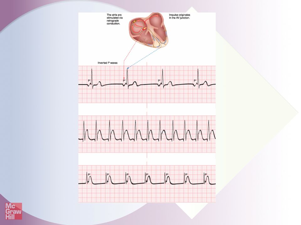
Inverted P’ Waves Produced when a P’ wave arises from the lower right atrium near the AV node, in the left atria or the AV junction Results in retrograde depolarization of the atria Instructional point: The term retrograde means that the impulse conducts upward (backward) through the heart instead of downward. I
 through the heart instead of downward. I.")
16
Inverted P’ Waves May immediately proceed, occur during or follow the QRS complex Associated with dysrhythmias that originate from the AV junction Instructional point: When the impulse arises from these locations the atria are depolarized in a retrograde fashion. I

18
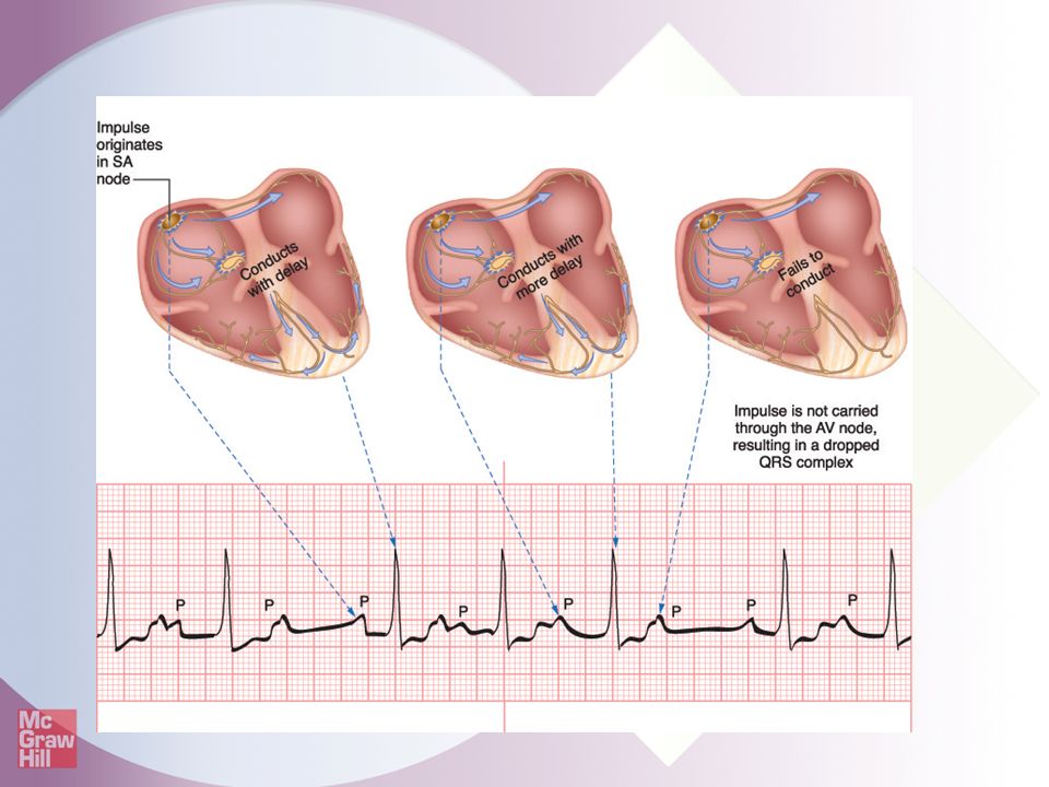
More P Waves Than QRS Complexes
Indicates the impulse was initiated in the SA node or atria but was blocked and did not reach the ventricles Instructional point: Normally there is just one P wave preceding each QRS complex. I

19
More P Waves Than QRS Complexes
Most common causes 2nd-degree AV heart block (Types I and II) 3rd-degree AV heart block Blocked premature atrial complexes (PACs)
 3rd-degree AV heart block. Blocked premature atrial complexes (PACs)")
21
Practice Makes Perfect
Determine the type of atrial waveforms Answer: normal P waves Ask the student to identify the rate and regularity as well as the type of atrial waveform. I

22
Practice Makes Perfect
Determine the type of atrial waveforms Answer: biphasic P waves Ask the student to identify the rate and regularity as well as the type of atrial waveform. I

23
Practice Makes Perfect
Determine the type of atrial waveforms Answer: absent P waves Ask the student to identify the rate and regularity as well as the type of atrial waveform. I

24
Practice Makes Perfect
Determine the type of atrial waveforms Answer: more P waves than QRS complexes Ask the student to identify the rate and regularity as well as the type of atrial waveform. I

25
Practice Makes Perfect
Determine the type of atrial waveforms Answer: F waves are seen instead of P waves Ask the student to identify the rate and regularity as well as the type of atrial waveform. I

26
Summary Third step of analyzing an ECG rhythm is examining the P waves. The P wave is the first deflection from the baseline at the beginning of the cardiac cycle. The amplitude of a normal P wave is 0.5 to 2.5 mm and the duration is 0.06 to 0.10 seconds.

27
Summary P waves that look different may originate in the SA node but conduct abnormally through altered, damaged atria or may result from a pacemaker site that occurs outside the SA node. Impulses that arise from the atria produce P Prime (P’) that look different than the sinus P waves.
 that look different than the sinus P waves.")
28
Summary An impulse that arises from the lower right atrium near the AV node or in the left atria results in retrograde atrial and an inverted P’ wave. The P’ wave in premature atrial complexes (PACs) have a different morphology than the other normal beats.
 have a different morphology than the other normal beats.")
29
Summary The P’ waves associated with atrial tachycardia look different than normal beats and are often buried in the T wave of the preceding beat. P waves that continuously change in their appearance indicate that the site of impulse origin is moving from site to site in the atria.

30
Summary Flutter waves (F waves) are seen when the impulse arises from atria at a rate of 250 to 350 BPM. These are often described as a “saw-toothed pattern.” There is an absence of discernable P waves when the impulses arise from many different sites in the atria at a rate greater than 350 BPM. Instead there is a chaotic looking baseline of “f” waves preceding the QRS complexes.

31
Summary Impulses that arise from the AV junction or ventricles produce an inverted P’ wave that may immediately proceed, occur during or follow the QRS. More P waves than QRS complexes indicate that the impulse was initiated in the SA node or atria but was blocked and did not reach the ventricles.

Similar presentations