Download presentation
Presentation is loading. Please wait.

1
Lab # 3 Gram and Acid Fast stain Medgar Evers College Biology 261 Prof. Santos

2
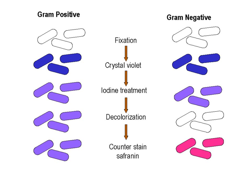
The Gram stain is a differential stain used to distinguish between gram positive and gram negative bacteria. This is base on the biochemistry of the cell wall. Gram – bacteria have a thin layer of peptidoglycan while gram + bacterial have a thicker layer of peptidoglycan.

4
Steps Step 1 make a smear of the sample and heat fix Step 2 the primary stain, crystal violet for 20 seconds. Gram – and gram + cells will pick up this stain and appear purple.

5
Step 3 Apply the mordant or Gram’s iodine for 1 minute. This forms a tight complex with the crystal violet in gram + cells. Both cells still appear purple. Step 4 the decolorizer is used to remove the crystal violet/Iodine from the gram – cells. The decolorizer, ethyl alcohol, is applied for 20 seconds. The gram + cells continue to appear purple while the others have become colorless.

6
Step 5 the counter stain safranin is applied for 1 minute. This is used to stain the gram – cells. At this point they will appear pink.

7
Things to consider when doing Gram stain When doing the Gram stain, it is important to use fresh cultures to minimize false results such as a gram + staining pink due to the fact that it’s so old it has problems picking up the crystal violet. Also keep in mind that gram – never convert to gram +.

8
It is critical to prepare a thin smear to allow you to see single cells instead of layers of cells superimposed on top of each other.

9
Acid Fast Stain The reason we do the acid fast stain is because some members of the genus Mycobacterium and some members of the genus Nocardia have a layer of mycolic acid that prevents them from being properly stained. Mycolic acid is a waxy material in their cell wall.

10
The important thing is that the primary stain used is Carbolfuchsin is applied over heat. This allows the stain to penetrate the layer of mycolic acid. The counter stain used is methylene blue. The acid fast cells tend to appear red or pink and the non acid fast cells appear blue.

12
Steps 1- prepare smear of Mycobacterium smegmatis and S. aureus and cover smear with carbolfuchsin. 2- steam over boiling water for 5 minutes 3- after slide has cooled, decolorize with acid alcohol for 15-20 seconds 4- rinse briefly with water 5- Counter stain with methylene blue for 30 seconds

13
6- briefly rinse with water 7- blot dry 8-Look under microscope

Similar presentations


 Abdelraheem BA>")


: Sharpie will wash off with alcohol! Be sure to resuspend broth cultures completely! Exercise.>")


 Acid fast stain Spore stain.>")

>")
