Download presentation
Presentation is loading. Please wait.

1
Tissues

2
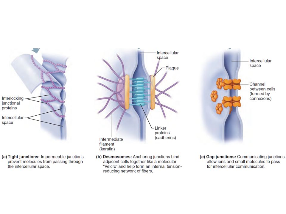
Impermeable junctions
Cell Junctions Tight Junctions Desmosomes Gap Junctions Impermeable junctions Anchoring junctions bind to adjacent cells like Velcro Allow for intercellular communication Prevent molecules from passing through intercellular space Form internal tension-reducing network of fibers; plaques on surface of membrane attach to protein filaments Allow ions and small molecules to pass through channels formed by connexon protein cylinders Example: Lining of the digestive tract Found in tissues subject to stress like skin; heart muscle Found in electrically excitable tissue (heart; smooth muscle) to synchronize
 to synchronize.")
4
Intercellular Junctions
Tight junctions Close space between cells Located among cells that form linings Cell membrane Tight junction Desmosomes Form anchors between cells Located among outer skin cells Cell membrane Desmosome Gap junctions Tubular channels between cells Located in cardiac muscle cells Cell membrane Gap junction

5
Four Primary Tissue Types:
Tissue = a group of similar cells that function together to carry out specialized activities. Histology = the study of microscopic anatomy (tissue). The body has different kinds of tissues, some highly organized and some not. *Form (structure) fits function. Four Primary Tissue Types: Epithelium Connective Muscle Nervous
. The body has different kinds of tissues, some highly organized and some not. *Form (structure) fits function. Four Primary Tissue Types: Epithelium. Connective. Muscle. Nervous.")
6
Epithelium: Basic Info.
Cover internal and external body surfaces Lines body cavities & hollow organs. 1. Protection (cells tightly packed) 2. Absorption 3. Secretion 4. Excretion 5. Sensory reception Epithelium: Basic Info.
 2. Absorption. 3. Secretion. 4. Excretion. 5. Sensory reception. Epithelium: Basic Info.")
7
Epithelium: General Characteristics
Consists mostly or entirely of closely packed cells that are arranged in continuous sheets. Has an apical surface (one side is always exposed to the external or internal environment), and a basement membrane. Basement membrane connects epithelium to connective tissue and other structures It’s a thin matrix of fibers that anchor the tissue. Avascular (no blood supply), but innervated. Nutrients must diffuse into the tissue from the blood vessels of the connective tissue. High capacity for renewal by cell division. Epithelium: General Characteristics
, and a basement membrane. Basement membrane connects epithelium to connective tissue and other structures. It’s a thin matrix of fibers that anchor the tissue. Avascular (no blood supply), but innervated. Nutrients must diffuse into the tissue from the blood vessels of the connective tissue. High capacity for renewal by cell division. Epithelium: General Characteristics.")
8
Epithelial tissue Basement membrane One side of the tissue is exposed to the outside and the tissue is connected by a basement membrane.

9
Epithelium: Nomenclature
Epithelial tissue is categorized according to the shape of the cells and number of layers. Three basic shapes: Squamous Cuboidal Columnar Layers: Single layer = “simple” Multiple layers = “stratified”

10
Squamous cells are flat.

11
Cuboidal epithelium cells are shaped like cubes.

12
Columnar epithelium cells are rectangular in shape.

13
Simple squamous epithelium: look for a thin layer of flat (squamous) cells.
Common site for passive transport (filtration & diffusion). Found in the lungs, walls of capillaries and inside of blood and lymphatic vessels.
. Found in the lungs, walls of capillaries and inside of blood and lymphatic vessels.")
14
Stratified squamous epithelium: as the cells reach the surface, they flatten out. Many cell layers. Top cells = flat This is a slide of the most superficial layer of the skin known as the epidermis. Keratin is found at the top of the epidermis: protein that keeps cells from being very moist Stratified squamous is also found lining orifices, like the oral cavity (non-keratinized here so cells are relatively moist)
")
15
Simple Cuboidal Epithelium: only one layer
Commonly found in ovaries, kidney tubules, ducts. Stratified cuboidal epithelium also lines ducts. It can be found in the ducts of mammary glands, sweat glands, salivary glands, pancreas. (2-3 layers)
")
16
Simple columnar epithelium: can be ciliated or non-ciliated.
Ciliated is found in the female reproductive tract. Non-ciliated is found in the uterus and digestive tract. Columnar epithelium can also be stratified. This tissue is found in the vas deferens and pharynx. It provides a thicker lining for some tubular structures in the body.

17
A special type of cell known as a goblet cell is usually associated with columnar epithelium. The goblet cell secretes mucus.

18
Cilia and microvilli may also be present on columnar cells.
The cilia and goblet cells work together to move substances along the cells, like in the respiratory tract. Cilia

19
Stratified columnar: Top layer of elongated cells
Cube-shaped cells in deeper layers Line part of male urethra and part of pharynx (b) (a) Lumen Stratified columnar epithelium Connective tissue Basement membrane Free surface of tissue
 (a) Lumen. Stratified. columnar. epithelium. Connective. tissue. Basement. membrane. Free surface. of tissue.")
20
Epithelium: Exception to the Rule #1
Pseudostratified columnar epithelium Looks like it’s stratified (more than one layer), but it’s not. It looks stratified because the nuclei of the cells are at various levels, but there is really only one layer. May have cilia, line resp. passages
, but it’s not. It looks stratified because the nuclei of the cells are at various levels, but there is really only one layer. May have cilia, line resp. passages.")
21
Pseudostratified vs. Simple columnar epithelium
Nuclei don’t line up—pseudostratified. Nuclei all line up nicely in one row—simple. Pseudostratified vs. Simple columnar epithelium

22
Epithelium: Exception to the Rule #2
Transitional epithelium Looks somewhat like stratified squamous, but there is a difference. Many layers; cube-shaped & elongated cells; can stretch In transitional epithelium, the cells are rounded both at the base of the tissue and the section exposed to the outside. Urinary bladder: multiple layers allow for the bladder to distend and contract. Forms a barrier to help protect the bladder from infection.

23
The cells of this tissue are specialized to change shape in response to pressure.
When the bladder is empty, these cells are more or less cuboidal in shape. As the bladder fills, the cells become compressed and flattened. Transitional epithelium

24
Epithelium: Glandular
Glandular epithelium Can secrete substances into the bloodstream (endocrine glands), or into ducts (exocrine glands). Epithelium: Glandular
, or into ducts (exocrine glands). Epithelium: Glandular.")
25
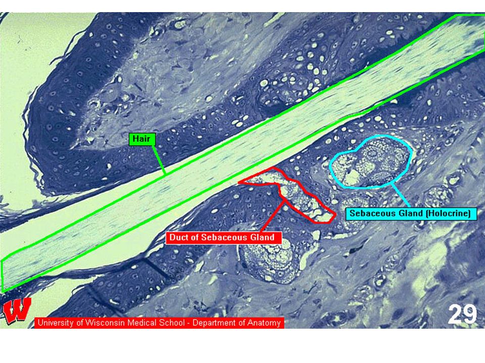
Exocrine Glands Can be classified by method of secretion:
Merocrine—release substance via exocytosis Also called “eccrine” Ex: salivary glands, pancreatic glands, some sweat glands Apocrine—lose small portion of cell body. Examples: mammary glands, some sweat glands Holocrine—release entire cell. Examples: sebaceous glands (hair follicles) Exocrine Glands
 Exocrine Glands.")
26
Duct Secretory portion Tissue surface Simple tubular Simple branched tubular alveolar Simple coiled Compound tubular Compound alveolar

27
Merocrine Glands Apocrine Glands Holocrine Glands Fluid product
Salivary glands Pancreas Sweat glands (also called eccrine) Apocrine Glands Cellular product Portions of cells Mammary glands Ceruminous glands Holocrine Glands Secretory products Whole cells Sebaceous glands (a) Merocrine gland (b) Apocrine gland (c) Holocrine gland Secretion Pinched off portion of cell (secretion) Intact cell Disintegrating cell and its contents New cell forming by mitosis and cytokinesis
 Apocrine Glands. Cellular product. Portions of cells. Mammary glands. Ceruminous glands. Holocrine Glands. Secretory products. Whole cells. Sebaceous glands. (a) Merocrine gland. (b) Apocrine gland. (c) Holocrine gland. Secretion. Pinched off. portion of cell. (secretion) Intact. cell. Disintegrating cell. and its contents. New cell. forming by. mitosis and. cytokinesis.")
Similar presentations


 Connective tissue Muscle.>")


