Download presentation
Presentation is loading. Please wait.

1
Staining of Blood Smear

2
Romanowsky stain Romanowsky stain→Eosin Y and Azure B)
Eosin:Acidic Dye bind to Basic groups (Hb,Granules) → reddish or orange color Azure B: Dye bind to nucleic acid & nucleoproteins →Blue-violet color Fixation →Methanol<4%water, with 1 hour Delay : Adherence of Pro. To slide → Blue background
 → reddish or orange color. Azure B: Dye bind to nucleic acid & nucleoproteins →Blue-violet color. Fixation →Methanol<4%water, with 1 hour. Delay : Adherence of Pro. To slide → Blue background.")
4
Romanowsky stain Wright Wright – Giemsa Lishman May- grunwald - Giemsa
jenner

5
Making blood film Blood film can be prepared from fresh blood without anticoagulant or from EDTA anticoagulanted blood. blood film should be made on clean glass . Clear without any dust

6
Wedge method The most commonly in routine lab Method
Thickness or thinness regulated by Amount of blood Speed of spreader Angle

7
Optimal blood smear characteristic
minimum 2.5 cm in length terminating at least 1 cm from the end of the slide Gradual Transition in thickness from thick to thin area ending in a Square or straight edge No streaks , waves , or troughs

11
Sources of error in preparation of a blood smear
problem Resolution Presence of crenated erythrocyte Dry smear quickly and thoroughly Thin smear due to anemia Increased spreader slide angle and increased push speed Thick smear due to polycythemia Decrease spreader slide angle and decrease push speed Presence of agglutinated erythrocytes associated with cold agglutinin disease Warm blood in 37°C for 15 min prior to preparing smear Increased viscosity associated with multiple myeloma

12
Spinner Blood films that combine the advantages of easy handling of the wedge slide & uniform distribution of cells of the coverglass reparation Method Advantages: minimal exposure to biohazardous , increased optimal counting area

13
Reference method Pure Azure B (260mg/100ml methanol)
Pure eosin y ( 130 mg/100ml methanol) 1 part Azure B + 1 part eosin y +10 part sorensens phosphate buffer 66mmol/l ph= 6.8 10 min washing
 1 part Azure B + 1 part eosin y +10 part sorensens phosphate buffer 66mmol/l ph= min. washing.")
14
Characteristic of aproperly stained blood smear
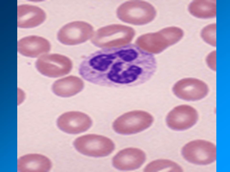
Type of evaluation Characteristic Macroscopic Smear is pinkish purple in color Microscopic Blood cells are evenly distributed Areas between cells are clear Erythrocytes are orange red Neutrophilic granules are lilac Eosinophilic granules are red orange Lymphocytes cytoplasm is blue Leukocytes nuclei are purple Precipitated stain is minimal or absent

15
problem Potential causes Excessively blue or dark stain
Prolonged staining Inadequate washing Too high an alkalinity of stain and / or buffer Thick blood smear Excessively pink or light stain Insufficient staining Prolonged washing Too high an acidity of stain and / or buffer Presence of precipitate Unclean slides Drying during staining process Inadequate filtration of stain

16
problem Potential causes Pale stainig Old stainig solution
overused staining solution Impure dyes High ambient temperature Blue Background Prolonged storage before fixation Blood collection into heparinas anticoagulant

17
Quality Control رنگ پس از تهيه از نظر آلودگی قارچی و ميکروبی وهر گونه رسوب و پارتيکل وهمچنين نحوه رنگ گرفتن سلول های خونی بررسی می گردد.رنگ آميزی گسترش های خونی روتين نيز هفته ای يک بار توسط مسئول فنی داخلی از نظر موارد فوق بررسی می گردد که بصورت مکتوب ومستند بايد در آزمایشگاه قرار گيرد.کيفيت رنگ آميزی مورد قبول سلول ها مطابق جدول زير می باشد .

18
هسته سيتوپلاسم گرانولها ساير انکلوژنها اجزاء سلولی رنگ کروماتين بنفش
هستک آبی روشن سيتوپلاسم اريتروبلاست آبی تيره ارتيروسيت صورتی تيره رتيکولوسيت خاکستری-آبی لنفوسيت آبی متاميلوسيت صورتی منوسيت نوتروفيل صورتی-نارنجی پروميلوسيت قرمز يا بنفش بازوفيل گرانولها پروميلوسيت (گرانولهای اوليه) بنفش تيره ائوزينوفيل قرمز - نارنجی توکسيک گرانول پلاکت ساير انکلوژنها آئور بادی کابوت رينگ هاول جولی بادی دوهل بادی
 بنفش تيره. ائوزينوفيل. قرمز - نارنجی. توکسيک گرانول. پلاکت. ساير انکلوژنها. آئور بادی. کابوت رينگ. هاول جولی بادی. دوهل بادی.")
19
GOOD LUCK !

Similar presentations


 and intercellular.>")
 4- body tube (carrying lens system) 5- light.>")
 Object – To study morphological structures of Plasmodia, to identify morphological structures of developing stages of erythrocytic.>")

