Download presentation
Presentation is loading. Please wait.

1
The Integumentary System and Body Membranes
Chapter 5 The Integumentary System and Body Membranes

2
Objectives Classify, compare the structure of, and give examples of each type of body membrane. Describe the structure and function of the epidermis and dermis. List and briefly describe each accessory organ of the skin. List and discuss the three primary functions of the integumentary system.

3
What are the four major types of membranes in the body?
Question What are the four major types of membranes in the body?

4
Membranes Epithelial membranes Mucous Membranes Serous membranes
Pleura Pericardium Peritoneum Connective Tissue Membranes One of the first parts of this chapter is about the different types of membranes in our body. This is because at the basic level, our skin is our body’s membrane. It separates us from the outer world. Membranes are coverings on or inside body surfaces. You find membranes over all surfaces – which means even your skin is a membrane! : ) We have 2 types of membranes that we discuss in this chapter and they are cutaneous and serous membranes. We discussed epithelial tissue in last weeks class. Serous membranes the ones that are the linings of the cavities inside the body cavities. Do you remember back in chapter 1, the definitions and locations of the body cavities? The cranial, spinal, thoracic, and abdominal? Well the serous membranes are the ones that are located in these cavities. They all the same structure. They are each two layers and are important because they secrete a thin, watery fluid. What do you think the name of the fluid would be? Right, how strange… that the fluid we find in serous membranes would be called serous fluid :-)
 We have 2 types of membranes that we discuss in this chapter and they are cutaneous and serous membranes. We discussed epithelial tissue in last weeks class. Serous membranes the ones that are the linings of the cavities inside the body cavities. Do you remember back in chapter 1, the definitions and locations of the body cavities The cranial, spinal, thoracic, and abdominal Well the serous membranes are the ones that are located in these cavities. They all the same structure. They are each two layers and are important because they secrete a thin, watery fluid. What do you think the name of the fluid would be Right, how strange… that the fluid we find in serous membranes would be called serous fluid :-)")
5
CLASSIFICATION OF BODY MEMBRANES
Epithelial membranes Cutaneous membrane—the skin More later!!

6
Classification of Body Membranes
Mucous membranes Line body surfaces that open directly to the exterior Produce mucus, a thick secretion that keeps the membranes soft and moist What are examples of mucous membranes?

7
Serous Membranes Line the cavities that do not open to the outside world Double layered Visceral Parietal Serous fluid is in the cavity between them The word root "viscer/o" means internal organ, so the visceral layer is the one that touches the internal organ. So next is the fluid. What is the purpose of having that serous fluid between the 2 layers? Those are all good answers! It is to reduce the friction when movement occurs. Friction creates heat, and heat building up between two body surfaces is not a good thing! The serous fluids prevent the heat from building up by preventing friction between surfaces. Where is the pleura located? Right, it is surrounding the lung. The parietal layer of the pleura for adheres to the wall of the chest and the visceral layer is attached to the lung. Every breath we take the two layers slide past each other with little friction, making it easier for us to breathe. How about the peritoneum? Where is it located? Great, it surrounds the abdominal and pelvic organs. Different sources and textbooks may refer to an additional serous membrane called the pericardium. Your text talks about this membrane in the heart chapter. This is the 2-layered membrane that surrounds the heart. It has the same structure as the pleura and the peritoneum. Ready for a tough question? What do you think mucous membranes do? They make mucous! This mucous is to catch debris, and not let it enter the body. It helps to keep out invading pathogens. Where do you think we find the most mucous membranes? One way to remember where they are is to think of all the places we have an opening to the outside. All of them have a mucous membrane. So our eyes, nose, mouth, vagina, anus are all mucous membranes. This mucus is in place to catch debris entering the body and is the body’s first line of defense against invading pathogens. So, now that you know all about membranes can you tell me what type of tissue they are? They are epithelial tissue! Good job. Epithelial tissue covers and lines structures.

8
What are the 2 most important serous membranes?
Right, the pleura and peritoneum. You can refer to the picture on page 101 for a picture. The pericardium is also an important membrane but isn’t always classified as a separate type. I want you to try a visual. Take a piece of paper or fabric and fold it in half. Then, wrap it around your hand, still folded. So , in my example, your hand is the organ, and the paper is the membrane. Can you see there are 2 layers? The one that is closest to your hand and one on the outside? Do you know the name of the inner layer, the one that touches the organ? Or in my example, the layer that is touching your hand? Good, right! The layer that touches your hand is the visceral layer. In the picture on page 101 it is the light blue line inside the purple one. The purple one is the one that is facing the outside is called the parietal layer. Does this make sense to you?

9
Connective tissue membranes
Synovial Membranes Connective tissue membranes Do not contain epithelial components Produce a lubricant called synovial fluid Examples are the synovial membranes in the spaces between joints and in the lining of bursal sacs

10
Disorders of Body Membranes
Diseases Pleurisy—inflammation of the serous membranes that line the chest cavity and cover the lungs Peritonitis—inflammation of the serous membranes in the abdominal cavity that line the walls and cover the abdominal organs

11
Integumentary System Integument = covering Consists of:
Skin Accessory Organs: Hair Nails Glands The largest organ of the body is the Skin Now that we have covered the basic membranes, lets move on to discuss skin!!! You might want to open your book to p 103 for a great picture. On the surface skin seems like it’s a fairly simple organ, but there’s a lot more that what you see on the surface PUN INTENDED there…. :-) Anatomy textbooks differ in how they present the skin. Your book lists the skin as having two distinct layers while many other books list three. Your textbook discusses the subcutaneous layer in the beginning of the chapter, but does not count it as a true ‘layer’. Either way is an acceptable way to describe the structure of the skin. The structure is still the same, no matter how we describe it . The two layers all books agree on are the two outside layers, the epidermis and dermis. Who has taken medical terminology already? Do you know what “epi” means?
 Anatomy textbooks differ in how they present the skin. Your book lists the skin as having two distinct layers while many other books list three. Your textbook discusses the subcutaneous layer in the beginning of the chapter, but does not count it as a true ‘layer’. Either way is an acceptable way to describe the structure of the skin. The structure is still the same, no matter how we describe it . The two layers all books agree on are the two outside layers, the epidermis and dermis. Who has taken medical terminology already Do you know what epi means")
12
What are the functions of the skin?
Question What are the functions of the skin?

13
Functions of Skin Protects from injuries
Acts as barrier and regulates what enters/leaves body. Regulates body temperature. Synthesizes, stores vitamins. Sensory functions

14
Functions of the Skin Sense organ activity
Skin functions as an enormous sense organ Receptors serve as receivers for the body, keeping it informed of changes in its environment—disorders of the skin (dermatoses)
")
15
Functions of the Skin Protection—first line of defense
Against infection by microbes Against ultraviolet rays from sun Against harmful chemicals Against cuts and tears Skin grafts

16
Structure of the Skin 3 main layers from superficial to deep
1) Epidermis Epi- derm/o -is above skin structure “structure above the skin” a. Thin cellular membrane layer Right, “epi” means above – so the “epidermis” translates as “pertaining to above the skin” The epidermis is above the dermis. It is what you see when you look at your own skin. Since it is a covering what kind of tissue do you think it might be? Right it is epithelial tissue. That was last chapter. In fact the epidermis is an entirely cellular layer. It is a layer of stratified squamous epithelium. It is squamous, which is what shape? scales? cubes? columns? Good job, it means that it is many layers of flat, scale-like cells. Please have your book handy with the picture on page 103. The epidermis has many layers. The deepest layer is where new cells are produced. These cells are constantly undergoing mitosis. As new cells are produced, they push the older cells toward the surface of the body. The layer on the surface is made up of “dead” cells. Cells are always being replaced, at a pretty rapid pace to replace the millions of skin cells we lose on a daily basis. It is funny to think that if “beauty is only skin deep” the top layer of cells, the one we see, is full of dead keratin-filled cells. We lose millions every day, many of them in bed! Does the epidermis have blood vessels or nerves within it? No it does not, it receives all of its nutrients from the dermis below. So as the cells are pushed toward the surface, they die and are filled with a hard protein. Do you know what this protein is called? Right, it is called keratin! Lets go a bit deeper with the structure of the skin. One the picture in your book, you can see a layer between the dermis and the epidermis that appears in your book as being blue. This is called the stratum germinativum. What is happening in this layer? Good job that is where mitosis is happening all the time. The layer at the most superficial aspect of the epidermis (closest to the outside) is the stratum corneum. This layer is full of those dead, hard cells that are filled with keratin. This layer is constantly flaking off, and being replaced. Have you ever noticed that different people have different colored skin? Even among Caucasions skin tones vary greatly. Some people have very light skin while others have darker skin. And of course, people of African or Asian descent have much darker skin. Those different colors are because of melanin. People with more melanin have darker skin. People with less melanin have lighter skin. But we all have the same number of melanocytes. Nifty huh? We just differ in the amount of melanin they produce. When your skin is exposed to UV rays, the melanocytes produce more pigment to protect us. This is the "suntan" effect.
 Epidermis. Epi- derm/o -is. above skin structure. structure above the skin a. Thin cellular membrane layer. Right, epi means above – so the epidermis translates as pertaining to above the skin The epidermis is above the dermis. It is what you see when you look at your own skin. Since it is a covering what kind of tissue do you think it might be Right it is epithelial tissue. That was last chapter. In fact the epidermis is an entirely cellular layer. It is a layer of stratified squamous epithelium. It is squamous, which is what shape scales cubes columns Good job, it means that it is many layers of flat, scale-like cells. Please have your book handy with the picture on page 103. The epidermis has many layers. The deepest layer is where new cells are produced. These cells are constantly undergoing mitosis. As new cells are produced, they push the older cells toward the surface of the body. The layer on the surface is made up of dead cells. Cells are always being replaced, at a pretty rapid pace to replace the millions of skin cells we lose on a daily basis. It is funny to think that if beauty is only skin deep the top layer of cells, the one we see, is full of dead keratin-filled cells. We lose millions every day, many of them in bed! Does the epidermis have blood vessels or nerves within it No it does not, it receives all of its nutrients from the dermis below. So as the cells are pushed toward the surface, they die and are filled with a hard protein. Do you know what this protein is called Right, it is called keratin! Lets go a bit deeper with the structure of the skin. One the picture in your book, you can see a layer between the dermis and the epidermis that appears in your book as being blue. This is called the stratum germinativum. What is happening in this layer Good job that is where mitosis is happening all the time. The layer at the most superficial aspect of the epidermis (closest to the outside) is the stratum corneum. This layer is full of those dead, hard cells that are filled with keratin. This layer is constantly flaking off, and being replaced. Have you ever noticed that different people have different colored skin Even among Caucasions skin tones vary greatly. Some people have very light skin while others have darker skin. And of course, people of African or Asian descent have much darker skin. Those different colors are because of melanin. People with more melanin have darker skin. People with less melanin have lighter skin. But we all have the same number of melanocytes. Nifty huh We just differ in the amount of melanin they produce. When your skin is exposed to UV rays, the melanocytes produce more pigment to protect us. This is the suntan effect.")
18
Structure of the Skin (cont’d)
2) Dermis Derm/o -is Skin structure “true skin” a. thick, contains connective tissue layer with collagen and elastic fibers, epithelial tissue, smooth muscle tissue, nervous tissue and blood The second layer of skin that your book talks about is the dermis. It is a much thicker layer than the epidermis and is made up of primarily connective tissues. What are the 2 main types of connective tissue that we find in the dermis? These connective tissues include collagen and elastin. Have you heard of “collagen injections”? People have them done to make their lips look more “plump” Hollywood glamour!! Collagen is a protein that gives the dermis its structure! The dermis has 2 layers as well. The top “papillary layer” and the thick Reticular layer. Take a look at the papillary layer that has those projections that push up into the epidermis, these fingers called “dermal papilla” bring nourishment to the epidermis. The reticular layer is the layer that is mostly connective layer, like collagen and elastin for the ability to stretch! It also houses all the other structures! The dermis is also important because it houses hair, glands, sensory receptors, and nails! Those all might seem like unimportant structures, but imagine what would happen if you didn’t have something like sensory receptors! Speaking of the sensory receptors, does anyone know what the two types of sensory receptors are in the skin? Good job, The receptors found in the dermis are the Pacinian corpuscles and Meissner’s corpuscles Which one senses touch? Right ! Meissner’s senses touch and Pacinian senses pressure! I want you to take a moment to locate them on the image on page 103, and see that Meissner’s is located closer to the surface and Pacinian is deeper in the dermis. The dermis also houses two types of glands…what are they? Good job, they are Sweat and sebaceous. The technical name for sweat gland is sudoriferous gland. They produce sweat! Did you know that sweat itself is colorless and odorless. When it comes into contact with bacteria on the skin surface, the bacteria breaks down and causes the smell. We have 2 types of sweat glands in the body, the primary type which we find all over the body and apocrine, which are larger and found primarily in the armpits and the genital region. What substance do sebaceous glands produce? They produce sebum – which we call oil. This oil helps to keep our skin moist, and waterproof. Have you ever “turned in to a prune” in the water? This happens when you take that oil off the skin surface. :-)
 Dermis. Derm/o -is. Skin structure. true skin a. thick, contains connective tissue layer with collagen and elastic fibers, epithelial tissue, smooth muscle tissue, nervous tissue and blood. The second layer of skin that your book talks about is the dermis. It is a much thicker layer than the epidermis and is made up of primarily connective tissues. What are the 2 main types of connective tissue that we find in the dermis These connective tissues include collagen and elastin. Have you heard of collagen injections People have them done to make their lips look more plump Hollywood glamour!! Collagen is a protein that gives the dermis its structure! The dermis has 2 layers as well. The top papillary layer and the thick Reticular layer. Take a look at the papillary layer that has those projections that push up into the epidermis, these fingers called dermal papilla bring nourishment to the epidermis. The reticular layer is the layer that is mostly connective layer, like collagen and elastin for the ability to stretch! It also houses all the other structures! The dermis is also important because it houses hair, glands, sensory receptors, and nails! Those all might seem like unimportant structures, but imagine what would happen if you didn’t have something like sensory receptors! Speaking of the sensory receptors, does anyone know what the two types of sensory receptors are in the skin Good job, The receptors found in the dermis are the Pacinian corpuscles and Meissner’s corpuscles Which one senses touch Right ! Meissner’s senses touch and Pacinian senses pressure! I want you to take a moment to locate them on the image on page 103, and see that Meissner’s is located closer to the surface and Pacinian is deeper in the dermis. The dermis also houses two types of glands…what are they Good job, they are Sweat and sebaceous. The technical name for sweat gland is sudoriferous gland. They produce sweat! Did you know that sweat itself is colorless and odorless. When it comes into contact with bacteria on the skin surface, the bacteria breaks down and causes the smell. We have 2 types of sweat glands in the body, the primary type which we find all over the body and apocrine, which are larger and found primarily in the armpits and the genital region. What substance do sebaceous glands produce They produce sebum – which we call oil. This oil helps to keep our skin moist, and waterproof. Have you ever turned in to a prune in the water This happens when you take that oil off the skin surface. :-)")
19
Sensory Structures of Dermis
Deep touch/pressure: Pacinian corpuscles Light touch/pressure: Meisner’s corpuscles Warm temperature: Free nerve endings Cold temperature: Free nerve endings Pain: Free nerve endings

20
Structure of the Skin (cont’d)
3) Subcutaneous tissue Sub- cutane/o -us Below skin structure “structure below the skin” a. thick, fat-containing tissue Ok so we talked about the 2 top layers, now I want to talk about the structure that lies under the dermis. Below the dermis is the subcutaneous layer. What is this layer primarily made of? Good job, it is mostly fat. It is right below the dermis and above the muscles. It is mostly fat for insulation and support of the skin.
 Subcutaneous tissue. Sub- cutane/o -us. Below skin structure. structure below the skin a. thick, fat-containing tissue. Ok so we talked about the 2 top layers, now I want to talk about the structure that lies under the dermis. Below the dermis is the subcutaneous layer. What is this layer primarily made of Good job, it is mostly fat. It is right below the dermis and above the muscles. It is mostly fat for insulation and support of the skin.")
21
Hypodermis (Subcutaneous)
Atlas of Human Anatomy in Cross Section: Section 2. Neck, Shoulders, Upper Arm, and Upper Thorax (Lungs) Key Figure 4a Ronald A. Bergman, Ph.D., Adel K. Afifi, M.D., Jean J. Jew, M.D., and Paul C. Reimann, B.S. Peer Review Status: Externally Peer Reviewed Hypodermis (Subcutaneous) Recognized by adipose tissue.
 Key Figure 4a. Ronald A. Bergman, Ph.D., Adel K. Afifi, M.D., Jean J. Jew, M.D., and Paul C. Reimann, B.S. Peer Review Status: Externally Peer Reviewed. Hypodermis (Subcutaneous) Recognized by adipose tissue.")
22
What are some of the appendages of the skin?
Question What are some of the appendages of the skin?

23
The Skin Appendages of the skin Hair
Soft hair of fetus and newborn called lanugo Hair growth requires epidermal tube-like structure called hair follicle Hair growth begins from hair papilla

24
The Skin Appendages of the skin Hair
Hair root lies hidden in follicle; visible part of hair called shaft Alopecia hair loss Arrector pili—specialized smooth muscle that produces “goose pimples” and causes hair to stand up straight

26
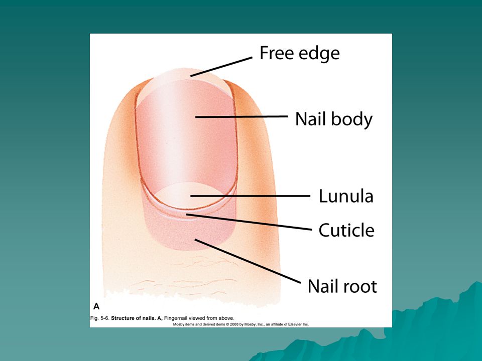
The Skin Nails Produced by epidermal cells over terminal ends of fingers and toes Visible part called nail body Root lies in a groove and is hidden by cuticle Crescent-shaped area nearest root called lunula Nail bed may change color with change in blood flow

29
The Skin Skin glands Types Sweat or sudoriferous Sebaceous

30
The Skin Skin glands Sweat or sudoriferous glands Types
Eccrine sweat gland Most numerous, important, and widespread of the sweat gland Produce perspiration or sweat, which flows out through pores on skin surface Function throughout life and assist in body heat regulation

31
The Skin Skin glands Sweat or sudoriferous glands Types
Apocrine sweat gland Found primarily in axilla and around genitalia Secrete a thicker, milky secretion quite different from eccrine perspiration Breakdown of secretion by skin bacteria produces odor

32
The Skin Skin glands Sweat or sudoriferous glands Types
Sebaceous gland Secrete oil or sebum for hair and skin Level of secretion increases during adolescence Amount of secretion regulated by sex hormones Sebum in sebaceous gland ducts may darken to form a blackhead Acne vulgaris inflammation of sebaceous gland ducts

33
Dermis Sweat gland Sebaceous gland Arrector pili muscle Blood vessels

34
The Skin Appendages of the skin Receptors
Specialized nerve endings—make it possible for skin to act as a sense organ Meissner’s corpuscle—capable of detecting light touch Pacinian corpuscle—capable of detecting pressure

35
Quick Quiz Which layer does not have a blood supply?
Ok, quick quiz. Which layer of the skin does not have a blood supply? Correct! The epidermis doesn’t have a blood supply. The epidermis and subcutaneous tissues do have a blood supply.

36
Quick Quiz Which layer has the pigment cells?
Which layer of the skin has the pigment cells? Correct! The epidermis again! Remember that it has many layers. The pigment cells begin development in the dermis and migrate to the epidermis as they mature.

37
Quick Quiz What is that pigment called?
What is that pigment that causes us to have different skin tones called? Correct! Melanin! It is produced by the melanocytes. Melatonin inhibits melanin production.

38
Quick Quiz Which layer has the fatty tissue?
Which layer of the skin contains the fatty tissue? Yes! The subcutaneous tissue.

39
Quick Quiz Which part of the epidermis is undergoing mitosis all the time? Which part of the epidermis is undergoing mitosis all the time? Yes! The germinativum.

40
Case Study Katie is a 15-year-old girl who is very upset because of the pimples on her face. She cannot understand why her little sister Kimberly, who is 7, doesn’t have acne.

41
Question What explanation can you give Katie concerning her skin?
A. In several years Kimberly also will have acne. B. Acne is present most likely because she doesn’t wash her face. C. Acne is an allergic reaction to certain creams used on the face. D. Acne in adolescence is a result of overactive sebaceous glands. D

42
Question Katie is embarrassed because of the pimples on her face. Her mother has decided to seek medical attention. What sort of doctor will she probably see? A. dermatologist B. cosmetic specialist C. plastic surgeon D. pediatrician A

43
Question Which of the following statements about hair follicles is true? A. Arrector pili muscles are associated with them. B. Sudoriferous glands empty into them. C. They arise directly from the epidermis layer of skin. D. All of the above. A

44
Burns Classification of burns
First-degree (partial-thickness) burns—only the surface layers of epidermis involved Second-degree (partial-thickness) burns—involve the deep epidermal layers and always cause injury to the upper layers of the dermis Third-degree (full-thickness) burns—characterized by complete destruction of the epidermis and dermis and subcutaneous tissues.
 burns—only the surface layers of epidermis involved. Second-degree (partial-thickness) burns—involve the deep epidermal layers and always cause injury to the upper layers of the dermis. Third-degree (full-thickness) burns—characterized by complete destruction of the epidermis and dermis and subcutaneous tissues.")
45
THE SKIN Burns Treatment and recovery or survival depend on total area involved and severity or depth of the burn Body surface area is estimated using the “rule of nines” (Figure 5-8) in adults Body is divided into 11 areas of 9% each Additional 1% located around genitals
 in adults. Body is divided into 11 areas of 9% each. Additional 1% located around genitals.")
46
Burns First-degree (partial-thickness) burns—only surface layers of epidermis involved Second-degree (partial-thickness) burns—involve the deep epidermal layers and always cause injury to the upper layers of the dermis

49
Burns Third-degree (full-thickness) burns
(Figure 6-14) characterized by complete destruction of the epidermis, dermis, and subcutaneous tissue May involve underlying muscle and bone (fourth-degree) Lesion is insensitive to pain because of destruction of nerve endings immediately after injury—intense pain is soon experienced
 characterized by complete destruction of the epidermis, dermis, and subcutaneous tissue. May involve underlying muscle and bone (fourth-degree) Lesion is insensitive to pain because of destruction of nerve endings immediately. after injury—intense pain is soon. experienced.")
51
Burns Estimating body surface area using the “rule of nines” in adults
Body divided into 11 areas of 9% each Additional 1% of body surface area around genitals

53
Skin Lesions Elevated lesions—cast a shadow outside their edges
Papule—small, firm raised lesion Plaque—large raised lesion Vesicle—blister Pustule—pus-filled lesion Crust—scab Wheal (hive)—raised, firm lesion with a light center
—raised, firm lesion with a. light center.")
54
Skin Lesions Flat lesions—do not cast a shadow
Macule—flat, discolored region Depressed lesions cast a shadow within their edges Excoriation—missing epidermis, as in a scratch wound Ulcer—craterlike lesion Fissure—deep crack or break

55
Skin Cancer Three common types Squamous cell carcinoma—the most
common type, characterized by hard, raised tumors Basal cell carcinoma—characterized by papules with a central crater; rarely spreads Melanoma—malignancy in a nevus (mole); the most serious type
; the most serious type.")
59
Skin Cancer The most important causative factor in common skin cancers is exposure to sunlight Kaposi sarcoma, characterized by purple lesions, is associated with AIDS and other immune deficiencies

61
Skin Infections Impetigo—highly contagious staphylococcal infection
Tinea—fungal infection (mycosis) of the skin; several forms occur Boils—furuncles; staphylococcal infection in hair follicles Scabies—parasitic infection
 of the. skin; several forms occur. Boils—furuncles; staphylococcal infection. in hair follicles. Scabies—parasitic infection.")
62
Vascular and Inflammatory Skin Disorders
Decubitus ulcers (bedsores) develop when pressure slows down blood flow to local areas of the skin Urticaria or hives—red lesions caused by fluid loss from blood vessels Scleroderma—disorder of vessels and connective tissue characterized by hardening of the skin; two types: localized and systemic
 develop when pressure slows down blood flow to local. areas of the skin. Urticaria or hives—red lesions caused by. fluid loss from blood vessels. Scleroderma—disorder of vessels and connective tissue characterized by hardening. of the skin; two types: localized and systemic.")
63
Vascular and Inflammatory Skin Disorders
Psoriasis—chronic inflammatory condition accompanied by scaly plaques Eczema—common inflammatory condition characterized by papules, vesicles, and crusts; not a disease itself but a symptom of an underlying condition

64
PSORIASIS

65
Let’s Review!

66
Which type of body membrane lines the digestive tract?
Cutaneous membrane Tympanic membrane Serous membrane Synovial membrane Mucous membrane Chapter: 5 Answer: E Discussion: Mucous membrane lines cavities that are open to the exterior, which includes the lumen of the digestive tract. This item presents a good opportunity to compare membrane types.

67
Which is the thickest part of the skin?
Epidermis Dermis Chapter: 5 Answer: B Discussion: Generally the connective dermis is much thicker than the epithelium of the epidermis. If you add “C. Hypodermis” that would be the best choice for thickest, in most areas of the body. But then, the hypodermis is not technically part of the skin, making for a good discussion point.

68
The gland that produces a “conditioning cream” for the hair and skin is the
Eccrine sweat gland Apocrine sweat gland Sebaceous gland Mammary gland Ceruminous gland Chapter: 5 Answer: C Discussion: Sebum is a conditioning “skin oil.” This item presents a good opportunity to mention that mammary glands are, anatomically speaking, skin structures. Ceruminous glands are not discussed in the chapter, but one could identify their role in producing ear wax here.

69
The skin plays important roles in maintaining a stable body temperature.
True False Chapter: 5 Answer: A Discussion: This item presents a good opportunity to discuss sweating and diversion of blood flow.

70
What is the largest sensory organ of the body?
Eye Ear Tongue Skin Nose Chapter: 5 Answer: D Discussion: All the sensory receptors in the largest organ of the body make skin the clear winner. This could be used just before introducing the skin as a topic, to stimulate thought and discussion.

71
What characterizes second-degree burns?
A. blisters B. swelling C. severe pain D. all of the above D

72
Questions? ?????????????????????????????????? So, how about any questions for the end of the integumentary system?

Similar presentations


 - The Integumentary System and Body Membranes>")