Download presentation
Presentation is loading. Please wait.

1
Windsor University School of Medicine
Gluteal region, thigh & leg The future belongs to those who believe in the beauty of their dreams. Eleanor Roosevelt Lecture Idara C. Eshiet

2
OBJECTIVES Be able to describe the bones of lower limb.
Be able to describe the muscles of the gluteal region, & thigh. Be able to describe the femoral triangle & popliteal fossa. Be able to describe the muscles of the leg.

3
Bones of lower limb Hip bone ( Ilium, Ischium and pubis) Femur Tibia
Fibula

4
Areas of transition/Bones & joints of the lower limb.

5
COXAL BONE /Acetabulum

6
Shaft /Proximal end of the femur

7
Femur vs. Tibia & Fibula

8
Proximal ends of tibia & fibula

9
Tibia & Fibula/ posteromedial view of distal ends.

10
Thigh Fascia Compartments : anterior, medial, posterior muscles

11
Fascia of the thigh Superficial is the continuity of the superficial fascia of anterior abdominal wall Deep fascia thickened laterally to form the illiotibial tact Has a gap called saphenous opening Divided into 3 compartments by 3 intermuscular septa

13
Muscle compartments in the thigh
In the thigh, there are medial (adductor), anterior (extensor), and posterior (flexor) compartments.
, anterior (extensor), and posterior (flexor) compartments.")
14
Anterior Thigh Muscles
Anterior thigh muscles are the flexors of the hip and extensors of the knee.

15
Iliacus Action: Chief flexor of the hip joint. Nerve Supply: Femoral nerve

16
Psoas Major Action: Flexes thigh on trunk Nerve Supply: femoral nerve

17
Pectineus Action: Flexion & adduction of hip joint Nerve supply: Femoral nerve

18
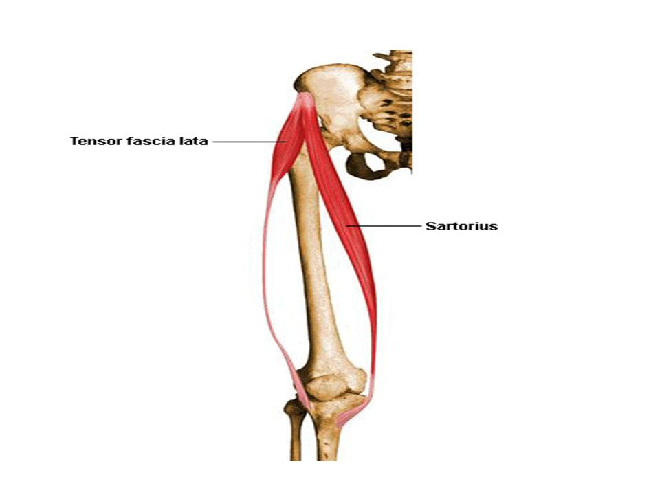
Sartorius Nerve Supply: Femoral nerve Action :
1.Flexion, abduction and Lateral rotation of thigh at hip joint. 2. Flexion leg at knee joint Nerve Supply: Femoral nerve

19
Tailor's muscle This combination of lateral rotation and flexion of the hip and flexion of the knee gave tailors particularly enlarged sartorius muscles. Looking at the bottom of one's foot, as if checking to see if one had stepped in gum, demonstrates all four actions of sartorius.

20
Quadriceps femoris Four muscles make up this group. They are:
rectus femoris, vastus lateralis, vastus medialis, vastus intermedialis Action: extension of knee Rectus femoris also flexes the hip joint as well. Nerve Supply: Femoral nerve

21
Quadriceps

22
Medial Compartment of Thigh
1. Gracilis Adduction of the the hip joint & flexion of knee joints 2. Adductor longus Adduction of the the hip joint. 3 2 1 4

23
3. Adductor brevis Adduction of the the hip joint. 4. Adductor magnus has 2 parts. Ant. Part is an adductor Post. Part is an extensor of the the hip joint 5. Obturator externus Lateral rotation of hip joint Obturator nerve but the post. part of adductor magnus is supplied by tibial nerve which is a branch of the sciatic nerve.

24
Adductor magnus & Obturator externus
Is innervated by the obturator nerve 2. Adductor magnus The adductor part is innervated by the obturator nerve & the hamstring part is innervated by the tibial division of the sciatic nerve 1 2

25
Pectineus, Adductors longus & brevis
Is innervated by the femoral nerve 2. Adductor longus Is innervated by the obturator nerve 3. Adductor brevis 1 3 2

26
Obturator nerve

27
Adductor Canal, subsartorial canal; Hunter canal
Approximately 15 cm. It extends from the apex of the femoral triangle, where the sartorius crosses over the adductor longus, to the adductor hiatus in the tendon of the adductor magnus. Contents: Femoral artery and vein, the saphenous nerve, and the nerve to vastus medialis.

30
Adductor Hiatus The adductor hiatus is an opening or gap between the aponeurotic distal attachment of the adductor part of the adductor magnus and the tendinous distal attachment of the hamstring part. The adductor hiatus transmits the femoral artery and vein from the adductor canal in the thigh to the popliteal fossa posterior to the knee.

31
3. Semimembranosus Extension of hip joint & flexion & medial rotation of knee joint. Nerve Supply: Tibial portion of sciatic nerve

32
Posterior Compartment of the Thigh
1. Biceps femoris Extension of the the hip joint & flexion & lateral rotation of the knee joint. Nerve supply: Long head: tibial portion of sciatic nerve Short head: common peroneal portion of sciatic nerve 2. Semitendinosus Extension of the the hip joint & flexion & medial rotation of the knee joint. Nerve supply: Tibial portion of sciatic nerve

34
Movements of Hip joint Flexion Extension Abduction Adduction
Circumduction Medial rotation Lateral rotation

35
Movements of Hip joints

36
Muscles responsible for movements of Hip joints.
Flexion : iliopsoas, rectus femoris Extension: Hamstrings, gluteus maximus Abductor: Gluteus medius & minimus Adduction: Gracilis & 3 Adductors

39
Muscles responsible for movements of Hip joints Cont’d.
Medial rotation: gluteus medius & minimus Lateral rotation: obturator internus & externus, piriformis, superior & inferior gemelli, quadratus femoris

40
Muscles responsible for movements of Knee Joints.
Flexion: Hamstrings Extension: Quadriceps Medial rotation: Semitendinosus & Semimembranosus. Lateral rotation : Biceps femoris.

42
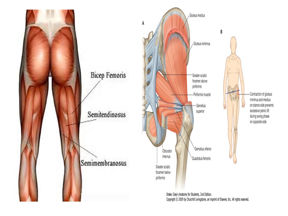
Muscles of the gluteal region

43
Muscles of the gluteal region
1. Gluteus maximus Extension of the the hip joint, lateral rotation, steadies the thigh, assists in rising from a sitting position Inferior gluteal nerve 3 1

44
44

45
Assists gluteus maximus in extending the knee joint
2. Tensor fasciae latae Assists gluteus maximus in extending the knee joint Superior gluteal nerve Tenses the fascia lata and iliotibial tract

46
3. Gluteus medius & minimus
Abduction and medial rotation of the thigh; tilts pelvis when walking to permit opp. Leg to clear the ground. Superior gluteal nerve

47
Positive Trendelenburg's sign
If right gluteus medius and minimus muscles are paralyzed, the unsupported left side of the pelvis falls (sags) instead of rising; normally, the pelvis rises.
 instead of rising; normally, the pelvis rises.")
51
Lateral rotation of the thigh@ hip joint nerve Supply: S1 & S2 nerves
4. Piriformis m. Lateral rotation of the hip joint nerve Supply: S1 & S2 nerves Important landmark of the gluteal region. 5. Obturator Internus Same function as piriformis Nerve supply: (L5-S1) nerves to obturator internus The piriformis muscle passes through the greater sciatic foramen. It divides in into a superior and inferior part.
 nerves to obturator internus. The piriformis muscle passes through the greater sciatic foramen. It divides in into a superior and inferior part.")
53
Muscles of the gluteal region Cont’d.
6. Superior Gemellus Lateral rotation of the the hip joint (L5-S1) nerves to obturator internus 7. Inferior Gemellus Same function as superior gamellus Nerve to quadratus femoris ( L5, S1)
 nerves to obturator internus. 7. Inferior Gemellus. Same function as superior gamellus. Nerve to quadratus femoris ( L5, S1)")
54
Lateral rotation of the thigh @ the hip joint
8. Quadratus Femoris Lateral rotation of the the hip joint (L5-S1) nerves to quadratus femoris 8
 nerves to quadratus femoris. 8.")
55
Femoral triangle & Popliteal fossa
Femoral triangle boundaries, floor, & contents Popliteal fossa boundaries, floor, & contents

56
The femoral triangle is bounded
Superiorly :Inguinal ligament that forms the base of the femoral triangle. Medially :Adductor longus. Laterally :Sartorius. Apex is where the lateral border of the sartorius crosses the medial border of the adductor longus. Roof :Fascia lata, subcutaneous tissue, and skin. Floor :Iliopsoas laterally and the pectineus medially.

57
Femoral Triangle Inguinal ligament Iliopsoas Sartorius Adductor longus
Pectineus Sartorius Adductor longus Adductor longus

58
Femoral Triangle Contents : (lat. to med.) Femoral nerve
Femoral artery Femoral vein Deep inguinal lymph nodes

60
Boundaries of Popliteal fossa
Superolateral : biceps femoris Superomedial: semimembranosus & semitendinosus Inferolateral: lateral head of gastrocnemius Inferomedial: medial head of gastrocnemius

62
Contents of the popliteal fossa
1. Termination of the small saphenous vein. 2. Popliteal arteries and veins. 3. Tibial and common fibular nerves. 4. Posterior cutaneous nerve of thigh 5. Popliteal lymph nodes and lymphatic vessels

63
Floor This is formed by: popliteal surface of the femur,
capsule of knee joint & popliteus muscle

64
LEG Fascia Muscles of anterior compartment
Muscles of lateral compartment Muscles of posterior compartment (superficial & deep layers) Main muscles responsible for ankle joint movements
 Main muscles responsible for ankle joint movements.")
65
Fascia of the Leg Deep fascia (crural fascia) .
Leg divided into 3 fascia compartments (anterior, posterior, lateral) by 3 intermuscular septa. In the region of the ankle the fascia forms retinacula : Superior & inferior extensor retinacula Flexor retinaculum Fibular retinaculum
 by 3 intermuscular septa. In the region of the ankle the fascia forms retinacula : Superior & inferior extensor retinacula. Flexor retinaculum. Fibular retinaculum.")
66
Cross-section through the left leg (post. View)
Muscles of the anterior compartment of leg dorsiflex the ankles, extend the toes, & invert the foot. (deep fibular nerve). Muscles in the posterior compartment plantarflex the ankle, flex the toe, & invert the foot. (tibial nerve). Muscles in the lateral compartment evert the foot. (superficial fibular nerve).
. Muscles in the posterior compartment plantarflex the ankle, flex the toe, & invert the foot. (tibial nerve). Muscles in the lateral compartment evert the foot. (superficial fibular nerve).")
69
Muscles of anterior compartment
1. Tibialis anterior Dorsiflexion & inversion of the foot at the ankle. 2. Extensor digitorum longus Extension of lateral 4 digits & dorsiflexion of the ankle 1 2

70
N. supply: Deep fibular nerve
3. Extensor hallucis longus Action: Extension of big toe & dorsiflexion of the ankle Deep fibular nerve from common fibular nerve 4. Fibularis Tertius Action :Dorsiflexion and eversion of foot N. supply: Deep fibular nerve

71
Extensor Hallucis longus

73
Muscles of lateral compartment
1. Fibularis longus Eversion & plantar flexion of foot. 2. Fibularis brevis Superficial fibular nerve from common fibular nerve 1 2

74
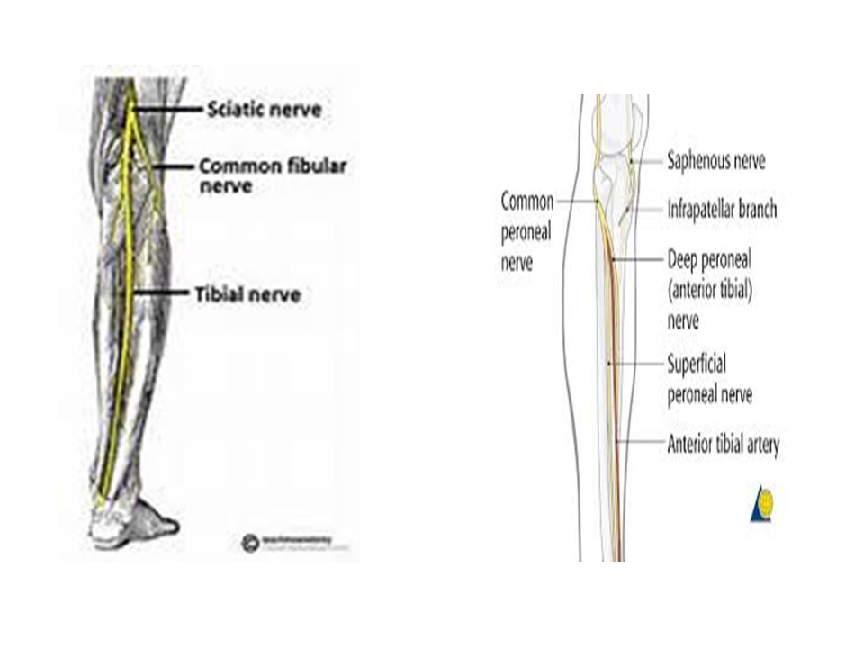
Injury to common fibular nerve
Footdrop and loss of eversion May cause sensory loss over lateral leg and dorsum of foot Causes Direct trauma as nerve passes superficially around neck of fibula

75
Foot drop Foot drop, sometimes called drop foot, is a general term for difficulty lifting the front part of the foot. The loss of dorsiflexion of the ankle causes footdrop.

76
Posterior compartment of leg
Muscles in the posterior (flexor) compartment of leg are organized into two groups; superficial and deep. Nerve supply: Tibial nerve
 compartment of leg are organized into two groups; superficial and deep. Nerve supply: Tibial nerve.")
77
Muscles of the Posterior Compartment of the Leg
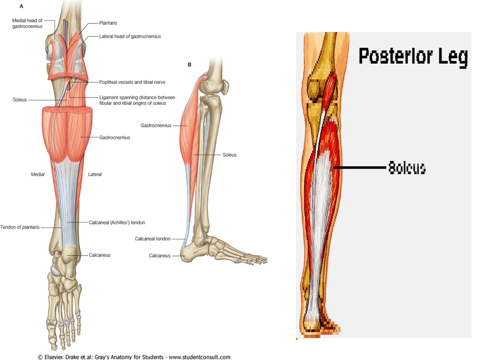
Superficial group of muscles 1.Gastrocnemius 2.Soleus 3.Plantaris Deep group of muscles 1.Popliteus 2.Tibialis posterior 3.Flexor digitorum longus 4.Flexor hallucis longus Muscles mainly plantarflex, invert the foot and flex the toes.

78
Superficial Group 1. Gastrocnemius Action: Plantar flexion of the foot the ankle), knee flexion Nerve Supply: Tibial nerve 2. Soleus Action: Plantarflexion of the foot
, knee flexion Nerve Supply: Tibial nerve 2. Soleus Action: Plantarflexion of the foot")
79
3. Plantaris: Action: plantarflexion of the foot Nerve Supply: Tibial nerve

80
Gastrocnemius has two heads; a medial and a lateral head
Gastrocnemius has two heads; a medial and a lateral head. The two heads of the Gastrocnemius muscle and the Soleus muscle are together called the Triceps Surae. Gastrocnemius and plantaris cross knee joint and thus also flex knee . All three contribute to calcaneal tendon

81
Gastrocnemius and plantaris

83
Muscles of posterior compartment (deep layer)
2. Popliteus Flexion of knee joint 3. Flexor digitorum longus Flexion of DIP of lateral 4 digits Tibial nerve 2 5 3 4

85
Plantarflexion & inversion of foot Nerve Supply: tibial nerve
4. Flexor hallucis longus Flexion of big toe 5. Tibialis posterior Plantarflexion & inversion of foot Nerve Supply: tibial nerve

87
Muscles responsible for ankle joint movements (fig.B)
Dorsiflexion : Tibialis anterior Extensor digitorum longus Extensor hallucis longus Plantarflexion : Triceps surae Tibialis posterior Flexor digitorium longus Flexor hallucis longus

88
Movements of knee & ankle

89
Surface Anatomy1.

90
Surface Anatomy2.

91
Surface Anatomy3.

92
POWER REVIEW1. 1. What are the 4 regions of the lower limb, and which bones are found in each region? Hip: ilium, Ischium, & pubis Thigh: Femur & patella Leg: Tibia & fibula Foot: Tarsal bones, metatarsal bones, & phalanges. 2. Name the 7 tarsal bones Talus, Calcaneus, Cuboid bone, Navicular bone, Cuneiform bones (3)
")
93
POWER REVIEW2. 3. what is the largest and most posterior tarsal bone?
The calcaneus 4. what structure inserts into the posterior surface of the calcaneus? The tendon calcaneus (Achilles tendon) 5.the calcaneus articulates with which 2 tarsal bones? The talus & the cuboid bone
 5.the calcaneus articulates with which 2 tarsal bones The talus & the cuboid bone.")
94
POWER REVIEW3. 6. The talus articulates with which 2 tarsal bones?
The calcaneus & the navicular bone 7. The navicular bone articulates with which 5 tarsal bones? The talus, the cuboid bone, and the 3 cuneiform bones. 8. which movements occur around the intertarsal joints? Inversion & eversion of the hindfoot

95
POWER REVIEW4. 9. which muscle is the major flexor at the hip joint?
Iliopsoas. 10. name the external rotators of the hip Piriformis, Gemellus superior, Obturator internus Gemellus inferior, Obturator externus, Quadratus femoris 11. Name the 5 ligaments that are associated with the hip joint. Iliofemoral ligament, ischiofemoral ligament, pubofemoral ligament Transverse acetabular ligament, ligament capitis femoris

96
POWER REVIEW5. 12. list the 4 muscles of the posterior thigh compartment. Semimembranous m., semitendinous m., biceps femoris m. (long & short head), adductor magnus m. (hamstring part) 13.what are the “hamstring” muscles? The semimembranous m., the tendinosus m., the long head of the biceps femoris m., and the adductor magnus m. (hamstring part)
, adductor magnus m. (hamstring part) 13.what are the hamstring muscles The semimembranous m., the tendinosus m., the long head of the biceps femoris m., and the adductor magnus m. (hamstring part)")
97
POWER REVIEW 6. 14. which of the medial thigh muscles contributes to the action of the hamstrings? The adductor magnus muscle has 2 portions with separate insertions & innervations, 1 of which contributes to the action of the hamstrings (flex the leg). 15. list the 6 muscles of the medial thigh compartment. Pectineus m., adductor longus m., Adductor magnus m., (adductor part), Adductor brevis m., Gracilis m., Obturator externus m.
. 15. list the 6 muscles of the medial thigh compartment. Pectineus m., adductor longus m., Adductor magnus m., (adductor part), Adductor brevis m., Gracilis m., Obturator externus m.")
98
POWER REVIEW 7. 16. list the 3 muscles of the anterior compartment of the thigh. Iliopsoas m., Sartorius m., Quadriceps femoris m. 17. which 4 muscles contribute to the quadriceps femoris muscles? Rectus femoris m., Vastus lateralis m., Vastus medialis m., Vastus intermedius m.,

99
Review Questions.1 1. Which of the following muscles is located in the posterior aspect of the thigh? 2. All of the following muscles are lateral rotators of the thigh EXCEPT 3. The deep fascia of the thigh is known as which of the following? 4. The medial and lateral malleoli articulate with which of the following bones? 5. Which of the following muscles is the strongest flexor of the hip joint?

100
Review Questions.2 6. The strongest dorsiflexor of the foot is which of the following muscles? 7. All of the following muscles are lateral rotators of the hip joint EXCEPT 8. Which of the following groups of muscles produce dorsiflexion of the ankle? 9. Which of the following muscles is a flexor of the knee joint? 10. All of the following muscles are located in the deep muscle group of the posterior compartment EXCEPT

101
Review Questions.3 11. Which of the following muscles is the strongest dorsiflexor and invertor of the foot? 12. Muscles that evert the foot include which of the following muscles? 13. Which of the following muscles dorsiflex the ankle? 14. All of the following statements concerning the popliteal fossa are correct EXCEPT 15. Which of the following muscles is located in the posterior aspect of the thigh?

102
Review Questions.4 16. All of the following muscles are lateral rotators of the thigh EXCEPT 17. All of the following statements concerning the gluteus medius and minimus are correct EXCEPT 18. All of the following statements concerning the gluteus maximus are correct EXCEPT 19. All of the following statements concerning the femoral triangle are correct EXCEPT 20. All of the following statements concerning the adductor magnus are correct EXCEPT 21. Which of the following statements concerning the gracilis muscle is correct?

103
T. S. Eliot No one can become really educated without having pursued some study in which he took no interest--for it is a part of education to learn to interest ourselves in subjects for which we have no aptitude. ………………..references…………………………. Dr. Bolgova PPt. Gray’s Anatomy for students, 2nd edition

Similar presentations


 by Susan J. Hall, Ph.D.>")
>")


>")

