Download presentation
Presentation is loading. Please wait.

1
Chapter 2. Radiation Radioactivity 2.Radiation interaction with Matter
3.Radiation Doses and hazard Assessment

2
2.1 Radioactivity Overview Types of Radioactive Decay Energetics of Radioactive Decay Characteristics of Radioactive Decay Decay Dynamics Naturally Occurring Radionuclides

3
c) Beta Decay Spectra and Neutrino
? Pauli: Neutrino with spin 1/2 is emitted simultaneously with beta, carrying the missing energy.

4
c) The mass of the neutrino is negligibly small.
 The mass of the neutrino is negligibly small.")
5
d) Positron Decay Energy
 Positron Decay Energy")
6
3)36CI decays into 36S (35. 967081 u) and 36Ar
3)36CI decays into 36S ( u) and 36Ar. If the energy release is MeV to 36S and MeV to 36Ar, calculate the masses of 36CI and 36Ar. Describe the modes of decay. 5) The radionuclide 41Ar decays by β- emission to an excited level of 41K that is MeV above the ground state. What is the maximum kinetic energy of the emitted β- particle?
36CI decays into 36S ( u) and 36Ar. If the energy release is MeV to 36S and MeV to 36Ar, calculate the masses of 36CI and 36Ar. Describe the modes of decay. 5) The radionuclide 41Ar decays by β- emission to an excited level of 41K that is MeV above the ground state. What is the maximum kinetic energy of the emitted β- particle")
7
Radioactive Decay Kinetics -exponential
Number of radioactive nuclei decrease exponentially with time as indicated by the graph here. As a result, the radioactivity vary in the same manner. Note l N = A l No = Ao

8
6) The activity of a radioisotope is found to decrease by 30% in one week. What are the values of its (a) decay constant, (b) half-life, and (c) mean life?
 decay constant, (b) half-life, and (c) mean life .")
9
b) Three Component Decay Chains
 Three Component Decay Chains")
10
Daughter Decays Faster than the Parent
λI < λ2, transient equilibrium: daughter's decay rate is limited by the decay rate of the parent. λI << λ2, The activity of the daughter approaches that of the parent. This extreme case is known as secular equilibrium(久期平衡).
.")
11
4)An initial number NA(0) of nuclei A decay into daughter nuclei B, which are also radioactive. The respective decay probabilities areλA and λB. IfλB = 2λA , calculate the time (in terms of λA)when NB is at its maximum. Calculate NB (max) in terms of NA(0)
when NB is at its maximum. Calculate NB (max) in terms of NA(0).")
12
2.2 Radiation interaction with Matter
overview Photon Interactions Neutron Interactions Interaction of Heavy Charged Particles with Matter Scattering of Electrons in a Medium

13
1) overview
 overview")
14
mean-free-path length Half-Thickness
I = Io e–μx mean-free-path length Half-Thickness

15
4) Interaction of Heavy Charged Particles with Matter
Fast moving protons, 4He, and other nuclei are heavy charged particles. Coulomb force dominates charge interaction. They ionize and excite (give energy to) molecules on their path. The Born-Bethe Formula for Energy Loss of Charged Particles.
 molecules on their path. The Born-Bethe Formula for Energy Loss of Charged Particles.")
16
能量损失

17
Range of Heavy Charged Particles in a Medium
Particles lose all their energy at a distance called range. source Shield

18
A material is found to have a tenth-thickness of 2. 3 cm for 1
A material is found to have a tenth-thickness of 2.3 cm for 1.25 MeV gamma rays, (a) What is the linear attenuation coefficient for this material? (b) What is the half-thickness? (c) What is the mean-free-path length for 1.25-MeV photons in this material? The specific rate of energy loss (-dE/ρdx) of a 5 MeV proton in silicon is 59 keV mg-1 cm2 and its range R' is 50 mg cm-2 . Calculate values of (-dE/ρdx) and range R' for deuterons, tritons, 3He and a particles, all of which have the same speed as the proton.
 What is the linear attenuation coefficient for this material (b) What is the half-thickness (c) What is the mean-free-path length for 1.25-MeV photons in this material The specific rate of energy loss (-dE/ρdx) of a 5 MeV proton in silicon is 59 keV mg-1 cm2 and its range R is 50 mg cm-2 . Calculate values of (-dE/ρdx) and range R for deuterons, tritons, 3He and a particles, all of which have the same speed as the proton.")
19
2.3 Radiation Doses and hazard Assessment
Historical Roots Dosimetric Quantities Natural Exposures for Humans Radiation Effects

20
Historical Roots Early workers exposed to X-rays developed dermatitis(皮炎). Uranium miners developed skin lesions. People working with radioactivity experienced illness. Researchers exposed to radioactivity suffered radiation sickness at advanced age. Manhattan project workers in Los Alamos, Oak Ridge, Hanford, and atomic worker in the former USSR suffered anorexia(厌食), fatigue, headache, nausea(反胃), vomiting, and diarrhea.
, fatigue, headache, nausea(反胃), vomiting, and diarrhea.")
21
Collective Response to Radiation Risk
In 1928, the International Committee on X-ray and Radium Protection was formed to look into the risk of radiation. It is now called International Commission on Radiological Protection, ICRP. In 1942, a group of health physicists had the responsibility to assess problems and implement safe operation procedures regarding radioactivity. After WW2, the (American) National Council of Radiation Protection (NCRP) was formed in 1946. Guidelines are given for radioactive material handling and applications. Today, safety committee is set up to deal with radiation risks.
 National Council of Radiation Protection (NCRP) was formed in Guidelines are given for radioactive material handling and applications. Today, safety committee is set up to deal with radiation risks.")
22
Mission Statement of the ICRP
The International Commission on Radiological Protection, ICRP, is an independent Registered Charity, established to advance for the public benefit the science of radiological protection, in particular by providing recommendations and guidance on all aspects of protection against ionising radiation. From check with ICRP for up-to-date guidance regarding radiation

23
Protection standards 辐射防护规定 环保局发布 GB4792-84 GB8703-88 GB 18871-2002
放射卫生防护基本标准 卫生部发布 GB 辐射防护规定 环保局发布 GB 电离辐射防护与辐射源安全基本标准 发布 实施 中华人民共和国国家质量监督检验检疫总局发布

24
2) Dosimetric quantities
Lord Kelvin 2) Dosimetric quantities ...When you can measure what you are speaking about, and express it in numbers, you know something about it... Lord Kelvin a physical measure correlated with a radiation effect.
 Dosimetric quantities. ...When you can measure what you are speaking about, and express it in numbers, you know something about it... Lord Kelvin. a physical measure correlated with a radiation effect.")
25
Radiation Absorption and Dosage
type units Radioactivity Bq, Ci Exposure dose Gy, rad (R) Quality factor Q Biological dose Sv, rem The amount of energy absorbed from exposure to radiation is called a dose. The radiation effect measured by a dosimeter reflects an equivalence of certain dosage of X-rays. The amounts are defined in certain units as shown here.
 Quality factor Q. Biological dose Sv, rem. The amount of energy absorbed from exposure to radiation is called a dose. The radiation effect measured by a dosimeter reflects an equivalence of certain dosage of X-rays. The amounts are defined in certain units as shown here.")
26
Units for Radiation Source (review)
The SI unit for radioactivity is Bq (1 becquerel = 1 dps). The decay is not necessary all absorbed unless it’s internal. 1 curie = 3.7e10 Bq. These units have nothing to do with energy, type (a, b, g, X-rays, neutrons, protons or particles), and effect of radiation. Commonly used units megacurie kilocurie millicurie microcurie nonocurie picocurie these modifiers are also used for other units. disintegrations per second the fluence is not closely enough related to most radiation effects to be a useful determinant.
. The decay is not necessary all absorbed unless it’s internal. 1 curie = 3.7e10 Bq. These units have nothing to do with energy, type (a, b, g, X-rays, neutrons, protons or particles), and effect of radiation. Commonly used units. megacurie. kilocurie. millicurie. microcurie. nonocurie. picocurie. these modifiers are also used for other units. disintegrations per second. the fluence is not closely enough related to most radiation effects to be a useful determinant.")
27
Dose Units - roentgen, rad, and gray
Amounts of absorbed energy are not the same as exposed. The amount of radiation energy absorbed is called a dose. A roentgen ( R) is a dose of X- or -rays that produce 1 esu charge at STP (negative and positive each or 2.1e9 ion pairs) in 1.0 L. 1 R = 352.1e = 7.35e10 eV (*1.6x10⁻¹² erg/eV) = 0.12 erg (per g air) = 1 rad (100 erg per g of any substance) 1 Gy = 1 J / kg (1 J per kg of any substance is a gray, Gy) = 1e7 erg / kg = 100(100 erg/g) ~ 100 rad average energy In air, the average energy required to produce an ion pair is 35 eV photons corpuscular radiation 1 Gy being equal to an imparted energy of 1 joule per kilogram.
 is a dose of X- or -rays that produce 1 esu charge at STP (negative and positive each or 2.1e9 ion pairs) in 1.0 L. 1 R = 352.1e9 = 7.35e10 eV (*1.6x10⁻¹² erg/eV) = 0.12 erg (per g air) = 1 rad (100 erg per g of any substance) 1 Gy = 1 J / kg (1 J per kg of any substance is a gray, Gy) = 1e7 erg / kg = 100(100 erg/g) ~ 100 rad. average energy. In air, the average energy required to produce an ion pair is 35 eV. photons. corpuscular radiation. 1 Gy being equal to an imparted energy of 1 joule per kilogram.")
28
A Dosage Evaluation Example
A 5-MeV particle is absorbed by 1 gram of water, estimate the dosage in rad and rem. The Q factor is 10 for particle, and thus the dose is 8e-7 rem or 8e-9 Sv. If the a particle is absorbed by a of 10-9 g cell, then the dose is 109 times higher (0.8 Gy, 8 Sv), exceeded lethal (致命) dose for most living beings.
, exceeded lethal (致命) dose for most living beings.")
29
Integral Dose Used in Radiation Therapy
Total energy absorbed by an organ called integral dose is gram-rad or g-rad or g-Gy total dosage received by an organ. g-Gy = dose * mass of the organ Accumulated dose is the dose received over a period, but g-Gy is the total dose received in a single time.

30
The Quality Factor QF and Dosage Units
The factor reflecting the relative harmfulness of various types of radiation is called the quality factor (QF) or relative biological effectiveness (rbe) Biological dose = QF * exposure dose
 or relative biological effectiveness (rbe) Biological dose = QF * exposure dose.")
31
Exposure and Biological Dosage
SI unit cgs unit Exposure unit 1 Gy = 100 rad (=100 R) Biological dose 1 Sv = 100 rem (= Qrad) Gy: gray, Sv: sievert, R: roentgen, rem: roentgen equivalent man
 Biological dose 1 Sv = 100 rem (= Qrad) Gy: gray, Sv: sievert, R: roentgen, rem: roentgen equivalent man.")
32
Summary of Units for Radioactive Dosage
Quantity Symbol SI unit cgs unit Conversion factor radioactivity A Bq Ci 1 Ci = 3.7e10 Bq exposure dose X C/kg R 1 C/kg = 3876 R absorbed dose D Gy (J/kg) rad 1 Gy = 100 rad =6.24 eV/g biological dose H Sv (QF*Gy) rem 1 Sv = 100 rem C/kg charge produced by exposure to radiation
 rad 1 Gy = 100 rad =6.24 eV/g biological dose H Sv (QF*Gy) rem 1 Sv = 100 rem. C/kg charge produced by exposure to radiation.")
33
Effective Dose Equivalent
In a human, different organs have different radiological sensitivities, To account for different organ sensitivities and the different doses received by the various organs a special dose unit, the effective dose equivalent HE, is used to describe better the hazard a human body experiences when placed in a radiation field.

34
Tissue weighting factors adopted by the ICRP [1977] for use in determining the effective dose equivalent.
![Tissue weighting factors adopted by the ICRP [1977] for use in determining the effective dose equivalent.](http://slideplayer.com/slide/4284502/14/images/34/Tissue+weighting+factors+adopted+by+the+ICRP+%5B1977%5D+for+use+in+determining+the+effective+dose+equivalent..jpg "Tissue weighting factors adopted by the ICRP [1977] for use in determining the effective dose equivalent.")
35
Naturally occurring radionuclides in the human body deliver
an annual dose to the various tissues and organs of the body as follows: lung 36 mrem, bone surfaces 110 mrem, red marrow 50 mrem, and all other soft tissues 36 mrem. What is the annual effective dose equivalent that a human receives?

36
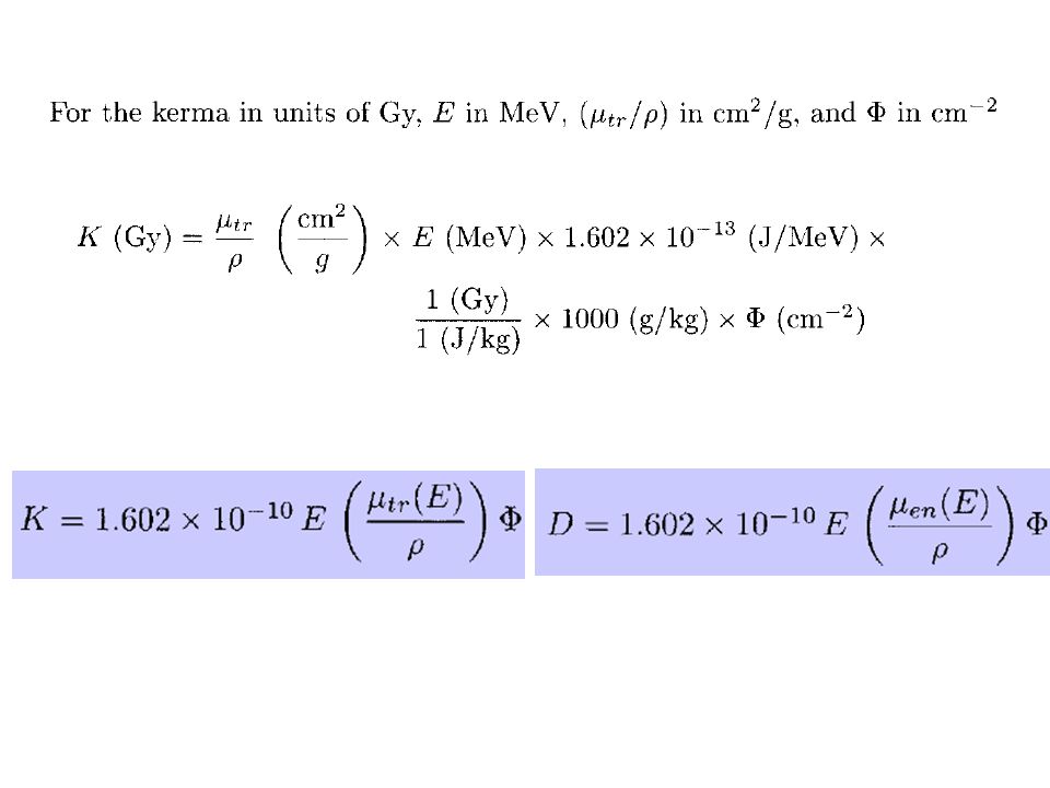
Kerma kinetic energy of radiation absorbed per unit mass
比释动能 indirectly ionizing (uncharged) radiation If Etr is the sum of the initial kinetic energies of all the charged ionizing particles released by interaction of indirectly ionizing particles in matter of mass m, then
 radiation. If Etr is the sum of the initial kinetic energies of all the charged ionizing particles released by interaction of. indirectly ionizing particles in matter of mass m, then.")
37
total moss interaction coefficient
the linear energy absorption coefficient μtri which account for fewer secondary photons escaping from the interaction site, are sometimes encountered. (a) Energy deposition for photon energy involved in the interactions in an incremental volume of material, (b) Formulas for the energy per unit mass of the material in the incremental volume, corresponding to the various energy increments in (a), (c) Linear coefficients defined by their proportionality to the mass energy relationships in diagrams (a) and (b).
 Energy deposition for photon energy involved in the interactions in an incremental volume of material, (b) Formulas for the energy per unit mass of the material in the incremental volume, corresponding to the various energy increments in (a), (c) Linear coefficients defined by their proportionality to the mass energy relationships in diagrams (a) and (b).")
38
Photon Kerma and Absorbed Dose
If, at some point of interest in a medium, the fluence of radiation with energy E is Ф, the kerma at that point is f(E) is the fraction of the fraction of the incident radiation article's energy E that is transferred to secondary charged particles μ(E)/ρ is the mass interaction coefficient for the detector material. μtr(E)/ρ for charged secondary particles and excludes the energy carried away from the interaction site by secondary photons一定物质对特定能量的间接致电离粒子的质量能量转移系数。
 is the fraction of the fraction of the incident radiation article s energy E that is transferred to secondary charged particles. μ(E)/ρ is the mass interaction coefficient for the detector material. μtr(E)/ρ for charged secondary particles and excludes the energy carried away from the interaction site by secondary photons一定物质对特定能量的间接致电离粒子的质量能量转移系数。")
40
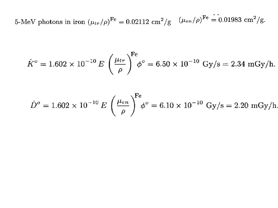
the total mass interaction coefficient for 5-MeV photons is found to .
Example What are the iron kerma and absorbed dose rates from uncollided photons 1 meter from a point isotropic source emitting MeV gamma rays per second into an water medium? the total mass interaction coefficient for 5-MeV photons is found to The uncollided flux density 1 meter from the source is,

42
neglect air attenuation over a distance of 15 m
Example : What is the dose equivalent 15 meters from a point source that emitted 1 MeV photons isotropically into an infinite air medium for 10 minutes at a rate of 109 photons per second? neglect air attenuation over a distance of 15 m QF = I 0.15 μSv

43
Dosimeters for Dosage Monitoring
Dosimeters are devices to measure exposed doses. Film-badges, electroscopes, ionization chambers, biological and chemical dosimeters have been used for radiation monitors. Plants, cells, bacteria, and viruses reacting to radiation are biological dosimeter candidates. Ferrous sulfate, FeSO4, solution is a chemical dosimeter due to the reaction: 4 Fe2+ + energy + O2 4 Fe3+(brown) + 2 O2- Some glasses and crystals serve as solid state dosimeters. Shelf life, linearity, stability, usage simplicity, easy-to-read, dose-rate and equal responses to various radiation are some considerations.
 + 2 O2- Some glasses and crystals serve as solid state dosimeters. Shelf life, linearity, stability, usage simplicity, easy-to-read, dose-rate and equal responses to various radiation are some considerations.")
44
Chemical 3-dimensional Dosimeter
Ferrous ions, Fe2+, are oxidized by ionizing radiation, and convert to ferric ions, Fe3+, which complexes with xylenol(二甲苯酚) orange dye to give an orange compound. When the sample is prepared in a gel form, it serves as a 3-dimensional dosimeter, because the complexes are localized in the gel. These dosimeters are useful for planning radiation medical treatments such as radiation cancer treatment.
 orange dye to give an orange compound. When the sample is prepared in a gel form, it serves as a 3-dimensional dosimeter, because the complexes are localized in the gel. These dosimeters are useful for planning radiation medical treatments such as radiation cancer treatment.")
45
2.3 Radiation Doses and hazard Assessment
Historical Roots Dosimetric Quantities Natural Exposures for Humans Radiation Effects

46
3) Natural Exposures for Humans
 Natural Exposures for Humans")
47
Radioactivity in Nature
222Rn is responsible for higher levels of background radiation in many parts of the world. because it is a gas and can easily seep out of the earth into unfinished basements and then into the house Radon The uranium decay series.

48
Summary of the annual effective dose equivalents from various sources of natural background radiation in the United States. Source: NCRP [1987].

49
Some Natural Occurring Radioactive Nuclides
Nuclides (t½ ~ y) Radiation 235, 238U, 232Th and offsprings , , 144Nd, 147, 148, 149Sm, 152Gd, 186Os, 190, 192Pt () 40K, 87Rb, 115In, 123Te, 138La, 176Lu, 187Re, 210Bi etc. +, , EC () Nuclides produced by cosmic rays 14C (5730 y), 3T (15 y), 7Be (53 d), 10Be (2.7×10⁶ y)
 Radiation. 235, 238U, 232Th and offsprings. , , 144Nd, 147, 148, 149Sm, 152Gd, 186Os, 190, 192Pt. () 40K, 87Rb, 115In, 123Te, 138La, 176Lu, 187Re, 210Bi etc. +, , EC () Nuclides produced by cosmic rays. 14C (5730 y), 3T (15 y), 7Be (53 d), 10Be (2.7×10⁶ y) ")
50
2.3 Radiation Doses and hazard Assessment
Historical Roots Dosimetric Quantities Natural Exposures for Humans Radiation Effects

51
Radiation Effects Somatic effects damages to cells passed on to succeeding cell generations, acute or chronic Genetic effects damages to genes that affect future generations. Genes are units of hereditary information that occupy fixed positions (locus) on a chromosome. Genes achieve their effects by directing the synthesis of proteins.
 on a chromosome. Genes achieve their effects by directing the synthesis of proteins.")
52
Somatic Effects Damages to cell membranes, mitochondria(线粒体) and cell nuclei result in abnormal cell functions, affecting their division, growth and general heath. Organs such as skin, lining of gastrointestinal tract(胃肠道), embryos, and bone marrow, whose cells proliferate rapidly are easily damaged. Bone marrow makes blood, and its damage leads to reduction of blood cell counts and anemia. Damage to germinal ( 幼体 tissues reduces cell division, and induces sterility.
 and cell nuclei result in abnormal cell functions, affecting their division, growth and general heath. Organs such as skin, lining of gastrointestinal tract(胃肠道), embryos, and bone marrow, whose cells proliferate rapidly are easily damaged. Bone marrow makes blood, and its damage leads to reduction of blood cell counts and anemia. Damage to germinal ( 幼体 tissues reduces cell division, and induces sterility.")
53
Is this change good or bad?
Cellular Effects Cell death Cell repair Cell change Is this change good or bad?

54
Dividing Cells are the Most Radiosensitive
Rapidly dividing cells are more susceptible to radiation damage. Examples of radiosensitive cells are; Blood forming Cells The intestinal lining Hair follicles(毛囊) A fetus Suggested narrative: When cells are dividing (or undergoing mitosis) they are more susceptible to radiation damage because the cells don’t have their full suite of repair mechanisms. Because of this, cells that are often dividing like The cells that create our blood or line our intestine, also Hair follicles, and, of course, fetal cells are more susceptible to radiation damage. This is why the fetus has a exposure limit (over gestation period) of 500 mrem (or 1/10th of the annual adult limit) Specialized or slowly dividing cells, like brain cells are radio-insensitive. Credit: The vedio (and voiceover) was Excerpted from DOE’s Transportation Emergency Preparedness Program (TEPP) This is why the fetus has a exposure limit (over gestation period) of 500 mrem (or 1/10th of the annual adult limit)
 A fetus. Suggested narrative: When cells are dividing (or undergoing mitosis) they are more susceptible to radiation damage because the cells don’t have their full suite of repair mechanisms. Because of this, cells that are often dividing like. The cells that create our blood or line our intestine, also. Hair follicles, and, of course, fetal cells. are more susceptible to radiation damage. This is why the fetus has a exposure limit (over gestation period) of 500 mrem (or 1/10th of the annual adult limit) Specialized or slowly dividing cells, like brain cells are radio-insensitive. Credit: The vedio (and voiceover) was Excerpted from DOE’s Transportation Emergency Preparedness Program (TEPP) This is why the fetus has a exposure limit (over gestation period) of 500 mrem (or 1/10th of the annual adult limit)")
55
Deterministic Effects in Organs and Tissues
Median effective absorbed doses D50 and threshold doses Dth for exposure of different organs and tissues in the human adult to gamma photons at dose rates < 0.06 Gy h-1.

56
Maximum permissible dosage of workers in radiation zone
Exposure Limit Maximum permissible dosage of workers in radiation zone Max. accumulated Max. dose/13 wk mSv mSv Whole body (age-18) 30 Hands and (750/y) forearms 1 Sv = 1000 mSv = 100 rem
 30. Hands and 250 (750/y) forearms. 1 Sv = 1000 mSv = 100 rem.")
57
We are facing many environmental toxic agents.
The risk estimation of these agents should be based on dose response curve. Response Dose The response in a low dose range could be extrapolated from high doses if it is a physical system. However, it is not true in biological systems. 57

58
Biological Response Dose Biological response to low dose radiation
is complicated. Bystander effect Biological Response ICRP (International Commission on Radiological Protection) Adaptive response Dose 58
 Adaptive response. Dose. 58.")
59
In the biological systems,
the dose response at low dose level cannot be extrapolated from high dose response. Instead, experimental as well as epidemiological studies are needed to clarify the dose response.

60
Bystander Effects The signals sent by the bystander cells may help repair the damaged cell, or it may trigger the cell to commit cell suicide. When a cell is damaged by radiation, it can send signals to bystander cells, which are the cells near the “hit” cell. With the microbeam, it is possible to expose individual cells and using the modern cellular and molecular techniques to study the changes that occur in the cell that is "hit" by the radiation and in neighboring cells that were not directly traversed by the radiation. It is possible to demonstrate that the "hit" cell communicates with its neighboring cells and triggers cellular and molecular changes in these cells. Bystander effects have been demonstrated in vitro using a number of different cell culture systems and a wide range of biological endpoints. It is critical to determine if this response increases or decreases the risk for production of late occurring disease. This is an area of current research and vigorous discussion. For the bystander effect to be of significance in terms of risk assessment it is important to determine if these effects are produced in vivo using experimental animals. The signals sent by the damaged cell may disrupt the normal function of it’s neighboring cells, or it may stimulate them to respond with additional signals back to the damaged cell or to other nearby cells. 60

61
Micronuclei Geard Cells were stained with two different dyes. Only the nuclei of the cells stained with pink dye were hit by alpha particles from a microbeam. The figures show the presence of broken chromosomes in the form of micronuclei (the smaller fragments of pink and blue). These micronuclei are present not only in the pink “hit” cells, but also in the blue non-exposed cells. Such studies provide direct evidence for bystander effects.
. These micronuclei are present not only in the pink hit cells, but also in the blue non-exposed cells. Such studies provide direct evidence for bystander effects.")
62
No bystander between organs exposed at low dose-rates
The site of deposition of the radioactive material is the site of cancer induction 90SR - bone cancer 144Ce – liver/bone cancer 239 PuO2 (inhaled)- lung cancer
- lung cancer.")
63
Lower half of lungs irradiated with 10 Gy
The influence of communication on radiation-induced micronuclei in lung Lung cells shielded from direct radiation showed a major increase in the production of micronuclei (one indicator of chromosome damage) when other cells in the lung tissue were irradiated, indicating some type of communication between cells. Shielded Cells 400 Micronuclei/1000 Cells 800 Lower half of lungs irradiated with 10 Gy Exposed Cells Khan et al 1998
 when other cells in the lung tissue were irradiated, indicating some type of communication between cells. Shielded Cells Micronuclei/1000 Cells Lower half of lungs irradiated with 10 Gy. Exposed Cells. Khan et al")
64
Why now? Standards have been set from high dose effects, but low dose effects have not been measurable until now New technological developments and biological discoveries have made it possible to study low dose effects Why now? It is important to understand that very extensive research has been conducted on the health effects of radiation. Because of this research we currently know more about the health effects of radiation than any other environmental pollution. The question is WHY NOW? Why start this new program? This program represents a merging of the newly developed techniques associated with the genome with new technology. This merging of techniques makes it possible to make measurements of radiation induced cellular and molecular changes that were not possible in the past. It is now possible to start to understand the impact of low doses of radiation on risk. This newly developed information will be essential for future development of models to predict radiation risk. 64

65
Single ion hit system FDSPM
A gradual deterioration due to accumulated radiation damage Transient malfunctions due to single particles hitting a sensitive node.

66
Does the bystander effect occur in animals as well as cell culture?
The bystander effect occurs in animal systems The bystander effect is limited to specific organs or tissues No bystander effects seen between organs at low dose rates

67
Genetic Effects Human cells contain 46 chromosomes(染色体). Germ or ovum cells contain 23. A chromosome contains a deoxyribonucleic acid (DNA) molecule. The double-helix DNA has two strands of phosphoric-acid and sugar linked bases of Adenine, Guanine Cytosine or Thymine. The A-T and G-C pairs stack on top of each other. The DNA codon transcripts mRNA, which directs the amino-acid sequences of protein. DNA Damages result in somatic and genetic effects. When DNA molecules replicate (pass on to next generation), they are sensitive to radiation damage. Joining wrong ends of broken DNA is called Translocation, which cause mutation and deformation at birth. Genetic effects increase frequency of mutation.
 molecule. The double-helix DNA has two strands of phosphoric-acid and sugar linked bases of Adenine, Guanine Cytosine or Thymine. The A-T and G-C pairs stack on top of each other. The DNA codon transcripts mRNA, which directs the amino-acid sequences of protein. DNA Damages result in somatic and genetic effects. When DNA molecules replicate (pass on to next generation), they are sensitive to radiation damage. Joining wrong ends of broken DNA is called Translocation, which cause mutation and deformation at birth. Genetic effects increase frequency of mutation.")
68
A simplified view of a portion of the DNA molecule, as well as the various types of damage
it can experience. Four building blocks or bases combine to form the DNA molecule: adenine (A)(腺嘌呤), guanine (G) (鸟嘌呤) , cytosine (C)(氧氨嘧啶), and thymine (T)(胸腺嘧啶).
(腺嘌呤), guanine. (G) (鸟嘌呤) , cytosine (C)(氧氨嘧啶), and thymine (T)(胸腺嘧啶).")
69
Genomic Instability Delayed Genetic Effects

70
What is Genomic Instability?
Often, after being damaged by radiation, cells are able to repair DNA damage and reproduce normally. However, sometimes damage may carry over for several generations before the unobserved damage causes the cell to lose control of its genome. At this point, cells may be unable to reproduce successfully. They may become genetically unstable, or become cancerous.

71
Genomic Instability New Paradigm
After a cell is exposed to radiation, biological changes are produced that, after many cell divisions, result in loss of genetic control. This is a frequent event that can be modified. Cell death Micronuclei Gene mutation Mitotic failure-aneuploidy( 非整倍的) Chromosome aberration
 Chromosome aberration.")
72
Early effects seen in “hit” cell
Chromosomal rearrangements Micronuclei Gene mutations Increased Reactive Oxygen Species (ROS) Inflammatory responses Change in gene expression
 Inflammatory responses. Change in gene expression.")
73
Effects seen in cell progeny
Chromosomal rearrangements Micronuclei Transformation Chromosome amplification Death inducing factors Gene mutations Cell death Change in gene expression

74
Radiation-related Gene Induction
It has been shown that certain genes are inappropriately induced, or “turned on” or “turned off” by radiation. The consequence of the gene alteration sometimes shows up more frequently several generations after the initial radiation exposure.

75
Genomic Instability can be demonstrated in some strains of mice
Hybrid Mouse Models After only a few generations of apparently normal breast cell division, the cells of the sensitive mice, BALBc, show increased chromosome aberrations and genomic instability, while cells of the radiation resistant mice, C57BL/6, remain stable. Cells of the sensitive BALBc mice are very sensitive to radiation-induced breast cancer. Other cells, such as those from the resistant C57BL/6 mice, are particularly resistance to this radiation-induced effect.

76
Genomic Instability can be demonstrated in cells of some strains of mice
0.35 Sensitive BALB/c mice Resistant C57BL/6 mice 0.3 0.25 0.2 Aberrations/Cell 0.15 0.1 0.05 4 8 12 16 20 24 28 Population Doublings B. Ponnaiya & R.L. Ullrich, 1998

77
Impact on Standards Genomic Instability
Provides a mechanism to explain how radiation can produce the multiple steps needed to transform a normal cell to a malignant cell Supports the LNTH if cellular genomic instability can be shown to increase cancer frequency

78
Summary Radiation-induced genomic instability is defined as detrimental effects that occur several cell generations after radiation exposure. This may be due to factors produced by inflammatory response or a failure of genes to turn on or off properly. Signaling factors involved in genomic instability may be similar to those involved in bystander effects. Increased Reactive Oxygen Species (ROS) may also interfere with normal cellular processes and produce genomic instability. 78
 may also interfere with normal cellular processes and produce genomic instability. 78.")
79
Dose Ranges Cancer Radiotherapy Experimental Radiobiology
(mSievert) Cancer Radiotherapy Total Body Therapy Total Tumor Dose Experimental Radiobiology Human LD50 A-bomb survivors Cancer Epidemiology Typical mission dose on Int. Space Station Significant cancer risk at > 200 mSv (UNSCEAR) DOE Low Dose Program Occupational Limit NRC, EPA Typical annual dose for commercial airline flight crews Medical Diagnostics Thyroid (I-123) Bone (Tc-99m) Natural background NRC Dose Limit for Public Site Decommissioning/License Termination Dental X-ray Chest X-ray EPA Clean-up Standards NRC Clean-up Standards Regulatory Standards ICRP Negligible Dose 3-Mile Island Ave Ind
 Cancer Radiotherapy. Total Body Therapy. Total Tumor Dose. Experimental Radiobiology. Human LD50. A-bomb survivors Cancer Epidemiology. Typical mission dose on Int. Space Station. Significant cancer risk at > 200 mSv (UNSCEAR) DOE Low Dose Program. Occupational Limit NRC, EPA Typical annual dose for commercial airline flight crews. Medical Diagnostics. Thyroid (I-123) Bone (Tc-99m) Natural background. NRC Dose Limit for Public. Site Decommissioning/License Termination. Dental X-ray Chest X-ray. EPA Clean-up Standards. NRC Clean-up Standards. Regulatory Standards. ICRP Negligible Dose Mile Island Ave Ind.")
80
920 mGy/min 1 mGy/min 12.5μGy/min 80

81
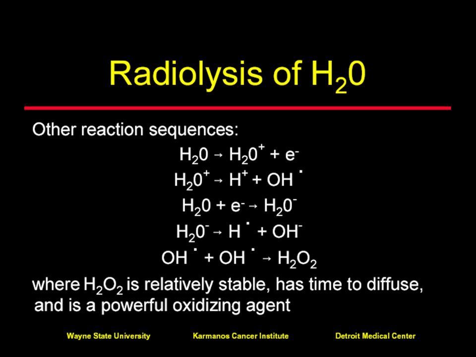
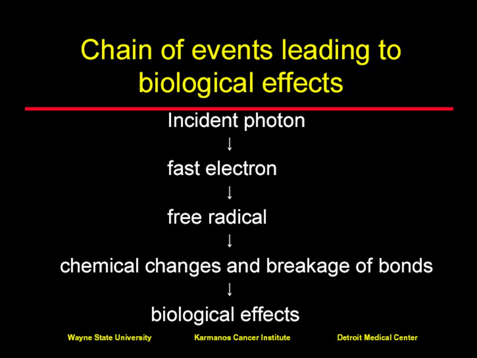
Direct and indirect action of radiation
Direct action: charged particle “directly” interacts with the target molecule, e.g. breaks bond in DNA molecule Indirect action: charged particle interacts with a water molecule producing “free radicals” which then interact with the target molecule For x and g radiations, indirect interactions cause about 80% of the biological damage

82
Direct and indirect action of radiation

87
Our Bodies Are Resilient
DNA damage is most important and can lead to cell malfunction or death. Our body has ~ 60 trillion cells Each cell takes “a hit” about every 10 seconds, resulting in tens of millions of DNA breaks per cell each year. BACKGROUND RADIATION causes only a very small fraction of these breaks (~ 5 DNA breaks per cell each year). Our bodies have a highly efficient DNA repair mechanisms Narrative The Cells in our body live in a continuing barrage of damaging events. About every 10 seconds, each cell in our body “takes a hit.” Sitting through this lecture, you will have been assaulted trillions of times! Call the police, Crime in {insert lecture location} is on the rise ;-) The VAST majority of these assaults is NOT from radiation, but from inescapable byproducts of the chemical processes in our bodies that enable us to live. Behind that is the natural or man-made toxins which we take into our body. Way below those effects are the 5 or so DNA breaks per cell each year that happen because of background radiation. (out of the 10s of millions total) Of course, if our bodies didn’t have extremely efficient DNA repair mechanisms, our breakfast would probably have already done us in. ====================== Notes ============== Most of this reference comes from: Smithsonian, V26, #9. December 1995, RISK, Part 2: Safeguarding our cells by James Trefil.
. Our bodies have a highly efficient DNA repair mechanisms. Narrative. The Cells in our body live in a continuing barrage of damaging events. About every 10 seconds, each cell in our body takes a hit. Sitting through this lecture, you will have been assaulted trillions of times! Call the police, Crime in {insert lecture location} is on the rise ;-) The VAST majority of these assaults is NOT from radiation, but from inescapable byproducts of the chemical processes in our bodies that enable us to live. Behind that is the natural or man-made toxins which we take into our body. Way below those effects are the 5 or so DNA breaks per cell each year that happen because of background radiation. (out of the 10s of millions total) Of course, if our bodies didn’t have extremely efficient DNA repair mechanisms, our breakfast would probably have already done us in. ====================== Notes ============== Most of this reference comes from: Smithsonian, V26, #9. December 1995, RISK, Part 2: Safeguarding our cells by James Trefil.")
88
2.3 Radiation Doses and hazard Assessment
Historical Roots Dosimetric Quantities Natural Exposures for Humans Radiation Effects

89
The three key rules of radiation protection: time, distance, and shielding.

90
ALARA principle: As Low As Reasonably Achievable
justification of practice optimization of radiation protectionwith (annual radiation) dose limits
 dose limits.")
91
With a frequency of 0.849 per decay
55-CESIUM-137 Radiations y(i) (Bq-s)-1 E(i) (MeV) y(i)×E(i) b- 1 9.47×10-01 1.743×10-01 * 1.65×10-01 b- 2 5.80×10-06 3.347×10-01 1.94×10-06 b- 3 5.30×10-02 4.163×10-01 2.21×10-02 g 1 2.835×10-01 1.64×10-06 g 2 8.51×10-01 6.617×10-01 5.63×10-01 With a frequency of per decay
 (Bq-s)-1. E(i) (MeV) y(i)×E(i) b × × * 1.65× b × × × b × × × g × × g × × × With a frequency of per decay.")
Similar presentations


 Ionizing Radiation>")


