Download presentation
Presentation is loading. Please wait.

1
THE SPECTRUM OF CONTINUOUS AIRWAY ASSESSEMENT AND MANAGEMENT
Credits should include Dr. Morgan Hillier who redesigned the Anatomy Lab presentation for the Sunnybrook RBH program and Halton EMS that freely shared presentation portions on the Bougie and intubation.

2
Learning Objectives: The following module covers:
Review of airway anatomy including pediatric differences. Acquiring and maintaining AW patency including manual and adjunct techniques. AW assessment and introduction to LEMON. Risk/benefit considerations. Problems and Rescue Procedures. Describe and identify normal airway anatomy Recognize physical exam signs suggestive of difficult BVM and difficult intubation Describe the spectrum of airway and ventilatory interventions from simple to invasive List the indications and contraindications of a surgical airway

3
Anterior View of the Larynx

4
Upper Airway Structures
Discus the major differences between adults and paediatric upper and lower anatomy: *** Important to remind: infants 0-6 months are obligatory nasal breathers !! Can’t breath thru their mouth. Large occipital part in peds – hyperextension can obstruct AW !! Tongue size Epiglottis size Anterior larynx in peds in comparison to adults Cricoid ring - the narrowest place in pediatric airway Trachea tend to collapse in severe SOB in peds. (Cartilage rings not fully developed) Smaller tidal volume and higher RR. Technical bullet: Cant obtain ETCO2 in children < 15kg (Zoll monitors), must use other methods (anatomical and physiological) to assess ventilation.
 Smaller tidal volume and higher RR. Technical bullet: Cant obtain ETCO2 in children < 15kg (Zoll monitors), must use other methods (anatomical and physiological) to assess ventilation.")
5
The Spectrum of Airway Management

6
Objectives Appreciate the spectrum of airway management from simple maneuvers to complex interventions Appreciate proper monitoring and need for further management during and after airway intervention What should the medic ask himself before deciding to manage patient airway? What is the patient most urgent problem? Cant maintain patent airway? Cant oxygenate? Cant ventilate? What tools are available to intervene, manage and treat patients problem? Can the problem be treated in the ambulance? What if everything goes wrong? Is there plan B?

7
Airway spectrum Alert/patent airway Simple manual maneuvers
Head-tilt, chin-lift, suction, left lateral recumbent, cricoid pressure (Sellick’s), chest compressions (FBOA) Simple and advanced adjuncts OPA, NPA, Bougie Supraglottic airways King-LT, LMA Give example of each that relate to another skill on the list to reinforce the concurrent relationship and the rationale for its potential effectiveness.
, chest compressions (FBOA) Simple and advanced adjuncts. OPA, NPA, Bougie. Supraglottic airways. King-LT, LMA. Give example of each that relate to another skill on the list to reinforce the concurrent relationship and the rationale for its potential effectiveness.")
8
Airway Spectrum Advanced airway
Direct laryngoscopy/intubation BURP (not Sellick’s) Failed airway , can’t oxygenate/can’t ventilate Needle cricothyrotomy McGill forceps
 Failed airway , can’t oxygenate/can’t ventilate. Needle cricothyrotomy. McGill forceps.")
9
Difficult Mask Ventilation
Obesity - BMI>26 kg/sq.m B Beard E Elderly S Snorer Edentulous Presence of 2 or more conditions – Difficult Bag Mask Ventilation

10
Difficult Face Mask Ventilation
Signs of inadequate face mask ventilation include absent or inadequate chest movement absent or inadequate breath sounds auscultatory signs of severe obstruction gastric air entry or dilatation decreasing or inadequate oxygen saturation (SpO2) absent or inadequate exhaled carbon dioxide hemodynamic changes associated with hypoxia, hypoxemia or hypercarbia (hypertension, tachycardia and arrhythmias, cyanosis) Compliance vs. resistance? The ease which lung and thorax expand vs. any mechanical factor that limits inspired air to reach the alveoli As the terms compliance and resistance may still confuse, describe each term separately: Compliance: Commonly described as the tendency of the lungs to expand due to any volume of air delivered/ inspired. A good example of decreased compliance is pulmonary edema or Pneumonia. An example for Increased Compliance is Emphysema. Resistance is easily described as the pressure you should apply in order to expand the lungs. An example to increased resistance is Cystic fibrosis or other restrictive pulmonary diseases. As in COPD (Bronchitis and Emphysema), in many cases, Decreased Compliance and increased resistance will appear together
 absent or inadequate exhaled carbon dioxide. hemodynamic changes associated with hypoxia, hypoxemia or hypercarbia (hypertension, tachycardia and arrhythmias, cyanosis) Compliance vs. resistance The ease which lung and thorax expand vs. any mechanical factor that limits inspired air to reach the alveoli. As the terms compliance and resistance may still confuse, describe each term separately: Compliance: Commonly described as the tendency of the lungs to expand due to any volume of air delivered/ inspired. A good example of decreased compliance is pulmonary edema or Pneumonia. An example for Increased Compliance is Emphysema. Resistance is easily described as the pressure you should apply in order to expand the lungs. An example to increased resistance is Cystic fibrosis or other restrictive pulmonary diseases. As in COPD (Bronchitis and Emphysema), in many cases, Decreased Compliance and increased resistance will appear together.")
11
Bag Mask Ventilation Important: Within the majority of patients, the correct positioning (sniffing position) is not about extending the head back (see upper example). The best positioning is to put a small pillow/padding under Pt’s occipital part and only then to extend the head back (See lower example). This manuver will inline the airway and provide the best position for proper ventilate.
 is not about extending the head back (see upper example). The best positioning is to put a small pillow/padding under Pt’s occipital part and only then to extend the head back (See lower example). This manuver will inline the airway and provide the best position for proper ventilate.")
12
Bag Mask Ventilation Key—ventilation volume: “enough to produce obvious chest rise” 1-Person difficult, less effective 2-Person easier, more effective

13
Cricoid Pressure (Sellick’s)
Posterior pressure on Cricoid Cartilage to occlude esophagus Purpose: prevent passive regurgitation of gastric contents **Can make intubation more difficult! Cricoid pressure should be applied constantly and firmly as long as the patient is ventilated with BVM. The medic should apply the pressure and avoid unnecessary movements, as the vagus branches that are localized just posteriorly to the trachea can stimulate nausea and vomiting (aspiration)
")
14
BURP maneuver Backward, upward, rightward pressure
Purpose: facilitate better laryngoscopic view Reinforce the medics to understand the differences between Sellick’s and BURP manuvers. If the PCP is not familiar with BURP, the ACP can apply the BURP while leading PCP’s hand to the specific location and ask him to continue the pressure

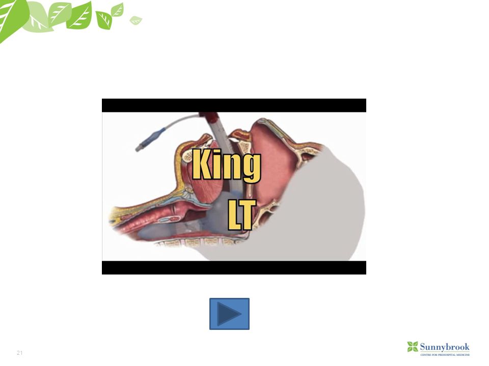
15
King LT

16
Supraglottic Airway King LT

17
Features of King SG Airways
The KING LTD is supplied clean, but is a non-sterile device It consists of a curved tube with ventilation apertures located between two inflatable cuffs. The distal cuff is designed to seal the esophagus Proximal cuff is intended to seal the oropharynx (King System, 2009). Cardiac arrest (PCP and ACP) Respiratory distress/arrest with GCS 3 (ACP only) (King System, 2009) As per medical directives, insertion on the king LT in unconscious patients(GCS=3) must be pre assessed for lack of gag reflex !!! 17
. Cardiac arrest (PCP and ACP) Respiratory distress/arrest with GCS 3 (ACP only) (King System, 2009) As per medical directives, insertion on the king LT in unconscious patients(GCS=3) must be pre assessed for lack of gag reflex !!! 17.")
18
King LT Sizing Chart

19
Advantages Emergency ventilation can take place within 15 seconds without a laryngoscope. Requires minimal movement of patient head. Requires minimal education to insert. The King laryngeal airway is designed to be inserted without direct visualization. Minimal risk of aspiration. The KING LTD provides a more secure, non-intubating emergency airway when direct laryngoscopy is not feasible (King System, 2009). (King System ,2009) The king LT has 3 major advance: Eliminate to need to keep patients head in a sniffing position by one medic as the other one is ventilating. The ability to ventilate with larger volumes and higher rates without a significant risk of stomach inflation. To reduce to significantly any risk of aspiration. Also can be utilized to ventilate patients with COPD exacerbation and severe asthma patients by attaching a PEEP device (Positive end expiratory pressure) to the king LT. ******* currently not in use in Ontario ******* 19
. (King System ,2009) The king LT has 3 major advance: Eliminate to need to keep patients head in a sniffing position by one medic as the other one is ventilating. The ability to ventilate with larger volumes and higher rates without a significant risk of stomach inflation. To reduce to significantly any risk of aspiration. Also can be utilized to ventilate patients with COPD exacerbation and severe asthma patients by attaching a PEEP device (Positive end expiratory pressure) to the king LT. ******* currently not in use in Ontario ******* 19.")
20
Disadvantages Only make limited sizes.
The KING LTD can be used in routine procedures only up to 8 hours. Unable to place medication down the tube. Trauma related to balloon in trachea (King System, 2009). Only minimizes risk of aspiration (aspiration can still occur) (King System, 2009 ). Note that large volumes and pressure of the cuffs, irritate the soft tissues of the hypo pharynx and esophagus stimulating the vomiting reflex, Bradycardia (stimulation of the vagal pathways ) and Ischemia to the soft palate and the esophagus. As such, ACP’s will remove the king LT as soon as possible and intubate the patient. A good sigh to assess patients gag reflex is to determine if the patient is trying to swallow (moving up and down his glottic structures). Reinforce medics to understand, that although the King LT provides a good protection from Aspiration and stomach content insufflations to the trachea, there will be a chance of aspiration without any external “evidence”. A good sign is to assess patients “fighting the tube”. As this occure, assume that the patient regain his gag reflex, and its time to consider removing the King LT 20
. Only minimizes risk of aspiration (aspiration can still occur) (King System, 2009 ). Note that large volumes and pressure of the cuffs, irritate the soft tissues of the hypo pharynx and esophagus stimulating the vomiting reflex, Bradycardia (stimulation of the vagal pathways ) and Ischemia to the soft palate and the esophagus. As such, ACP’s will remove the king LT as soon as possible and intubate the patient. A good sigh to assess patients gag reflex is to determine if the patient is trying to swallow (moving up and down his glottic structures). Reinforce medics to understand, that although the King LT provides a good protection from Aspiration and stomach content insufflations to the trachea, there will be a chance of aspiration without any external evidence . A good sign is to assess patients fighting the tube . As this occure, assume that the patient regain his gag reflex, and its time to consider removing the King LT. 20.")
22
Monitoring Airway Interventions
Ventilation: Chest wall excursion, ease of BVM vents, ETCO2 Oxygenation: SpO2, skin colour, heart rate As monitoring is performed, the two most reliable and early accurate parameters are ETCO2 and heart rate (SpO2 is a late sign). The heart will alter the rate with any slight rise in spinal H+ concentrations (occurring from changes in O2 and CO2 levels in the spinal cord). Thus, any rise in patients pulse during ventilation should be assessed also as a ventilation/oxygenation problem.
. The heart will alter the rate with any slight rise in spinal H+ concentrations (occurring from changes in O2 and CO2 levels in the spinal cord). Thus, any rise in patients pulse during ventilation should be assessed also as a ventilation/oxygenation problem.")
23
Management of the Difficult Airway

24
DEFINITION Difficult airway:
ANYTHING that interferes with ventilation or intubation Anatomic Traumatic Infectious (airway edema) Allergic Behavioral Elicit examples of each, which will lead into the following sections of the presentation.
 Allergic. Behavioral. Elicit examples of each, which will lead into the following sections of the presentation.")
25
Normal Adult Vocal Cords
Cords Abducted (open) The ideal picture, but….
 The ideal picture, but….")
26
Laryngoscopic View What we hope not to see...

27
LEMON Law Look at anatomy Examine the airway Mallampati Scale
Obstructions Neck Mobility By utilizing the LEMON mnemonic as a tool when evaluating an airway for potential difficulties during intubation you can anticipate what other techniques, tools may be required. Be aware that not always all the tools will be available or applicable. For example: One can’t evaluate neck mobility in trauma patients with suspected C-spine injury…

28
Look at Anatomy Obesity: rapid desaturation, short or thick neck-difficult intubation. Facial hair: hides small chin, can make BVM ventilation difficult. Teeth: hide airway, obscure tube passage, may lacerate balloon, dentures. Poor Neck Mobility: surgery, kyphosis Large Tongue:

29
Evaluate the 3-3-2 Rule The 3 – 3 – 2 rule: Mouth open: 3 fingers
Allows insertion of tube, laryngoscope Mentum (chin) to hyoid: 3 fingers Predicts ability to lift tongue into mandible Floor of mouth to thyroid cartilage: 2 fingers If high larynx, airway tucked under base of tongue, hard to visualize
 to hyoid: 3 fingers. Predicts ability to lift tongue into mandible. Floor of mouth to thyroid cartilage: 2 fingers. If high larynx, airway tucked under base of tongue, hard to visualize.")
30
Mallampati Score The Mallampati Score is based on the structures visualized with maximal mouth opening and tongue protrusion in the sitting position. The amount of the posterior pharynx you can visualize is important and correlates with the difficulty of intubation. Visualization of the pharynx is obscured by a large tongue (relative to the size of the mouth), which also interferes with visualization of the larynx on laryngoscopy. Originally, Malamppati score was developed for the Anaesthesiologists to assess patients AW prior to surgery and was performed while patient is sitting and opening his mouth as per physicians request. Although most of “our” patients will be prone and unconscious, yet it’s a great tool to utilize when assessing the potential difficulty of the AW.
, which also interferes with visualization of the larynx on laryngoscopy. Originally, Malamppati score was developed for the Anaesthesiologists to assess patients AW prior to surgery and was performed while patient is sitting and opening his mouth as per physicians request. Although most of our patients will be prone and unconscious, yet it’s a great tool to utilize when assessing the potential difficulty of the AW.")
31
Obstruction Evaluation for stridor, foreign bodies, and other forms of sub- and supraglottic obstruction should be performed in every patient prior to laryngoscopy

33
Food More common in children In adults there are typically co-factors
Foreign Bodies Food More common in children In adults there are typically co-factors 2 major groups in risk of FBAO: Children <5 - tend to put everything in their mouth Adults and geriatric patients receiving Tricyclic antidepressants – causing decrease in gag reflex

34
Foreign Body (food, toys) Trauma Edema Neoplasm Blood
Causes of Obstruction Foreign Body (food, toys) Trauma Edema Neoplasm Blood
 Trauma. Edema. Neoplasm. Blood.")
35
Neck Mobility Patients with degenerative or rheumatoid arthritis may have limited neck motion, and this should be assessed to assure the ability to adequately extend the neck during laryngoscopy and intubation.

36
Indications for Intubation
Based on three fundamental clinical assessments: Is there a failure of airway maintenance or protection? Is there a failure of ventilation or oxygenation? What is the anticipated clinical course? Ron Walls -

37
Failure of Airway Maintenance or Protection
Upper airway muscles and protective reflexes normally protect the airway from aspiration. Patent airway helps with ensuring adequate oxygenation. What devices do we carry to help maintain a patent airway? Not all airway devices protect the airway. In absence of immediately reversible condition, intubation should be considered. What is the most reliable method to assess protective reflexes? Having a patient swallow is most reliable, it is a complex reflex that requires ability to sense foreign material in hypopharynx and execution of a series of intricate and coordinated muscle actions to direct material or secretions down past the closed airway into the esophagus.

38
Failure of Oxygenation/Ventilation
Oxygenation/Ventilation provide oxygen to vital organs, remove waste carbon dioxide and help regulate pH. If oxygenation still does not improve despite efforts to improve ventilation, intubation should be considered. Eg. Asthma/CHF-these patients have patent airways and can protect them, but ventilatory failure will ultimately lead to inadequate oxygenation and death. Engage in discussion about what options medics have (ACP and PCP) to improve ventilation/oxygenation
 to improve ventilation/oxygenation.")
39
What is Anticipated Clinical Course?
Although airway and ventilation are adequate at this time, conditions may change in future require airway control or protection: stab wound to neck for example. Other examples? Other examples: Allergic reaction Epiglottitis Facial burns Localized trauma to the mouth (gunshot wound)
")
40
Tracheal Tube Introducer (Bougie)
GEB or “Bougie” is a device utilized to increase success when securing an airway. EMS and hospital trials have shown increased success rate, and decreased time to intubate with the use of the bougie. First used by Robert Macintosh in 1943 during a difficult intubation. Airway management, and specifically intubation is being de-emphasized in a number of field situations, and outright discouraged in many other field situations. This may translate the Paramedics doing a lower number of intubations in the field on average. With that in mind, and the fact that the majority of the airways that Paramedics intubate are considered “difficult airways”, it is important to provide adjuncts that increase success rates. The GEB has been shown to do just that.

41
Gum Elastic Bougie The Bougie is a long (60 cm) introducer with a 38 degree bend at the tip. Became popular among hospital staff about 30 years ago. Crept its way into EMS approximately 10 years ago. To date in the CPER region, Six Nations Ambulance Service as well as Dufferin County EMS have been trained and are utilizing the bougie. Halton EMS and PRPS are now using them as well.

42
Logistics to Consider It can be a challenge to store the device. It’s shape has a lot to do with its advantage as an airway adjunct so it must be stored in a way to not compromise that. It is a single use, disposable device. Applicable to ACP scope of practice. Does not require a specific Medical Directive as it is considered an adjunct under the ETI Medical Directive.

43
Inserting the Bougie You may choose to lubricate the tip of the Bougie with a water based solution, however it is generally not required. Perform laryngoscopy as per your normal technique. At this point instead of the usual ET tube with stylet, grasp the Bougie. Pass the Bougie through the glottic opening to the point of the 40 cm marking on the device, or until slight resistance is met. The resistance indicates the device contacting the carina or entering the bronchial tree. At this point, ideally it becomes a two person procedure. This discussion is to serve as an overview of the procedure for use of the device. It will become much clearer to the learner when the skill is actually performed at the skill station, so at this point in the presentation it is not imperative they know all the steps. Also, the fact that it is ideally a two person procedure does not mean one practitioner can not do it. You can relate it to utilizing a BVM. Ideally BVM ventilations should be done with 2 person technique, but it is not always practical or possible, so there is also one person technique. Be sure the PCPs in the group are engaged in the discussions around what their role will be in the 2 person technique.

44
Bougie Insertion

45
Confirming the Tube As you advance the device, you get two additional indications of correct placement, both tactile in nature. i) Clicking sensation as the device passes over the tracheal rings. Studies indicate these are felt up to 90% of the time. ii) Slight resistance as the device advances to the carina or into the bronchi (generally around 40 cm in an adult). The device is marked at this point. Other methods of tube confirmation should still be utilized and remain unchanged i.e.) ETCO2, chest rise and fall, auscultation, misting etc. While the studies indicate that clicking along tracheal rings is felt up to 90% of the time, subjective feedback from individuals that use the device tends to be much lower. So it they feel the clicking it can be helpful in validating proper position of ET tube, but if they don’t feel the sensation, they should still generally proceed with the intubation and use other accepted tube confirmation techniques. The slight resistance around the 40cm mark tends to be more commonly felt than the tracheal rings. That being said if the angle was such that the device enters the right main stem with just a glance on the carina, the sensation could be lost. In this situation, a slight twisting of the bougie would indicate entry into one of the main stems.
 Clicking sensation as the device passes over the tracheal rings. Studies. indicate these are felt up to 90% of the time. ii) Slight resistance as the device advances to the carina or into the. bronchi (generally around 40 cm in an adult). The device is marked at. this point. Other methods of tube confirmation should still be utilized and remain unchanged i.e.) ETCO2, chest rise and fall, auscultation, misting etc. While the studies indicate that clicking along tracheal rings is felt up to 90% of the time, subjective feedback from individuals that use the device tends to be much lower. So it they feel the clicking it can be helpful in validating proper position of ET tube, but if they don’t feel the sensation, they should still generally proceed with the intubation and use other accepted tube confirmation techniques. The slight resistance around the 40cm mark tends to be more commonly felt than the tracheal rings. That being said if the angle was such that the device enters the right main stem with just a glance on the carina, the sensation could be lost. In this situation, a slight twisting of the bougie would indicate entry into one of the main stems.")
46
SpO2 & ETCO2 SpO2 – Reflects the percentage of Oxyhemoglobin in the blood. SpO2 less than 90 = SpO2 less than 60 = Hypoxia SpO2 80 = PaO2= 45 !! Goal: To keep Saturation between 94-98% The Affinity of the Hb to O2 depends on DPG production, which differs and depends on Body temp., aerobic conditions (enough glucose in the system) and diseases. High concentration of DPG will decrease the affinity of Hb to O2 and release more O2 to the tissue (fever, blood acidity) and vise versa (Hypothermia, alkalosis). The medic should always remember that SpO2 is a relatively slow (up to 2 minutes delay) sign of the real O2 concentration in Plasma. For example: Patients can stop breathing for more than 90 sec, and SpO2 will remain high (even higher than usual) for 90 sec up to 2 minutes !! Try in class if you have a monitor: attach SpO2 and ask one of the medics to take 6-7 deep breaths and then stop breathing as long as he can… saturation will remain high for a long period of time….
 and diseases. High concentration of DPG will decrease the affinity of Hb to O2 and release more O2 to the tissue (fever, blood acidity) and vise versa (Hypothermia, alkalosis). The medic should always remember that SpO2 is a relatively slow (up to 2 minutes delay) sign of the real O2 concentration in Plasma. For example: Patients can stop breathing for more than 90 sec, and SpO2 will remain high (even higher than usual) for 90 sec up to 2 minutes !! Try in class if you have a monitor: attach SpO2 and ask one of the medics to take 6-7 deep breaths and then stop breathing as long as he can… saturation will remain high for a long period of time….")
47
SpO2 & ETCO2 ETCO2: The highest measurable point of CO2 concentration during patient exhalation cycle. Always reflects the current amount of exhaled CO2: Provides the most reliable sign of patients cardiopulmonary performance. Normal physiological range: 35-45 >45 Hypercarbia (R. Acidosis) <35 Hypocarbia (R. Alkalosis)
 <35 Hypocarbia (R. Alkalosis)")
48
SpO2 & ETCO2 Points to remember:
There is no correlation between SpO2 and ETCOs2 (Pt. can present high O2SAT and low ETCO2 and vise versa). SpO2 can’t be measured accurately in hemodynamicaly compromised pt’s (levels lower than 90 may not be accurate). ETCO2 will reflect the patients current cardiopulmonary physiological condition. Discuss the common misconceptions that ETCO2 and SpO2 are dependant on each other.
. SpO2 can’t be measured accurately in hemodynamicaly compromised pt’s (levels. lower than 90 may not be accurate). ETCO2 will reflect the patients current cardiopulmonary physiological condition. Discuss the common misconceptions that ETCO2 and SpO2 are dependant on each other.")
49
SpO2 & ETCO2 Displacement (tube, king LT) Obstruction (kink, blood)
In case of sudden loss of ETCO2 waveform, check and correct: Displacement (tube, king LT) Obstruction (kink, blood) Pneumothorax (usually tension) Equipment malfunction (probe, cable, connections) Bronchconstriction Pulmonary embolism And remember… Its always a great idea to check Pt’s pulse No waves (Loss of wave morphology) Apnea (N/A in ventilated patients) Cardiac Arrest Loss of BP More options?
 Obstruction (kink, blood) Pneumothorax (usually tension) Equipment malfunction (probe, cable, connections) Bronchconstriction. Pulmonary embolism. And remember… Its always a great idea to check Pt’s pulse. No waves (Loss of wave morphology) Apnea (N/A in ventilated patients) Cardiac Arrest. Loss of BP. More options")
50
Needle Cricothyrotomy
Replacing Portex as of May 4th Only 13 have been done in a 5 year period (PRPS and TEMS) 1 outright successful 1 was a tracheal tube replacement 50% were successfully intubated in the field post surgical airway attempt Extremely expensive for training / stocking (for the number of uses)
 1 outright successful. 1 was a tracheal tube replacement. 50% were successfully intubated in the field post surgical airway attempt. Extremely expensive for training / stocking (for the number of uses)")
51
Indications for Cricothyrotomy
Unconscious patient with a complete airway obstruction unrelieved by all other means appropriate to the situation Unconscious patient with a complete airway obstruction unrelieved by all other means appropriate to the situation Instructor: Stress the importance of attempting other means of airway control. Discuss with the class common techniques to improve ventilation (specific to different situations)
")
52
Causes Causes may include: Include:
Foreign body aspiration Severe facial trauma Infections Swelling of the airway External swelling from injury Foreign body aspiration Sever facial trauma Infections Swelling of the airway External Swelling from injury Unconscious patient with a complete airway obstruction unrelieved by all other means appropriate to the situation

53
Suspected laryngeal fracture Unable to identify landmarks
Contraindications: Suspected laryngeal fracture Unable to identify landmarks Reinforce: If there is so much swelling or deformity, that the paramedic cannot identify the landmarks, the procedure should not be attempted.

54
The procedure step by step
As the resistance of the canule is high (a great example for resistance), ventilating the patient will be almost impossible. Yet significant amounts of Oxygen could be delivered via Cricothyrotomy needle. Medics should expect to see ETCO2 rising to very high levels, but also SpO2 would rise, buying the patient some minutes until he reaches the hospital.
, ventilating the patient will be almost impossible. Yet significant amounts of Oxygen could be delivered via Cricothyrotomy needle. Medics should expect to see ETCO2 rising to very high levels, but also SpO2 would rise, buying the patient some minutes until he reaches the hospital.")
55
Cricothyroid Membrane
Is located between the thyroid and cricoid cartilage on the anterior neck

56
If no trauma- extend the neck
Procedure If no trauma- extend the neck

57
Procedure continued… Identify the superior aspect of the thyroid cartilage prominence, midline, anterior neck

58
Procedure continued… Follow midline down to the soft cricothyroid membrane (approximately 1 cm) feels like a slight dip in the neck
 feels like a slight dip in the neck.")
59
Cleanse the site with an alcohol wipe
Procedure continued… Cleanse the site with an alcohol wipe

60
Procedure continued… Stabilize the larynx by holding the cartilage between your fingers Direct the needle at a 45o angle to the patient Slowly advance the needle 1/2” - 3/4” with plastic catheter

61
Procedure continued… Attempt to aspirate free air as you advance
if unable to aspirate free air, back the needle up about 1cm at a time while aspirating you may have inserted the needle too far and entered the esophagus

62
Procedure continued… Advance the catheter over the needle until you reach the hub Remove the needle Attach BVM by using: ETT #3 adaptor OR 3 ml syringe with a #7 ETT adaptor Secure catheter in place

63
Procedure continued… Assess the patient’s ABCs
do not expect to see significant rise and fall of the chest wall if the patient begins spontaneous breathing, time your oxygenation with inhalation As the resistance of the canule is high (a great example for resistance), ventilating the patient will be almost impossible. Yet significant amounts of Oxygen could be delivered via Cricothyrotomy needle. Medics should expect to see ETCO2 rising to very high levels, but also SpO2 would rise, buying the patient some minutes until he reaches the hospital.
, ventilating the patient will be almost impossible. Yet significant amounts of Oxygen could be delivered via Cricothyrotomy needle. Medics should expect to see ETCO2 rising to very high levels, but also SpO2 would rise, buying the patient some minutes until he reaches the hospital.")
64
Final thoughts and considerations:
Every AW is a difficult AW in the prehospital environment. Numerous tools in the arsenal for comprehensive, effective AW management. No one adjunct tool does it all. Resistance is not futile and it is often correctable. Airway management requires constant surveillance and frequent reassessment. Most management techniques are applied concurrently to achieve maximum airway access for improved oxygenation and ventilation. Always consider risk/benefit for the patient and how to maximize the talents at the scene.

Similar presentations


 ภาควิชาวิสัญญีวิทยา วิทยาลัยแพทยศาสตร์ กรุงเทพมหานครและวชิรพยาบาล.>")

