Download presentation
Presentation is loading. Please wait.

1
OSCE RHTSK A&E 4th September 2013

2
Case 1 F/81 PHx: Dementia/ Parkinsonism/ HT Decrease GC for few days
Vital signs stable ECG done

3
ECG

4
Questions The doctor considers the rhythm as VT. Do you agree? Give 1 reason. NO. Presence of regular, normal looking QRS on anterior leads unlikely VT What is your diagnosis and what can be done to confirm this? Motion artefact. Repeat the ECG with limb restraint

5
Suggest 3 ECG features that suggest VT as the cause of wide-complex tachycardia.
Other ECG features suggestive of VT: Capture beats Fusion beats AV dissociation Very broad QRS complexes (>0.16 second) Brugada’s sign Josephson’s sign
 Brugada’s sign. Josephson’s sign.")
6
ECG after the “maneuver”

7
“WCT” – differential diagnosis
VT SVT with aberrant conduction due to BBB SVT with abnormal accessory pathway (e.g. WPW)
")
8
Brugada Algorithm

9
DDx of WCT origin – Wellen’s Criteria
VT SVT plus aberrancy Clinical Features Age >50 Hx of MI, CHF, CABG or ASHD Previous Hx of VT Age < 30 None Previous Hx of SVT P/E Cannon A wave Variation in arterial pulse Variable 1st HS All absent ECG Fusion beats; AV dissociation QRS >0.14 sec; Extreme LAD (< -30 degree) No response to vagal maneuvers No fusion, P preceding QRS; QRS usu < 0.14 sec; axis normal Slow or terminate with vagal maneuvers Specific QRS pattern V1 : R, qR or RS V6 : S, rS or qR Identical to previous VT tracing Concordance of positivity or negativity V1 : rSR’ V6 : qRs Identical to previous SVT tracing
 No response to vagal maneuvers. No fusion, P preceding QRS; QRS usu < 0.14 sec; axis normal. Slow or terminate with vagal maneuvers. Specific QRS pattern. V1 : R, qR or RS V6 : S, rS or qR. Identical to previous VT tracing. Concordance of positivity or negativity. V1 : rSR’ V6 : qRs. Identical to previous SVT tracing.")
10
Brugada’s sign

11
Josephson’s sign

12
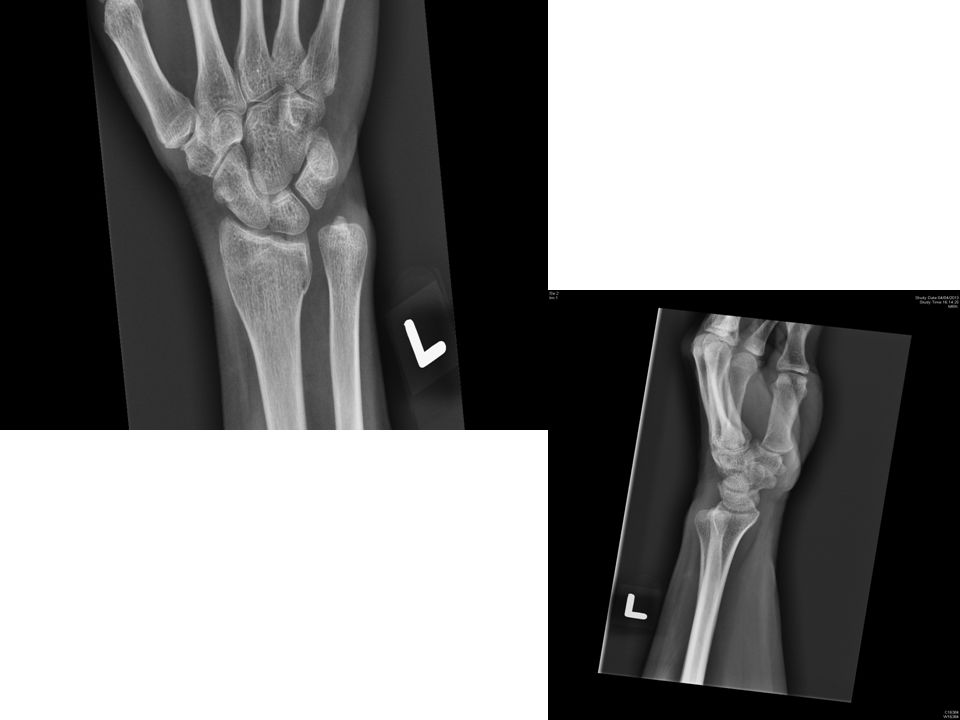
Case 2 F/53 PHx: Good S/F resulting in left wrist painful swelling
X-ray left wrist done

15
Question Suggest 2 x-ray abnormalities.
Dorsal distal RUJ dislocation, fracture left distal radius What is the major pathophysiology of the deformity shown on the x-ray? Damage to TFCC (triangular fibrocartilage complex)
")
16
Suggest 1 image modality helpful to further delineate the injury
CT (to evaluate suspected fracture, degenerative change, DRUJ subluxation) What is the management? CR with digital pressure on ulnar head and supination, followed by long arm splint if position is stable; ORIF if unstable or irreducible
 What is the management CR with digital pressure on ulnar head and supination, followed by long arm splint if position is stable; ORIF if unstable or irreducible.")
17
Case 3 M/22 PHx: Good c/o palpitation
Vital signs stable except tachycardia ECG done

18
ECG

19
Question What are the ECG findings? What is the diagnosis?
Regular WCT with QRS 0.14s, RBBB, LAD What is the diagnosis? Fascicular VT Which class of drug should be used to terminate the rhythm? Calcium channel blocker (e.g. Verapamil, Herbesser)
")
20
What is the underlying pathophysiology of the rhythm?
Calcium-dependent re-entry circuit at Purkinje fibers within LV Suggest 3 more investigations helpful to delineate the diagnosis Cardiac MRI, Echo, Electrophysiology Study What is the definitive treatment? Radio-frequency ablation

21
After Herbesser

22
Fascicular VT The 2nd most common idiopathic VT
Due to re-entry circuit within left ventricle Most episodes occur at rest, but may be triggered by stress or exercise May be misdiagnosed as SVT with RBBB Treatment: Calcium channel blocker (ATP/ vagal maneuver ineffective)
")
23
Case 4 F/41 Good past health, on OCP
Brought to AED after 1 episode of convulsion Regained consciousness few minutes later c/o headache in the past few days P/E: Fully conscious, no focal neurological sign, vital signs stable, h’stix 6.8 No scalp wound Another episode of convulsion at AED, lasting for 1 minute

24
Case 4

25
Question Name 2 differential diagnosis.
ICH/ Cerebral sinus thrombosis Name 1 investigation to confirm the diagnosis CT venogram/ MRI

26
Suggest 2 risk factors. What is the treatment?
Risk factors include: thrombophilia, nephrotic syndrome, pregnancy, OCP, infection (e.g. meningitis/ mastoiditis), chronic inflammatory diseases What is the treatment? Anticoagulation/ (Thrombolytic therapy may be considered if anticoagulation fails + deteriorating condition)
, chronic inflammatory diseases. What is the treatment Anticoagulation/ (Thrombolytic therapy may be considered if anticoagulation fails + deteriorating condition)")
27
CT venogram

28
Cerebral vein thrombosis
Thrombosis of the dural sinuses Frequently involving: Sup. Sagittal sinus, transverse/sigmoid sinus and cavernous sinus Symptoms: Headache, visual loss, convulsion, weakness/focal deficits

29
Cord Sign homogeneous, hyperattenuating, cordlike appearance on a unenhanced transverse computed tomographic (CT) scan of the brain.
 scan of the brain.")
30
Dense Triangular Sign

31
Empty Delta Sign

32
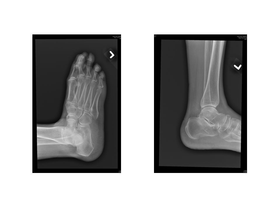
Case 5 F/83 PHx: Dementia Found painful swelling and bruise at left foot for 2 days, after falling from bed X-ray done

34
Question Suggest 2 radiological abnormalities.
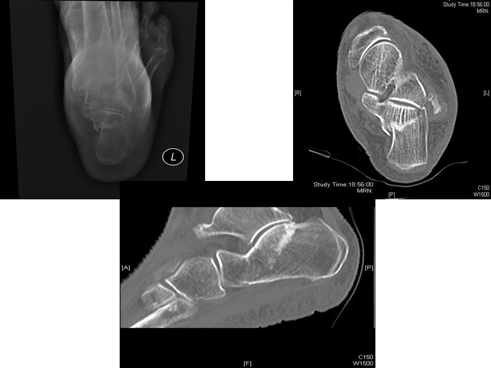
Fracture calcaneum/ Depressed Bohler angle/ Widened Gissane angle What other x-ray view can be done to confirm diagnosis? Axial view of calcaneum Suggest 1 investigation that helps confirming diagnosis and guiding management Plain CT scan

35
Name 1 classification based on this imaging modality.
Sanders classification Suggest 3 indications of operative management of this injury Displaced or comminuted intra-articular fracture/ displaced posterior avulsion fracture/ open fracture/ fracture-dislocation/ displaced fracture of calcaneal tuberosity

37
THE END

Similar presentations


 58% Cardiac Disease (arrhythmias) 23% Neurologic or.>")


 Heart Blocks>")


