Download presentation
Presentation is loading. Please wait.

1
Nonlinear dynamics and generalized synchronization: clinical applications in epilepsy and dementia
C.J. Stam Department of clinical neurophysiology VU University Medical Center Amsterdam Oscillations and Instability; control, near and far from equilibrium in biology Leiden,

2
Nonlinear dynamics and generalized synchronization: clinical applications in epilepsy and dementia
Introduction Functional connectivity Synchronization likelihood Applications Seizure detection Cognition Normal disturbed Small-world networks in Alzheimer’s disease

3
Mechanisms of higher brain functions (cognition)
The brain shows local specialization Complex tasks require cooperation between multiple brain areas Synchronization is a key mechanism for functional integration Synchronization results in the formation of functional networks with temporal and spatial structure

4
Functional integration in the brain:
- synchronous networks (‘binding’) - dynamic changes Cognitive dysfunction: ‘breakdown of binding’ tijd
 - dynamic changes. Cognitive dysfunction: ‘breakdown of binding’ tijd.")
5
? ‘Functional connectivity’
How do distributed systems in the brain integrate their activity under normal and pathological conditions? A ? B ‘Functional connectivity’ Dynamics of Synchronization: Diminished: Dysconnection / Cognitive dysfunction Excessive: seizures Normal: ‘fragile binding’

6
Synchronization of oscillators
Christiaan Huygens /

7
Synchronization: Adjustment of rhythms of (self sustained)
oscillating objects through weak interactions

8
Synchronization of chaotic oscillators
Complete / identical synchronization Synchronization of chaos refers to a process wherein two (or many) systems (either equivalent or nonequivalent) adjust a given property of their motion to a common behavior due to a coupling or to a forcing (periodical or noisy) S. Boccaletti e.a. Physics reports 2002; 366: (intermittent) lag synchronization (intermittent) phase synchronization Generalized synchronization
 systems (either equivalent or nonequivalent) adjust a given property of their motion to a common behavior due to a coupling or to a forcing (periodical or noisy) S. Boccaletti e.a. Physics reports 2002; 366: (intermittent) lag synchronization. (intermittent) phase synchronization. Generalized synchronization.")
9
Characterization of interdependencies between time series

10
Synchronization likelihood: an unbiased measure of generalized synchronization in multivariate data sets C.J. Stam1, B.W. van Dyk2 Physica D, 2002; 163: 1 department of clinical neurophysiology, VU University Medical Centre 2 MEG Centre, VU University Medical Centre

11
time-delay embedding L L x(t) x(t+L) x(t+2*L) x(t+2*L) x(t+L) x(t)
Time series x(t) x(t+L) x(t+2*L) x(t+2*L) Trajectory in state space x(t+L) x(t)
 x(t+L) x(t+2*L) x(t+2*L) Trajectory in state space. x(t+L) x(t)")
12
Generalized synchronization
State of the response system Is a (non linear) function of the state of the driver system X Y Y=F(X)
 function of the state of the driver system. X. Y. Y=F(X)")
13
Synchronization likelihood
Measure of the synchronization between two signals X Y Y=F(X)
")
14
Synchronization likelihood
SL between X and Y at time i is the likelihood that Ya,b resembles Yi, given that Xa,b resembles Xi Xi Xa Xb X Yi Ya Yb Y t=i

15
Synchronization likelihood
rx X Xi Pref = ry Yi Y SL =

16
Nonlinearly coupled non-identical Henon systems

17
Linear and nonlinear components of coupling:
multichannel surrogate data testing

18
The influence of different noise levels
on synchronization estimate

19
Bias in synchronization estimates due to filtering
5 Hz low pass unfiltered

22
Nonlinear dynamics and generalized synchronization: clinical applications in epilepsy and dementia
Introduction Functional connectivity Synchronization likelihood Applications Seizure detection Cognition Normal disturbed Small-world networks in Alzheimer’s disease

23
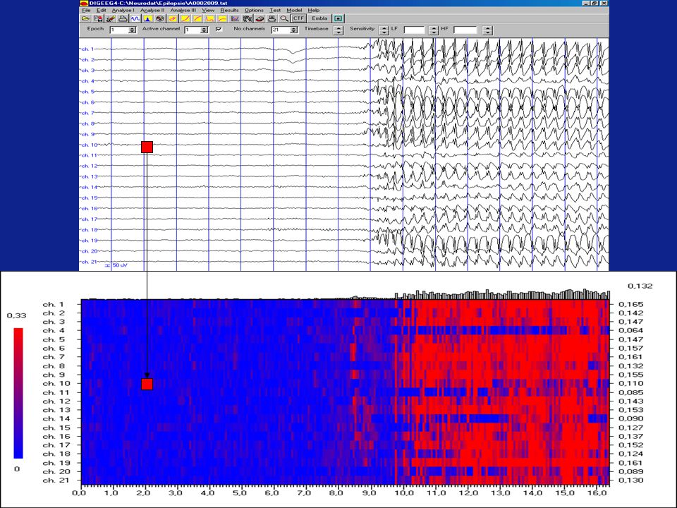
Seizure detection in the neonatal intensive care unit
Seizure occur frequently in neurologically compromized neonates Up to 85% of the seizures are subclinical Current methods for seizure detection have limitations: Gotman CFM

25
Seizure detection in neonates with synchronization likelihood
Altenburg et al., Clin Neurophysiol. 2003;114: 50-5. Smit et al., Neuropediatrics 2004; 35: 1-7.

26
Towne et al., Neurology 2000 236 coma patients
no clinical symptoms of seizures EEG: 8% of these patients is in non convulsive status epilepticus (NCSE) NCSE: “silent epidemic” in intensive care patients
 NCSE: silent epidemic in intensive care patients.")
27
oogknipperen

28
propofol

29
Visual Working Memory Task Response: items remembered

30
synchronization likelihood during retention interval:
increase in 2-6 Hz synchronization decrease of 6-10 Hz synchronization 2-6 Hz: “theta” working memory 6-10 Hz: lower alpha attention

31
Changes in synchronization entropy during working memory task

32
Nonlinear synchronization in EEG and whole-head MEG recordings of healthy subjects Stam CJ, Breakspear M, van Cappellen van Walsum AM, van Dijk BW. Human Brain Mapping 2003; 19:

33
Alzheimer’s disease: a dysconnection syndrome?

34
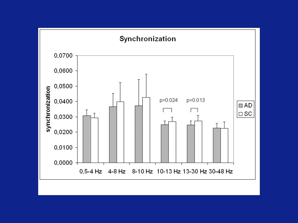
Generalized synchronization in Alzheimer’s disease
Subjects: 20 AD patients MMSE: 21.3 20 healthy controls Recording: 151 channel MEG Condition: eyes closed, no task

36
Control gamma band (20-50 Hz)
synchronous neural networks

37
Alzheimer gamma band (20-50 Hz)
")
38
Dynamics of functional connectivity in Alzheimer’s disease
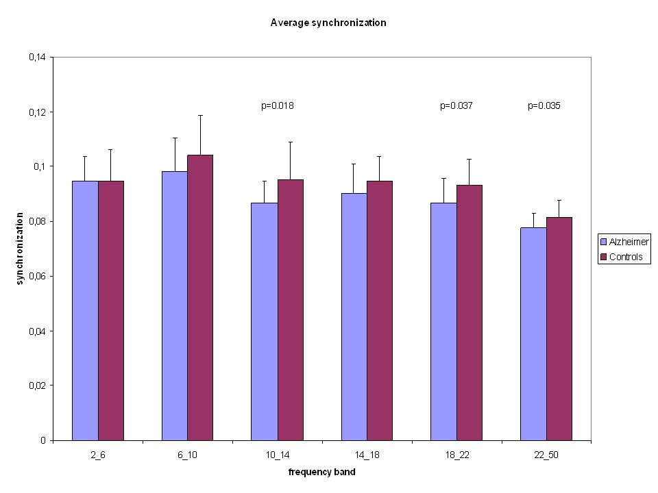
Alzheimer patients (N = 24) Control subjects (N = 19) 21 channel EEG, no-task, eyes-closed Synchronization likelihood: mean level of synchronization Synchronization rate: rate of change of synchronization * * * *
 Control subjects (N = 19) 21 channel EEG, no-task, eyes-closed. Synchronization likelihood: mean level of synchronization. Synchronization rate: rate of change of synchronization. * * * *")
39
Dynamics of functional connectivity Control subject Alzheimer patient

40
Are fluctuations of global synchronization levels scale-free?

41
Detrended fluctuation analysis (DFA)
Plot of Log(fluctuation) / Log(timescale) Time series integration Fluctuation at timescale t Scaling (self similarity) exponent: slope of linear fit through Log(fluctuation) / Log(timescale)
 / Log(timescale) Time series. integration. Fluctuation. at. timescale t. Scaling (self similarity) exponent: slope of linear fit through. Log(fluctuation) / Log(timescale)")
42
Detrended fluctuation analysis of synchronization likelihood
SL 8-13 Hz DFA 8-13 Hz SL Hz DFA Hz

43
Detrended fluctuation analysis

44
Disturbed fluctuations of resting state EEG synchronization in Alzheimer’s disease
C.J. Stam, T. Montez, B.F. Jones, S.A.R.B. Rombouts, Y. van der Made, Y.A.L. Pijnenburg, Ph. Scheltens Clin Neurophysiol, 2005; 116:

47
Interim conclusions: Results so far: Questions:
Synchronisation analysis can detect and characterize functional networks Networks change: Cognitive tasks Brain pathology Questions: What is an ‘optimal’ network? How can we detect / characterize an optimal network?

48
Nonlinear dynamics and generalized synchronization: clinical applications in epilepsy and dementia
Introduction Functional connectivity Synchronization likelihood Applications Seizure detection Cognition Normal disturbed Small-world networks in Alzheimer’s disease

49
How to analyze a complex system as the brain?
Graph theory Chaos theory Information theory Self-organized criticality

50
The ‘Kevin Bacon’ game

51
Cp: Cluster coefficient Lp: Pathlength : vertex : edge
Fig. 1 Graph F E D C A B Cp: Cluster coefficient Lp: Pathlength : vertex : edge

52
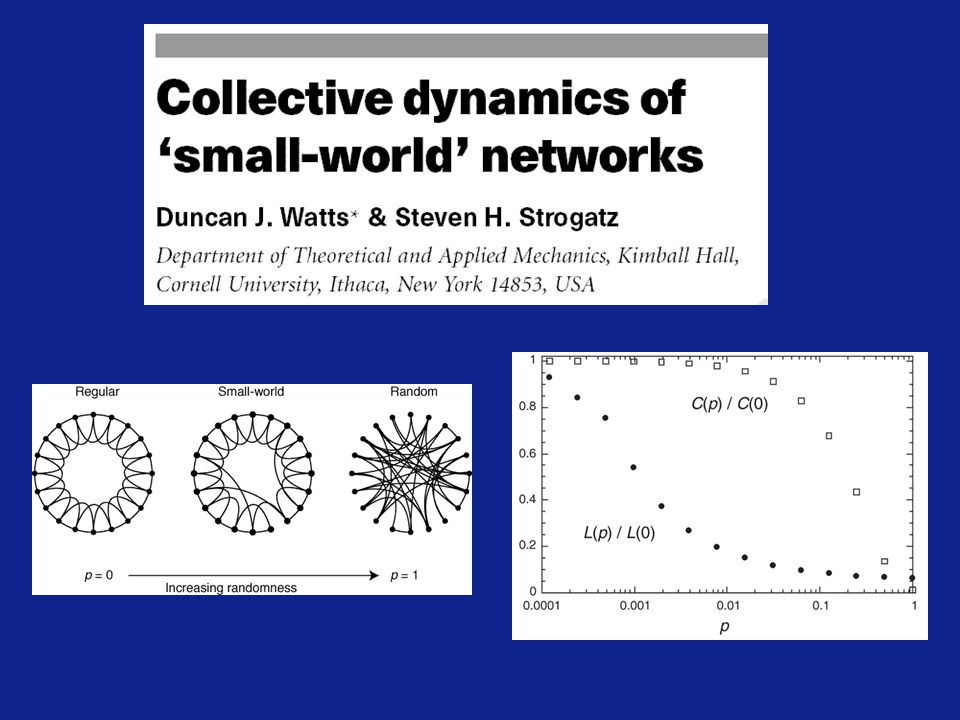
The enigma of the ‘small-world’ phenomenon
Most networks are sparsely connected Most connections are local (high Cluster coefficient) The distance between any two network elements is small: how is this possible? Example: 1011 neurons 104 synapses / neuron Typically any two neurons are only 2 to 3 synapses away
 The distance between any two network elements is small: how is this possible Example: 1011 neurons. 104 synapses / neuron. Typically any two neurons are only 2 to 3 synapses away.")
54
‘small-world’ networks:
High cluster coefficient C Short path length L Realistic model real complex networks ‘optimal configuration’: Sparse connectivity Maximal communication between all parts of the network Balance local specialisation / global integration

55
Neuro anatomical networks:
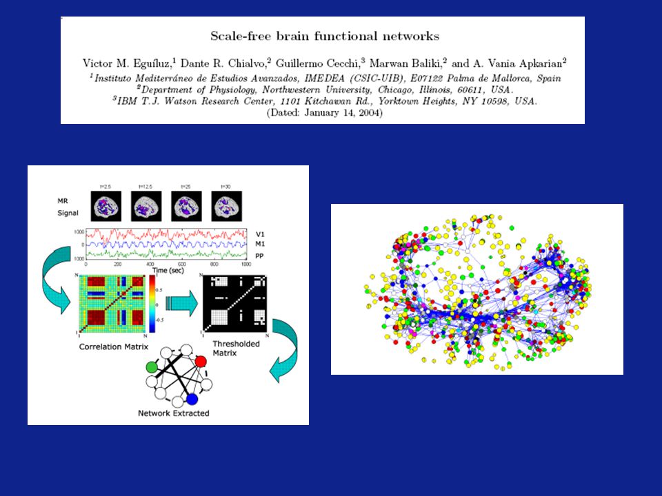
Experimental evidence for the existence of ‘small-world’ networks in the brain: Neuro anatomical networks: C. Elegans (Watts and Strogatz, 1998) Visual cortex cat (Scannell et al., 1994) Animal model / database (Hilgetag et al., 2000) Functional neural networks: Animal model / strychnine (Stephan et al., 2000) fMRI (Dodel et al., 2002; Eguiluz et al., 2004) MEG (Stam, 2004)
 Visual cortex cat (Scannell et al., 1994) Animal model / database (Hilgetag et al., 2000) Functional neural networks: Animal model / strychnine (Stephan et al., 2000) fMRI (Dodel et al., 2002; Eguiluz et al., 2004) MEG (Stam, 2004)")
56
C/Crandom = 2.08 L/Lrandom = 1.09

59
Questions: Is it possible to detect functional networks with EEG ?
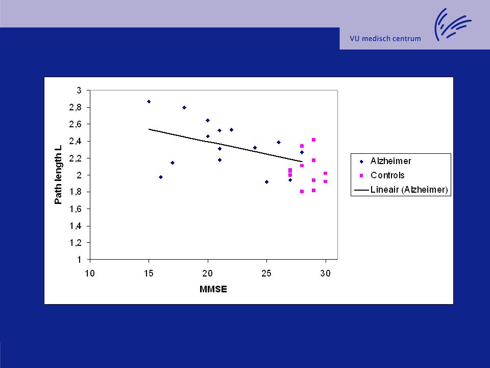
Can these networks be characterized with graph theoretical measures? What changes occur in Alzheimer’s disease ? Loss of ‘clustering’ (cluster coefficient C) ? Loss of ‘integration’ (path length L) ? How does this correlate with cognitive dysfunction ?
 Loss of ‘integration’ (path length L) How does this correlate with cognitive dysfunction")
60
‘Small-world’ networks in Alzheimer’s disease
69.6 (7.9) MMSE = 21.4 (4.0) Controls (subjective complaints) N = 13 70.6 (7.7) MMSE = 28.4 (1.1) EEG 21 channels Beta band (13-30 Hz) Rest / eyes closed
 MMSE = 21.4 (4.0) Controls (subjective complaints) N = (7.7) MMSE = 28.4 (1.1) EEG. 21 channels. Beta band (13-30 Hz) Rest / eyes closed.")
62
Application of graph analysis to EEG:
1 2 3 4 C threshold L

63
Synchronization matrix
Alzheimer patients Control subjects

64
Synchronization matrix converted to ‘graph’
Alzheimer patients Control subjects

67
Graph splitting and fragmentation
B C T=0.029 T=0.034 T=0.045 Fully connected Splitting off Fragmentation

69
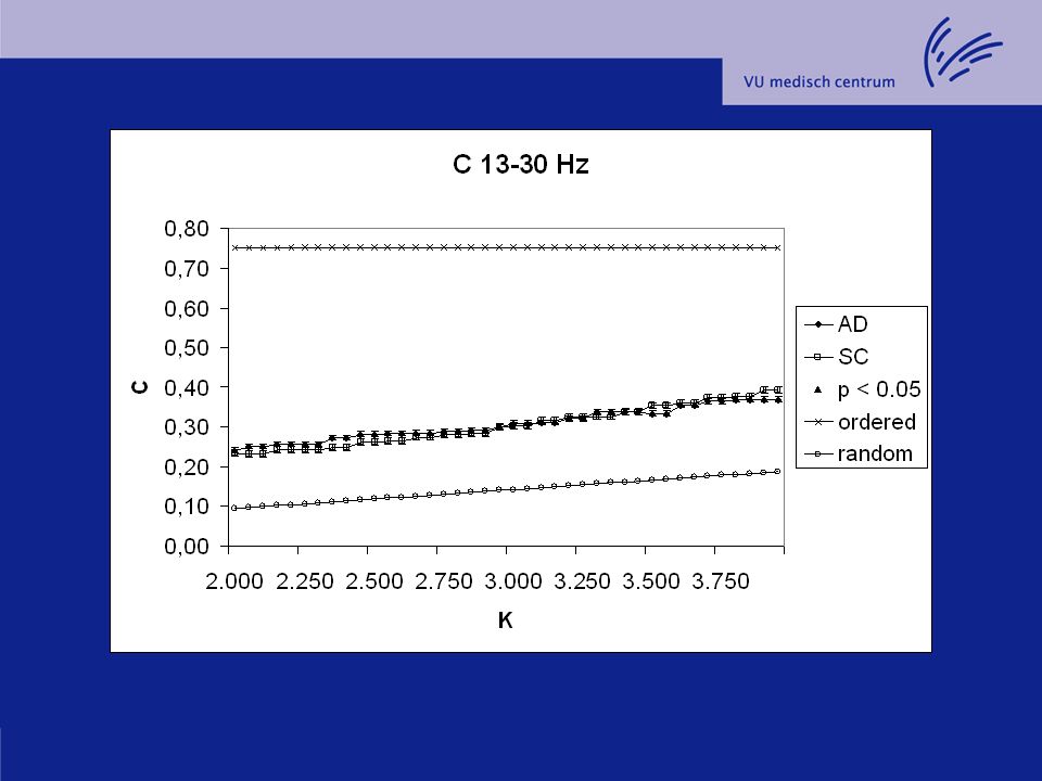
Problem: Mean synchronisation is lower in AD than controls
Applying the same threshold means that AD networks will have less connections Increased path length in Ad might be a trivial consequence of the smaller number of supra threshold connections Solution: compute C and L as a function of K (edges / vertex)
")
70
Networks Normalized for K (edges / vertex)
Alzheimer patients Control subjects

73
‘small-world’ networks?
C/Crandom L/Lrandom Present study AD 1.93 0.97 * Controls 2.13 0.89 Stam, 2004 1.89 1.19 Salvador, 2005 2.08 1.09 Hilgetag, 2000 Macaque visual ctx 1.85 1.02 Cat whole ctx 1.99 1.07 Watts & Strogatz, 1998 C. Elegans 5.6 1.18

74
Conclusions: Synchronization likelihood analysis can track ‘fragile binding’ in EEG and MEG Healthy subjects: Frequency specific changes in synchronization in working memory task Scale-free fluctuations of SL Alzheimer patients: Lower synchronization Disturbed fluctuations of SL Disturbed spatial patterns

75
Acknowledgements: Afdeling KNF Afdeling neurologie MEG centrum
R.L.M. Strijers E.M. Vriens H.E. Ronner W. de Rijke L.S. Smit laboranten Afdeling neurologie H.W. Berendse Y.A.L. Pijnenburg Ph. Scheltens M.C. Visser MEG centrum B.W. van Dijk T. Montez J.C. de Munck J. Verbunt K. Cover Kinderneurologie R.J. Vermeulen J. Altenburg Neonatale IC W.P.F. Fetter Intensive care A.R.J. Girbes J.J. Spijkstra Neurochirurgie W.P. VanderTop UMC F.S.S. Leijten W Spetgens Overige R. Ferri S. Micheloyannis M. Breakspear G. Nolte J. Terry

Similar presentations


 signal using different mathematical models in Matlab to predict.>")


