Download presentation
Presentation is loading. Please wait.

1
CHAPTER 17 FROM GENE TO PROTEIN
Section A: The Connection Between Genes and Proteins 1. The study of metabolic defects provided evidence that genes specify proteins 2. Transcription and translation are the two main processing linking gene to protein: an overview 3. In the genetic code, nucleotide triplets specify amino acids 4. The genetic code must have evolved very early in the history of life Copyright © 2002 Pearson Education, Inc., publishing as Benjamin Cummings

2
Transcription and Translation
What do you know already? List steps of Trxn and Tlxn in order (manipulative)
")
7
transcribe and translate game

8
OVERVIEW of the following process:
transcription translation video

9
SO….. We finally figured out that DNA was the inheritable material
BUT….what does it do AND How is it related to protein that actually governs your characteristics?

10
Genes indirectly control phenotype through enzymes
Gerrod Genes indirectly control phenotype through enzymes

11
Introduction The information content of DNA is in the form of specific sequences of nucleotides along the DNA strands. The DNA inherited by an organism leads to specific traits by dictating the synthesis of proteins. Proteins are the links between genotype and phenotype. For example, Mendel’s dwarf pea plants lack a functioning copy of the gene that specifies the synthesis of a key protein, gibberellins. Gibberellins stimulate the normal elongation of stems. Copyright © 2002 Pearson Education, Inc., publishing as Benjamin Cummings

12
1. The study of metabolic defects provided evidence that genes specify proteins
In 1909, Archibald Gerrod was the first to suggest that genes dictate phenotype through enzymes that catalyze specific chemical reactions in the cell. The symptoms of an inherited disease reflect a person’s inability to synthesize a particular enzyme. Gerrod speculated that alkaptonuria, a hereditary disease, was caused by the absence of an enzyme that breaks down a specific substrate, alkapton. Research conducted several decades later supported Gerrod’s hypothesis. Copyright © 2002 Pearson Education, Inc., publishing as Benjamin Cummings

13
Beadle and Tatum ONE gene-One enzyme

14
Progress in linking genes and enzymes rested on the growing understanding that cells synthesize and degrade most organic molecules in a series of steps, a metabolic pathway. In the 1930s, George Beadle and Boris Ephrussi speculated that each mutation affecting eye color in Drosophila blocks pigment synthesis at a specific step by preventing production of the enzyme that catalyzes that step. However, neither the chemical reactions nor the enzymes were known at the time. Copyright © 2002 Pearson Education, Inc., publishing as Benjamin Cummings

15
Beadle and Edward Tatum were finally able to establish the link between genes and enzymes in their exploration of the metabolism of a bread mold, Neurospora crassa. They mutated Neurospora with X-rays and screened the survivors for mutants that differed in their nutritional needs. Wild-type Neurospora can grow on a minimal medium of agar, inorganic salts, glucose, and the vitamin biotin. Most nutritional mutants can survive on a complete growth medium which includes all 20 amino acids. Copyright © 2002 Pearson Education, Inc., publishing as Benjamin Cummings

16
One type of mutant required only the addition of arginine to the minimal growth medium.
Beadle and Tatum concluded that this mutant was defective somewhere in the biochemical pathway that normally synthesizes arginine. They identified three classes of arginine deficient mutants, each apparently lacking a key enzyme at a different step in the synthesis of arginine. They demonstrated this by growing these mutant strains in media that provided different intermediate molecules. Their results provided strong evidence for the one gene - one enzyme hypothesis. Copyright © 2002 Pearson Education, Inc., publishing as Benjamin Cummings

17
Fig. 17.1 Copyright © 2002 Pearson Education, Inc., publishing as Benjamin Cummings

18
Later research refined the one gene - one enzyme hypothesis.
First, it became clear that not all proteins are enzymes and yet their synthesis depends on specific genes. This tweaked the hypothesis to one gene - one protein. Copyright © 2002 Pearson Education, Inc., publishing as Benjamin Cummings

19
Later research demonstrated that many proteins are composed of several polypeptides, each of which has its own gene. Therefore, Beadle and Tatum’s idea has been restated as the one gene - one polypeptide hypothesis.

20
Genes provide the instructions for making specific proteins.
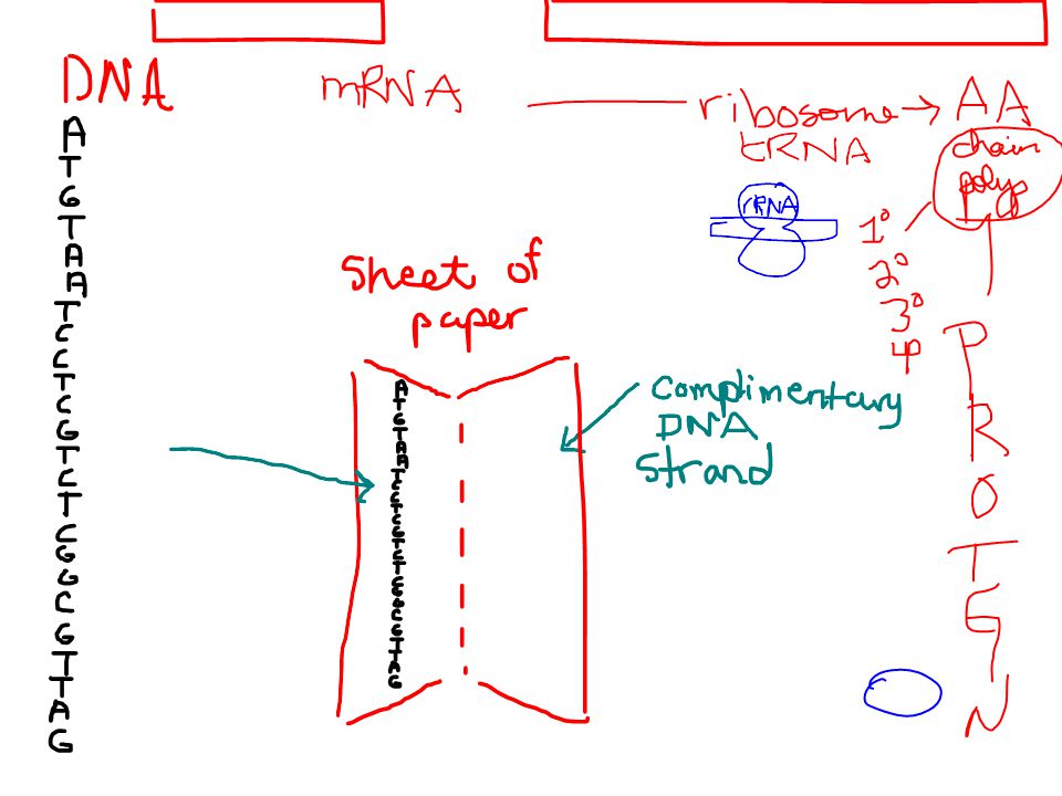
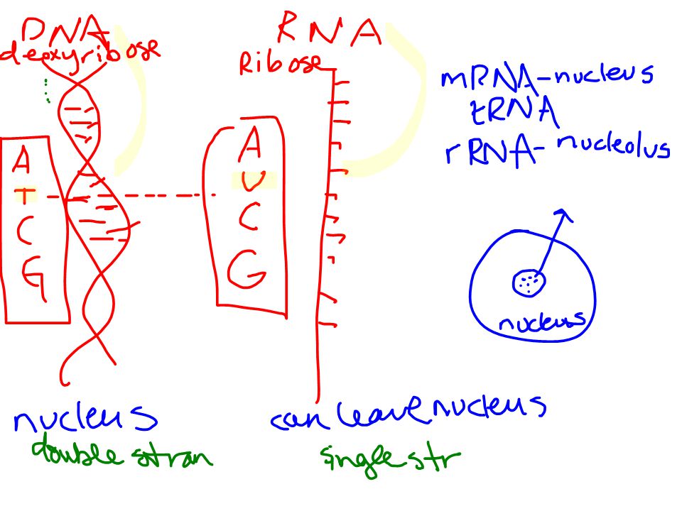
2. Transcription and translation are the two main processes linking gene to protein: an overview Genes provide the instructions for making specific proteins. The bridge between DNA and protein synthesis is RNA. RNA is chemically similar to DNA, except that it contains ribose as its sugar and substitutes the nitrogenous base uracil for thymine. An RNA molecules almost always consists of a single strand. Copyright © 2002 Pearson Education, Inc., publishing as Benjamin Cummings

21
Structure of RNA- what do you know

23
In DNA or RNA, the four nucleotide monomers act like the letters of the alphabet to communicate information. The specific sequence of hundreds or thousands of nucleotides in each gene carries the information for the primary structure of a protein, the linear order of the 20 possible amino acids. To get from DNA, written in one chemical language, to protein, written in another, requires two major stages, transcription and translation. Copyright © 2002 Pearson Education, Inc., publishing as Benjamin Cummings

24
Fill out chart as we go Rep/Transcrip/Transla Location Start molecule
Process End molecule

25
Transcription and Translation
What happens? Where does it happen? When does it happen?

26
Transcription of a gene produces a messenger RNA (mRNA) molecule.
During transcription, a DNA strand provides a template for the synthesis of a complementary RNA strand-in the nucleus This process is used to synthesize any type of RNA from a DNA template. Transcription of a gene produces a messenger RNA (mRNA) molecule. Copyright © 2002 Pearson Education, Inc., publishing as Benjamin Cummings
 molecule. Copyright © 2002 Pearson Education, Inc., publishing as Benjamin Cummings.")
27
During translation, the information contained in the order of nucleotides in mRNA is used to determine the amino acid sequence of a polypeptide. Translation occurs at ribosomes.

28
Prokaryotes and Eukaryotes perform the process differently
WHY? HOW?

29
The basic mechanics of transcription and translation are similar in eukaryotes and prokaryotes.
Because bacteria lack nuclei, transcription and translation are coupled. Ribosomes attach to the leading end of a mRNA molecule while transcription is still in progress. Fig. 17.2a Copyright © 2002 Pearson Education, Inc., publishing as Benjamin Cummings

30
In a eukaryotic cell, almost all transcription occurs in the nucleus and translation occurs mainly at ribosomes in the cytoplasm. In addition, before the primary transcript can leave the nucleus it is modified in various ways during RNA processing before the finished mRNA is exported to the cytoplasm. Fig. 17.2b Copyright © 2002 Pearson Education, Inc., publishing as Benjamin Cummings

31
The molecular chain of command in a cell is DNA is the template for
To summarize, genes program protein synthesis via genetic messenger RNA. The molecular chain of command in a cell is DNA is the template for m RNA which is the triplet code for the amino acid sequence in a Polypeptide chain which folds into or combines with other chains to make a protein. Copyright © 2002 Pearson Education, Inc., publishing as Benjamin Cummings

32
ACTUALLY ISNT DNA (PER SE)
THE CODE ACTUALLY ISNT DNA (PER SE)
")
33
3. In the genetic code, nucleotide triplets specify amino acids
If the genetic code consisted of a single nucleotide or even pairs of nucleotides per amino acid, there would not be enough combinations (4 and 16 respectively) to code for all 20 amino acids. Triplets of nucleotide bases are the smallest units of uniform length that can code for all the amino acids. In the triplet code, three consecutive bases specify an amino acid, creating 43 (64) possible code words. The genetic instructions for a polypeptide chain are written in DNA as a series of three-nucleotide words. Copyright © 2002 Pearson Education, Inc., publishing as Benjamin Cummings
 to code for all 20 amino acids. Triplets of nucleotide bases are the smallest units of uniform length that can code for all the amino acids. In the triplet code, three consecutive bases specify an amino acid, creating 43 (64) possible code words. The genetic instructions for a polypeptide chain are written in DNA as a series of three-nucleotide words. Copyright © 2002 Pearson Education, Inc., publishing as Benjamin Cummings.")
34
During transcription, one DNA strand, the template strand, provides a template for ordering the sequence of nucleotides in an RNA transcript. The complementary RNA molecule is synthesized according to base-pairing rules, except that uracil is the complementary base to adenine. During translation, blocks of three nucleotides, codons, are decoded into a sequence of amino acids. Fig. 17.3 Copyright © 2002 Pearson Education, Inc., publishing as Benjamin Cummings

36
During translation, the codons are read in the 5’->3’ direction along the mRNA.
Each codon specifies which one of the 20 amino acids will be incorporated at the corresponding position along a polypeptide. Because codons are base triplets, the number of nucleotides making up a genetic message must be three times the number of amino acids making up the protein product. It would take at least 300 nucleotides to code for a polypeptide that is 100 amino acids long. Copyright © 2002 Pearson Education, Inc., publishing as Benjamin Cummings

37
Nirenberg THE code

38
The task of matching each codon to its amino acid counterpart began in the early 1960s.
Marshall Nirenberg determined the first match, that UUU coded for the amino acid phenylalanine. He created an artificial mRNA molecule entirely of uracil and added it to a test tube mixture of amino acids, ribosomes, and other components for protein synthesis. This “poly(U)” translated into a polypeptide containing a single amino acid, phenyalanine, in a long chain. Other more elaborate techniques were required to decode mixed triplets such a AUA and CGA. Copyright © 2002 Pearson Education, Inc., publishing as Benjamin Cummings
 translated into a polypeptide containing a single amino acid, phenyalanine, in a long chain. Other more elaborate techniques were required to decode mixed triplets such a AUA and CGA. Copyright © 2002 Pearson Education, Inc., publishing as Benjamin Cummings.")
39
By the mid-1960s the entire code was deciphered.
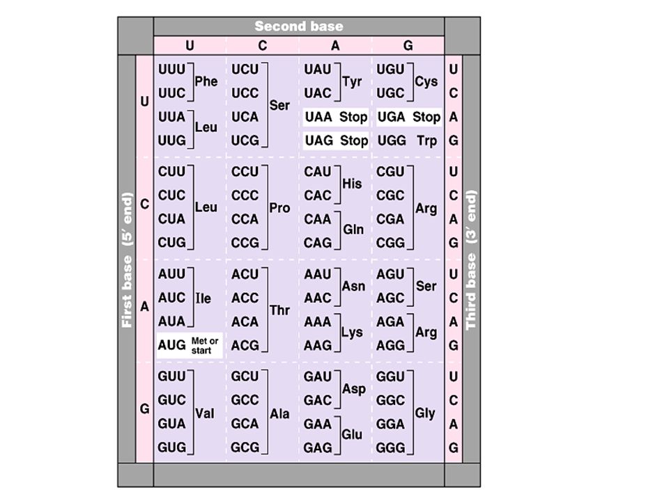
61 of 64 triplets code for amino acids. The codon AUG not only codes for the amino acid methionine but also indicates the start of translation. Three codons do not indicate amino acids but signal the termination of translation. Fig. 17.4 Copyright © 2002 Pearson Education, Inc., publishing as Benjamin Cummings

41
The genetic code is redundant but not ambiguous.
There are typically several different codons that would indicate a specific amino acid. However, any one codon indicates only one amino acid. [If you have a specific codon, you can be sure of the corresponding amino acid, but if you know only the amino acid, there may be several possible codons.] Both GAA and GAG specify glutamate, but no other amino acid. Codons synonymous for the same amino acid often differ only in the third codon position. Copyright © 2002 Pearson Education, Inc., publishing as Benjamin Cummings

42
To extract the message from the genetic code requires specifying the correct starting point.
This establishes the reading frame and subsequent codons are read in groups of three nucleotides. The cell’s protein-synthesizing machinery reads the message as a series of nonoverlapping three-letter words. In summary, genetic information is encoded as a sequence of nonoverlapping base triplets, or codons, each of which is translated into a specific amino acid during protein synthesis. Copyright © 2002 Pearson Education, Inc., publishing as Benjamin Cummings

43
Is every organism’s code the same?
If I put a fruit fly DNA sequence into a plant and transcribe it..will I get the same protein?

44
4. The genetic code must have evolved very early in the history of life
The genetic code is nearly universal, shared by organisms from the simplest bacteria to the most complex plants and animals. In laboratory experiments, genes can be transcribed and translated after they are transplanted from one species to another. This tobacco plant is expressing a transpired firefly gene. Fig. 17.5 Copyright © 2002 Pearson Education, Inc., publishing as Benjamin Cummings

45
glow in dark bunny

46
This has permitted bacteria to be programmed to synthesize certain human proteins after insertion of the appropriate human genes. This and other similar applications are exciting developments in biotechnology. Exceptions to the universality of the genetic code exist in translation systems where a few codons differ from standard ones. These occur in certain single-celled eukaryotes like Paramecium. Other examples include translation in certain mitochondria and chloroplasts. Copyright © 2002 Pearson Education, Inc., publishing as Benjamin Cummings

47
The near universality of the genetic code must have been operating very early in the history of life. A shared genetic vocabulary is a reminder of the kinship that bonds all life on Earth. Copyright © 2002 Pearson Education, Inc., publishing as Benjamin Cummings

48
CHAPTER 17 FROM GENE TO PROTEIN
Section B: The Synthesis and Processing of RNA 1. Transcription is the DNA-directed synthesis of RNA: a closer look 2. Eukaryotic cells modify RNA after transcription Copyright © 2002 Pearson Education, Inc., publishing as Benjamin Cummings

49
1. Transcription is the DNA-directed synthesis of RNA: a closer look
Messenger RNA is transcribed from the template strand of a gene. RNA polymerase separates the DNA strands at the appropriate point and bonds the RNA nucleotides as they base-pair along the DNA template. Like DNA polymerases, RNA polymerases can add nucleotides only to the 3’ end of the growing polymer. Genes are read 3’->5’, creating a 5’->3’ RNA molecule. Copyright © 2002 Pearson Education, Inc., publishing as Benjamin Cummings

50
3’ to 5’ read gene

51
Where is the gene?

52
Specific sequences of nucleotides along the DNA mark where gene transcription begins and ends.
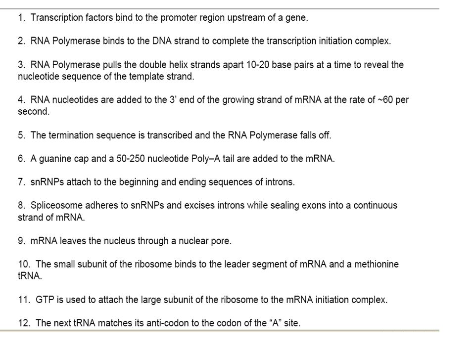
RNA polymerase attaches and initiates transcription at the promotor, “upstream” of the information contained in the gene, the transcription unit. The terminator signals the end of transcription. Bacteria have a single type of RNA polymerase that synthesizes all RNA molecules. In contrast, eukaryotes have three RNA polymerases (I, II, and III) in their nuclei. RNA polymerase II is used for mRNA synthesis. Copyright © 2002 Pearson Education, Inc., publishing as Benjamin Cummings
 in their nuclei. RNA polymerase II is used for mRNA synthesis. Copyright © 2002 Pearson Education, Inc., publishing as Benjamin Cummings.")
53
3 stages of Transcription
Initiation Elongation Termination

54
Fig. 17.6a Copyright © 2002 Pearson Education, Inc., publishing as Benjamin Cummings

55
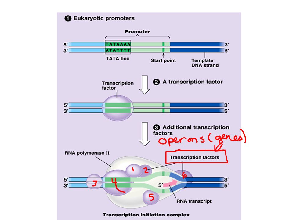
INITIATION The presence of a promotor sequence determines which strand of the DNA helix is the template. Within the promotor is the starting point for the transcription of a gene. The promotor also includes a binding site for RNA polymerase several dozen nucleotides upstream of the start point. In prokaryotes, RNA polymerase can recognize and bind directly to the promotor region. Copyright © 2002 Pearson Education, Inc., publishing as Benjamin Cummings

56
RNA polymerase then starts transcription.
In eukaryotes, proteins called transcription factors recognize the promotor region, especially a TATA box, and bind to the promotor. After they have bound to the promotor, RNA polymerase binds to transcription factors to create a transcription initiation complex. RNA polymerase then starts transcription. Fig. 17.7 Copyright © 2002 Pearson Education, Inc., publishing as Benjamin Cummings

58
ELONGATION

59
The enzyme adds nucleotides to the 3’ end of the growing strand.
As RNA polymerase moves along the DNA, it untwists the double helix, 10 to 20 bases at time. The enzyme adds nucleotides to the 3’ end of the growing strand. Behind the point of RNA synthesis, the double helix re-forms and the RNA molecule peels away. Fig. 17.6b Copyright © 2002 Pearson Education, Inc., publishing as Benjamin Cummings

60
A growing strand of RNA trails off from each polymerase.
A single gene can be transcribed simultaneously by several RNA polymerases at a time. A growing strand of RNA trails off from each polymerase. The length of each new strand reflects how far along the template the enzyme has traveled from the start point. The congregation of many polymerase molecules simultaneously transcribing a single gene increases the amount of mRNA transcribed from it. This helps the cell make the encoded protein in large amounts. Copyright © 2002 Pearson Education, Inc., publishing as Benjamin Cummings

61
termination

62
Transcription proceeds until after the RNA polymerase transcribes a terminator sequence in the DNA.
In prokaryotes, RNA polymerase stops transcription right at the end of the terminator. Both the RNA and DNA is then released. In eukaryotes, the polymerase continues for hundreds of nucleotides past the terminator sequence, AAUAAA. At a point about 10 to 35 nucleotides past this sequence, the pre-mRNA is cut from the enzyme. Copyright © 2002 Pearson Education, Inc., publishing as Benjamin Cummings

63
FINISHING G-Cap Poly A-Tail Splicing

64
2. Eukaryotic cells modify RNA after transcription
Enzymes in the eukaryotic nucleus modify pre-mRNA before the genetic messages are dispatched to the cytoplasm. At the 5’ end of the pre-mRNA molecule, a modified form of guanine is added, the 5’ cap. This helps protect mRNA from hydrolytic enzymes. It also functions as an “attach here” signal for ribosomes. Copyright © 2002 Pearson Education, Inc., publishing as Benjamin Cummings

65
At the 3’ end, an enzyme adds 50 to 250 adenine nucleotides, the poly(A) tail.
In addition to inhibiting hydrolysis and facilitating ribosome attachment, the poly(A) tail also seems to facilitate the export of mRNA from the nucleus. The mRNA molecule also includes nontranslated leader and trailer segments. Fig. 17.8 Copyright © 2002 Pearson Education, Inc., publishing as Benjamin Cummings
 tail also seems to facilitate the export of mRNA from the nucleus. The mRNA molecule also includes nontranslated leader and trailer segments. Fig Copyright © 2002 Pearson Education, Inc., publishing as Benjamin Cummings.")
66
The most remarkable stage of RNA processing occurs during the removal of a large portion of the RNA molecule during RNA splicing. Most eukaryotic genes and their RNA transcripts have long noncoding stretches of nucleotides. Noncoding segments, introns, lie between coding regions. The final mRNA transcript includes coding regions, exons, that are translated into amino acid sequences, plus the leader and trailer sequences. Copyright © 2002 Pearson Education, Inc., publishing as Benjamin Cummings

67
Fig. 17.9 RNA splicing removes introns and joins exons to create an mRNA molecule with a continuous coding sequence. Copyright © 2002 Pearson Education, Inc., publishing as Benjamin Cummings

68
This splicing is accomplished by a spliceosome.
spliceosomes consist of a variety of proteins and several small nuclear ribonucleoproteins (snRNPs). Each snRNP has several protein molecules and a small nuclear RNA molecule (snRNA). Each is about 150 nucleotides long. Copyright © 2002 Pearson Education, Inc., publishing as Benjamin Cummings
. Each snRNP has several protein molecules and a small nuclear RNA molecule (snRNA). Each is about 150 nucleotides long. Copyright © 2002 Pearson Education, Inc., publishing as Benjamin Cummings.")
69
(2) Within the spliceosome,
(1) Pre-mRNA combines with snRNPs and other proteins to form a spliceosome. (2) Within the spliceosome, snRNA base-pairs with nucleotides at the ends of the intron. (3) The RNA transcript is cut to release the intron, and the exons are spliced together; the spliceosome then comes apart, releasing mRNA, which now contains only exons. Fig Copyright © 2002 Pearson Education, Inc., publishing as Benjamin Cummings
 Pre-mRNA combines with snRNPs and other proteins to form a spliceosome. (2) Within the spliceosome, snRNA base-pairs with nucleotides at the ends of the intron. (3) The RNA transcript is cut to release the intron, and the exons are spliced together; the spliceosome then comes apart, releasing mRNA, which now contains only exons. Fig Copyright © 2002 Pearson Education, Inc., publishing as Benjamin Cummings.")
70
In this process, the snRNA acts as a ribozyme, an RNA molecule that functions as an enzyme.
Like pre-mRNA, other kinds of primary transcripts may also be spliced, but by diverse mechanisms that do not involve spliceosomes. In a few cases, intron RNA can catalyze its own excision without proteins or extra RNA molecules. The discovery of ribozymes rendered obsolete the statement, “All biological catalysts are proteins.” Copyright © 2002 Pearson Education, Inc., publishing as Benjamin Cummings

71
The presence of introns increases the probability of potentially beneficial crossing over between genes. Introns increase the opportunity for recombination between two alleles of a gene. This raises the probability that a crossover will switch one version of an exon for another version found on the homologous chromosome. There may also be occasional mixing and matching of exons between completely different genes. Either way, exon shuffling could lead to new proteins through novel combinations of functions. Copyright © 2002 Pearson Education, Inc., publishing as Benjamin Cummings

72
CHAPTER 17 FROM GENE TO PROTEIN Section C: The Synthesis of Protein
1. Translation is the RNA-directed synthesis of a polypeptide: a closer look 2. Signal peptides target some eukaryotic polypeptides to specific destinations in the cell 3. RNA plays multiple roles in the cell: a review 4. Comparing protein synthesis in prokaryotes and eukaryotes: a review 5. Point mutations can affect protein structure and function 6. What is a gene? revisiting the question Copyright © 2002 Pearson Education, Inc., publishing as Benjamin Cummings

73
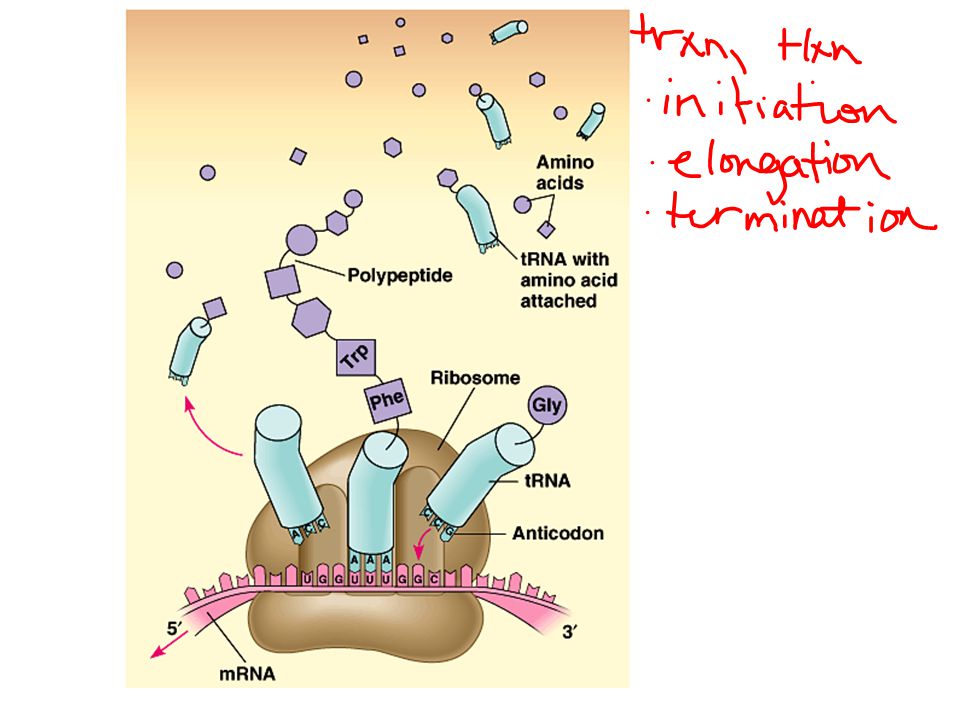
1. Translations is the RNA-directed synthesis of a polypeptide: a closer look
In the process of translation, a cell interprets a series of codons along a mRNA molecule. Transfer RNA (tRNA) transfers amino acids from the cytoplasm’s pool to a ribosome. The ribosome adds each amino acid carried by tRNA to the growing end of the polypeptide chain. Fig Copyright © 2002 Pearson Education, Inc., publishing as Benjamin Cummings
 transfers amino acids from the cytoplasm’s pool to a ribosome. The ribosome adds each amino acid carried by tRNA to the growing end of the polypeptide chain. Fig Copyright © 2002 Pearson Education, Inc., publishing as Benjamin Cummings.")
74
Translation stages Initiation Elongation Termination
FOLDING or FINISHING

76
Structure, Function, and Location
mRNA structure/location/function tRNA structure/location/function rRNA structure/location/function Amino Acids

77
Location tRNA Like other types of RNA, tRNA molecules are transcribed from DNA templates in the nucleus. Once it reaches the cytoplasm, each tRNA is used repeatedly to pick up its designated amino acid in the cytosol, to deposit the amino acid at the ribosome, and to return to the cytosol to pick up another copy of that amino acid. Copyright © 2002 Pearson Education, Inc., publishing as Benjamin Cummings

78
Structure tRNA

79
A tRNA molecule consists of a strand of about 80 nucleotides that folds back on itself to form a three-dimensional structure. It includes a loop containing the anticodon and an attachment site at the 3’ end for an amino acid. Fig Copyright © 2002 Pearson Education, Inc., publishing as Benjamin Cummings

80
Function of tRNA During translation, each type of tRNA links a mRNA codon with the appropriate amino acid. Each tRNA arriving at the ribosome carries a specific amino acid at one end and has a specific nucleotide triplet, an anticodon, at the other. Copyright © 2002 Pearson Education, Inc., publishing as Benjamin Cummings

81
The anticodon base-pairs with a complementary codon on mRNA.
If the codon on mRNA is UUU, a tRNA with an AAA anticodon and carrying phenyalanine will bind to it. Codon by codon, tRNAs deposit amino acids in the prescribed order and the ribosome joins them into a polypeptide chain.

82
The anticodons of some tRNAs recognize more than one codon.
If each anticodon had to be a perfect match to each codon, we would expect to find 61 types of tRNA, but the actual number is about 45. The anticodons of some tRNAs recognize more than one codon. This is possible because the rules for base pairing between the third base of the codon and anticodon are relaxed (called wobble). At the wobble position, U on the anticodon can bind with A or G in the third position of a codon. Some tRNA anticodons include a modified form of adenine, inosine, which can hydrogen bond with U, C, or A on the codon. Copyright © 2002 Pearson Education, Inc., publishing as Benjamin Cummings
. At the wobble position, U on the anticodon can bind with A or G in the third position of a codon. Some tRNA anticodons include a modified form of adenine, inosine, which can hydrogen bond with U, C, or A on the codon. Copyright © 2002 Pearson Education, Inc., publishing as Benjamin Cummings.")
83
Examples of Wobble Why would evolution select for “wobbling”?

84
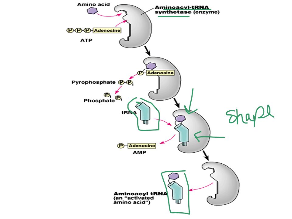
The 20 different synthetases match the 20 different amino acids.
Each amino acid is joined to the correct tRNA by aminoacyl-tRNA synthetase. The 20 different synthetases match the 20 different amino acids. Each has active sites for only a specific tRNA and amino acid combination. The synthetase catalyzes a covalent bond between them, forming aminoacyl-tRNA or activated amino acid. Fig Copyright © 2002 Pearson Education, Inc., publishing as Benjamin Cummings

86
Structure/Function/Location rRNA

87
Structure of Ribosomes (rRNA)
Ribosomes facilitate the specific coupling of the tRNA anticodons with mRNA codons. Each ribosome has a large and a small subunit. These are composed of proteins and ribosomal RNA (rRNA), the most abundant RNA in the cell. Fig a Copyright © 2002 Pearson Education, Inc., publishing as Benjamin Cummings
, the most abundant RNA in the cell. Fig a. Copyright © 2002 Pearson Education, Inc., publishing as Benjamin Cummings.")
88
Each ribosome has a binding site for mRNA and three binding sites for tRNA molecules.
The P site holds the tRNA carrying the growing polypeptide chain. The A site carries the tRNA with the next amino acid. Discharged tRNAs leave the ribosome at the E site. Fig b &c Copyright © 2002 Pearson Education, Inc., publishing as Benjamin Cummings

89
Recent advances in our understanding of the structure of the ribosome strongly supports the hypothesis that rRNA, not protein, carries out the ribosome’s functions. RNA is the main constituent at the interphase between the two subunits and of the A and P sites. It is the catalyst for peptide bond formation Fig Copyright © 2002 Pearson Education, Inc., publishing as Benjamin Cummings

90
Location of rRNA

91
The subunits exit the nucleus via nuclear pores.
After rRNA genes are transcribed to rRNA in the nucleus, the rRNA and proteins form the subunits in the nucleolus. The subunits exit the nucleus via nuclear pores. The large and small subunits join to form a functional ribosome only when they attach to an mRNA molecule. While very similar in structure and function, prokaryotic and eukaryotic ribosomes have enough differences that certain antibiotic drugs (like tetracycline) can paralyze prokaryotic ribosomes without inhibiting eukaryotic ribosomes. Copyright © 2002 Pearson Education, Inc., publishing as Benjamin Cummings
 can paralyze prokaryotic ribosomes without inhibiting eukaryotic ribosomes. Copyright © 2002 Pearson Education, Inc., publishing as Benjamin Cummings.")
92
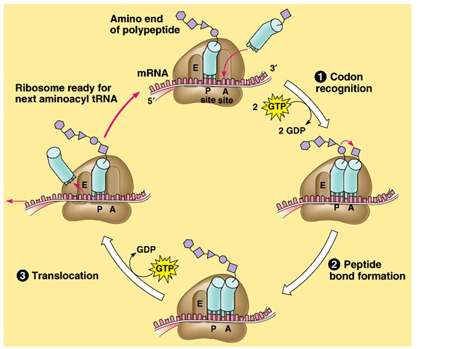
Translation can be divided into three stages: initiation elongation
Translation can be divided into three stages: initiation elongation peptide bond formation translocation termination All three phase require protein “factors” that aid in the translation process. Both initiation and chain elongation require energy provided by the hydrolysis of GTP. Its like stringing dental floss through your teeth Copyright © 2002 Pearson Education, Inc., publishing as Benjamin Cummings

93
Create three positions on the floor with pieces of tape
I need four volunteers Three on the tape One at the desk holding the markers or legos

94
Initiation brings together mRNA, a tRNA with the first amino acid, and the two ribosomal subunits.
First, a small ribosomal subunit binds with mRNA and a special initiator tRNA, which carries methionine and attaches to the start codon. Initiation factors bring in the large subunit such that the initiator tRNA occupies the P site. Fig Copyright © 2002 Pearson Education, Inc., publishing as Benjamin Cummings

95
Elongation consists of a series of three step cycles as each amino acid is added to the proceeding one. During codon recognition, an elongation factor assists hydrogen bonding between the mRNA codon under the A site with the corresonding anticodon of tRNA carrying the appropriate amino acid. This step requires the hydrolysis of two GTP. Copyright © 2002 Pearson Education, Inc., publishing as Benjamin Cummings

96
During peptide bond formation, an rRNA molecule catalyzes the formation of a peptide bond between the polypeptide in the P site with the new amino acid in the A site. This step separates the tRNA at the P site from the growing polypeptide chain and transfers the chain, now one amino acid longer, to the tRNA at the A site. Copyright © 2002 Pearson Education, Inc., publishing as Benjamin Cummings

97
During translocation, the ribosome moves the tRNA with the attached polypeptide from the A site to the P site. Because the anticodon remains bonded to the mRNA codon, the mRNA moves along with it. The next codon is now available at the A site. The tRNA that had been in the P site is moved to the E site and then leaves the ribosome. Translocation is fueled by the hydrolysis of GTP. Effectively, translocation ensures that the mRNA is “read” 5’ -> 3’ codon by codon. Copyright © 2002 Pearson Education, Inc., publishing as Benjamin Cummings

98
The three steps of elongation continue codon by codon to add amino acids until the polypeptide chain is completed. Fig Copyright © 2002 Pearson Education, Inc., publishing as Benjamin Cummings

100
This frees the polypeptide and the translation complex disassembles.
Termination occurs when one of the three stop codons reaches the A site. A release factor binds to the stop codon and hydrolyzes the bond between the polypeptide and its tRNA in the P site. This frees the polypeptide and the translation complex disassembles. Fig Copyright © 2002 Pearson Education, Inc., publishing as Benjamin Cummings

101
Multiple ribosomes, polyribosomes, may trail along the same mRNA.
Typically a single mRNA is used to make many copies of a polypeptide simultaneously. Multiple ribosomes, polyribosomes, may trail along the same mRNA. A ribosome requires less than a minute to translate an average-sized mRNA into a polypeptide. Fig Copyright © 2002 Pearson Education, Inc., publishing as Benjamin Cummings

102
Chaperone proteins may aid correct folding.
During and after synthesis, a polypeptide coils and folds to its three-dimensional shape spontaneously. The primary structure, the order of amino acids, determines the secondary and tertiary structure. Chaperone proteins may aid correct folding. In addition, proteins may require posttranslational modifications before doing their particular job. This may require additions like sugars, lipids, or phosphate groups to amino acids. Enzymes may remove some amino acids or cleave whole polypeptide chains. Two or more polypeptides may join to form a protein. Copyright © 2002 Pearson Education, Inc., publishing as Benjamin Cummings

103
Write the mRNA and the AA chain
TATAAATTTACCCGGGGAAAATATACGGCGATTCA

104
2. Signal peptides target some eukaryotic polypeptides to specific destinations in the cell
Two populations of ribosomes, free and bound, are active participants in protein synthesis. Free ribosomes are suspended in the cytosol and synthesize proteins that reside in the cytosol. Bound ribosomes are attached to the cytosolic side of the endoplasmic reticulum. They synthesize proteins of the endomembrane system as well as proteins secreted from the cell. Copyright © 2002 Pearson Education, Inc., publishing as Benjamin Cummings

106
This consists of a sequence of about 20 amino acids.
While bound and free ribosomes are identical in structure, their location depends on the type of protein that they are synthesizing. Translation in all ribosomes begins in the cytosol, but a polypeptide destined for the endomembrane system or for export has a specific signal peptide region at or near the leading end. This consists of a sequence of about 20 amino acids. A signal recognition particle (SRP) binds to the signal peptide and attaches it and its ribosome to a receptor protein in the ER membrane. The SRP consists of a protein-RNA complex. Copyright © 2002 Pearson Education, Inc., publishing as Benjamin Cummings
 binds to the signal peptide and attaches it and its ribosome to a receptor protein in the ER membrane. The SRP consists of a protein-RNA complex. Copyright © 2002 Pearson Education, Inc., publishing as Benjamin Cummings.")
107
Fig Copyright © 2002 Pearson Education, Inc., publishing as Benjamin Cummings

108
After binding, the SRP leaves and protein synthesis resumes with the growing polypeptide snaking across the membrane into the cisternal space via a protein pore. An enzyme usually cleaves the signal polypeptide. Secretory proteins are released entirely into the cisternal space, but membrane proteins remain partially embedded in the ER membrane. Copyright © 2002 Pearson Education, Inc., publishing as Benjamin Cummings

109
Other kinds of signal peptides are used to target polypeptides to mitochondria, chloroplasts, the nucleus, and other organelles that are not part of the endomembrane system. In these cases, translation is completed in the cytosol before the polypeptide is imported into the organelle. While the mechanisms of translocation vary, each of these polypeptides has a “postal” code that ensures its delivery to the correct cellular location. Copyright © 2002 Pearson Education, Inc., publishing as Benjamin Cummings

110
3. RNA plays multiple roles in the cell: a review
The cellular machinery of protein synthesis and ER targeting is dominated by various kinds of RNA. The diverse functions of RNA are based, in part, on its ability to form hydrogen bonds with other nucleic acid molecules (DNA or RNA). It can also assume a specific three-dimensional shape by forming hydrogen bonds between bases in different parts of its polynucleotide chain. DNA may be the genetic material of all living cells today, but RNA is much more versatile. Copyright © 2002 Pearson Education, Inc., publishing as Benjamin Cummings
. It can also assume a specific three-dimensional shape by forming hydrogen bonds between bases in different parts of its polynucleotide chain. DNA may be the genetic material of all living cells today, but RNA is much more versatile. Copyright © 2002 Pearson Education, Inc., publishing as Benjamin Cummings.")
111
The diverse functions of RNA range from structural to informational to catalytic.
Copyright © 2002 Pearson Education, Inc., publishing as Benjamin Cummings

112
4. Comparing protein synthesis in prokaryotes and eukaryotes: a review
Although bacteria and eukaryotes carry out transcription and translation in very similar ways, they do have differences in cellular machinery and in details of the processes. Eukaryotic RNA polymerases differ from those of prokaryotes and require transcription factors. They differ in how transcription is terminated. Their ribosomes are also different. Copyright © 2002 Pearson Education, Inc., publishing as Benjamin Cummings

113
The new protein quickly diffuses to its operating site.
In one big differences, prokaryotes can transcribe and translate the same gene simultaneously. The new protein quickly diffuses to its operating site. Fig Copyright © 2002 Pearson Education, Inc., publishing as Benjamin Cummings

114
In eukaryotes, the nuclear envelope segregates transcription from translation.
In addition, extensive RNA processing is inserted between these processes. This provides additional steps whose regulation helps coordinate the elaborate activities of a eukaryotic cell. In addition, eukaryotic cells have complicated mechanisms for targeting proteins to the appropriate organelle. Copyright © 2002 Pearson Education, Inc., publishing as Benjamin Cummings

115
Types of Mutations Point Base Pair Substitution Missense Nonsense
Frame Shift Deletion Insertion

116
5. Point mutations can affect protein structure and function
Mutations are changes in the genetic material of a cell (or virus). These include large-scale mutations in which long segments of DNA are affected (for example, translocations, duplications, and inversions). A chemical change in just one base pair of a gene causes a point mutation. If these occur in gametes or cells producing gametes, they may be transmitted to future generations. Copyright © 2002 Pearson Education, Inc., publishing as Benjamin Cummings
. These include large-scale mutations in which long segments of DNA are affected (for example, translocations, duplications, and inversions). A chemical change in just one base pair of a gene causes a point mutation. If these occur in gametes or cells producing gametes, they may be transmitted to future generations. Copyright © 2002 Pearson Education, Inc., publishing as Benjamin Cummings.")
117
Write the mRNA and the AA chain
ATTTACCCGGGGAAAATATACGGCGATTCA-original ATTTACCGGGGGAAAATATACGGCGATTCA-point mutation

118
For example, sickle-cell disease is caused by a mutation of a single base pair in the gene that codes for one of the polypeptides of hemoglobin. A change in a single nucleotide from T to A in the DNA template leads to an abnormal protein. Fig Copyright © 2002 Pearson Education, Inc., publishing as Benjamin Cummings

119
A point mutation that results in replacement of a pair of complimentary nucleotides with another nucleotide pair is called a base-pair substitution. Some base-pair substitutions have little or no impact on protein function. In silent mutations, alterations of nucleotides still indicate the same amino acids because of redundancy in the genetic code. Other changes lead to switches from one amino acid to another with similar properties. Still other mutations may occur in a region where the exact amino acid sequence is not essential for function. Copyright © 2002 Pearson Education, Inc., publishing as Benjamin Cummings

120
Other base-pair substitutions cause a readily detectable change in a protein.
These are usually detrimental but can occasionally lead to an improved protein or one with novel capabilities. Changes in amino acids at crucial sites, especially active sites, are likely to impact function. Missense mutations are those that still code for an amino acid but change the indicated amino acid. Nonsense mutations change an amino acid codon into a stop codon, nearly always leading to a nonfunctional protein. Copyright © 2002 Pearson Education, Inc., publishing as Benjamin Cummings

121
Fig Copyright © Pearson Education, Inc., publishing as Benjamin Cummings

122
Insertions and deletions are additions or losses of nucleotide pairs in a gene.
These have a disastrous effect on the resulting protein more often than substitutions do. Unless these mutations occur in multiples of three, they cause a frameshift mutation. All the nucleotides downstream of the deletion or insertion will be improperly grouped into codons. The result will be extensive missense, ending sooner or later in nonsense - premature termination. Copyright © 2002 Pearson Education, Inc., publishing as Benjamin Cummings

123
Fig Copyright © 2002 Pearson Education, Inc., publishing as Benjamin Cummings

124
Mutations can occur in a number of ways.
Errors can occur during DNA replication, DNA repair, or DNA recombination. These can lead to base-pair substitutions, insertions, or deletions, as well as mutations affecting longer stretches of DNA. These are called spontaneous mutations. Copyright © 2002 Pearson Education, Inc., publishing as Benjamin Cummings

125
Chemical mutagens may operate in several ways.
Mutagens are chemical or physical agents that interact with DNA to cause mutations. Physical agents include high-energy radiation like X-rays and ultraviolet light. Chemical mutagens may operate in several ways. Some chemicals are base analogues that may be substituted into DNA, but that pair incorrectly during DNA replication. Other mutagens interfere with DNA replication by inserting into DNA and distorting the double helix. Still others cause chemical changes in bases that change their pairing properties. Copyright © 2002 Pearson Education, Inc., publishing as Benjamin Cummings

126
Researchers have developed various methods to test the mutagenic activity of different chemicals.
These tests are often used as a preliminary screen of chemicals to identify those that may cause cancer. This make sense because most carcinogens are mutagenic and most mutagens are carcinogenic. Copyright © 2002 Pearson Education, Inc., publishing as Benjamin Cummings

127
6. What is a gene? revisiting the question
The Mendelian concept of a gene views it as a discrete unit of inheritance that affects phenotype. Morgan and his colleagues assigned genes to specific loci on chromosomes. We can also view a gene as a specific nucleotide sequence along a region of a DNA molecule. We can define a gene functionally as a DNA sequence that codes for a specific polypeptide chain. Copyright © 2002 Pearson Education, Inc., publishing as Benjamin Cummings

128
Transcription, RNA processing, and translation are the processes that link DNA sequences to the synthesis of a specific polypeptide chain. Fig Copyright © 2002 Pearson Education, Inc., publishing as Benjamin Cummings

129
Even the one gene-one polypeptide definition must be refined and applied selectively.
Most eukaryotic genes contain large introns that have no corresponding segments in polypeptides. Promotors and other regulatory regions of DNA are not transcribed either, but they must be present for transcription to occur. Our definition must also include the various types of RNA that are not translated into polypeptides. A gene is a region of DNA whose final product is either a polypeptide or an RNA molecule. Copyright © 2002 Pearson Education, Inc., publishing as Benjamin Cummings

130
Practice ’90/’94 In DNA replication, DNA polymerase catalyzes the reaction in which A. The double helix unwinds B. The sugar-phosphate bonds of each strand are broken C.A phosphate group is added to the 3’-carbon or 5’-carbon of ribose D.A nucleotide with a base complementary of the base on the template stand is added to the new DNA strand E. The two nucleotide strands come together and intertwine to form a double helix

131
Practice The relative location of four genes on a chromosome can be mapped from the following data on crossover frequencies. GenesFrequency of crossover B and D 5% C and A % A and B % C and B % C and D 50% Which of the following represents the relative positions of these four genes on the chromosome? A. ABCD B. ADCB C.CABD D.CBAD E. DBCA

132
The illustrations above represent homologous pairs of chromosomes as they appear in various stages of mitosis and meiosis (2n=2). 1. At which stage do the chromosomes have the LEAST amount of DNA per cell? 2. Which diagram represents anaphase of meiosis I?

133
Shading indicates the presence of sickle cell anemia.
The phenotype of individual C is best explained by the fact that this individual received an allele for sickle cell anemia from A. an autosomal chromosome of each parent B. the Y chromosome contributed by the father C.the X chromosome contributed by the mother D.the X chromosome contributed by the father E. the Y chromosome contributed by the mother Shading indicates the presence of sickle cell anemia.

134
What is the probability that the next child of parents A and B would have had sickle cell anemia?

135
The most reasonable explanation for the fact that the offspring of C and D do not have sickle cell anemia is that each received a A. sickle allele from the mother B. normal allele from the father C.sickle allele from each parent D.normal allele from each parent E. pair of normal alleles from the father

136
Which of the following statements is correct about the four offspring of C an D?
A.Only the females are carriers of the sickle cell trait. B.Only the males are carriers of the sickle cell trait. C.Only the females are heterozygous for the sickle cell trait. D.All are homozygous for the sickle cell trait. E.All are carriers of the sickle cell trait.

137
Achondroplastic dwarfism is a dominant genetic trait that causes severe malformation of the skeleton. Homozygotes for this condition are spontaneously aborted (hence, the homozygous condition is lethal) but heterozygotes will develop to be dwarfed. Matthew has a family history of the condition, although he does not express the trait. Jane is an achondroplastic dwarf. Matthew and Jane are planning a family of several children and want to know the chances of producing a child with achondroplastic dwarfism.

138
The genotypes of Matthew and Jane are best represented as
Matthew Jane (A) AA Aa (B) Aa aa (C) aa aa (D) aa Aa (E) Aa Aa
 AA Aa. (B) Aa aa. (C) aa aa. (D) aa Aa. (E) Aa Aa.")
139
The probability that Matthew and Jane’s first child will be an achondroplastic dwarf is

140
If three children are born to Matthew and Jane, what are the chances that the first two children will not express the trait but that the third child will be an achondroplastic dwarf? (A) 5/8 (B) 4/8 (C) 3/8 (D) 1/8 (E) 1/16
 5/8. (B) 4/8. (C) 3/8. (D) 1/8. (E) 1/16.")
Similar presentations


 hypothesized that the symptoms of an inherited disease reflect a person’s inability to make a particular enzyme. The breakthrough.>")


 and phenotype (physical expression)>")


 Chapter 17.>")