Download presentation
Presentation is loading. Please wait.

1
Motion Analysis Summer Course
Speaker: Yi-Jung Tsai Date: 2011/07/13 Motion Analysis Laboratory

2
Outline Part I: Introduction of motion analysis Basic introduction
Research methods in motion analysis Instrumentation Data collection Data analysis Part II : Application Gait analysis Clinical application Sports medicine

3
Part I: Introduction of motion analysis
Basic introduction Research methods in motion analysis Instrumentation Data collection Data analysis

4
Introduction What is motion analysis ?
When and why do we need to analyze motion? What knowledge do we need before research?

5
Introduction Kinematics (運動學)
Kinematics is concerned with the geometry of motion and deal with relationships among displacement, velocity, acceleration, and time without any reference to the cause of motion To describe the motions we see Kinematics: what we see

6
Introduction Kinetic (力動學)
Kinetics deal with relationships among forces, mass, and motion of the body, it is concerned with the cause of motion To understand why the motions occur force / torque

7
Introduction Anthropometry (人體測量學)
Involving body and limb measurements mass of segment location of mass center segment length center of rotation angle of muscles mass and cross-sectional area of muscles moment of inertia Important For kinetic

8
BM = Total Body Mass; BH = Body Height
Segment L (%BH) Mass (%BM) Child < 14 y Mass (kg) %LCoM (%BH) (from distal joint) Child < 14y %LCoM (%BH) %I * (in kg.m2) Head 9.6 7.8 *age 50 49.5 Torso 31.6 46.84 *age *age 50.3 Upper arm 16.4 2.7 0.084*age+2.2 56.4 -0.028*age+55.7 32.2 Forearm 13.7 2.3 0.015*age+1.2 55 0.19*age+56.1 30.3 Hand 8.2 0.6 29.7 Thigh 25.4 9.9 0.364*age+6.634 56.7 *age 32.3 Shank 23.3 4.6 0.122*age+3.809 57 *age 30.2 Foot 11.7 1.4 0.015*age+1.87 *age 47.5 BM = Total Body Mass; BH = Body Height I = %I*Segment Mass*Segment Length2
 Mass (%BM) Child < 14 y Mass (kg) %LCoM (%BH) (from distal joint) Child < 14y %LCoM (%BH) %I * (in kg.m2) Head *age Torso *age *age Upper arm *age *age Forearm *age *age Hand Thigh *age *age Shank *age *age Foot *age *age BM = Total Body Mass; BH = Body Height. I = %I*Segment Mass*Segment Length2.")
9
Introduction Muscle and joint biomechanics
Characteristics of muscle and joint - length-tension relationship - force-velocity relationship - joint type

10
Introduction Electromyography (肌電圖)
- the study of muscle electrical activities providing information about the control and execution of voluntary movement

11
Steps of motion analysis
Setting the purpose Choosing the appropriate instrumentation Data collection Data analysis Results and interpretation

13
Instrument_ kinematics
(Electro) goniometers (量角器) - a device for measuring joint angles
 goniometers (量角器) - a device for measuring joint angles.")
14
Instrument_ kinematics
Accelerometer (加速規) - a device that measures acceleration types: strain gauge piezoresistive piezoelectric
 - a device that measures acceleration. types: strain gauge. piezoresistive. piezoelectric.")
15
Instrument_ kinematics
Imaging system Cinematograph digital video charge-couple device (CCD) cameras: - Motion analysis, VICON, Qualisys system
 cameras: - Motion analysis, VICON, Qualisys system.")
17
Eagle Digital RealTime System
Hz selectable frame rates Passive (retroreflective) markers
 markers.")
18
EVa Real-Time Software (EVaRT)
3D Display XYZ Graphs Analog Graphs

19
Instrument_ kinetic Force transducers - measure the applied forces
types: Piezoresistive Piezoelectric

20
Instrumentation_ kinetic
Force plate - most commonly used type of force transducer - measuring ground reaction force (GRF) type: Strain gauge Piezoelectric
 type: Strain gauge. Piezoelectric.")
21
Instrumentation_ kinetic
Kistler force plate

22
Instrumentation_ kinetic
Pressure sensor

23
Instrument_ EMG Types: - Surface EMG - Wire EMG - Needle EMG

24
Steps of motion analysis
Setting the purpose Choosing the instrumentation (kinematics, kinetic, EMG…..) Data collection Data analysis Results and interpretation
 Data collection. Data analysis. Results and interpretation.")
25
Data collection Calibration - to define the global coordinate system

26
Data collection Preparation - measurements of basic data
- placement of EMG electrodes - marker attachment on the landmark (marker set) - others
 - others.")
27
Data collection Placement of EMG electrodes
Others: setting the appropriate mode - sampling rate - collection time - amplify….

28
Steps of motion analysis
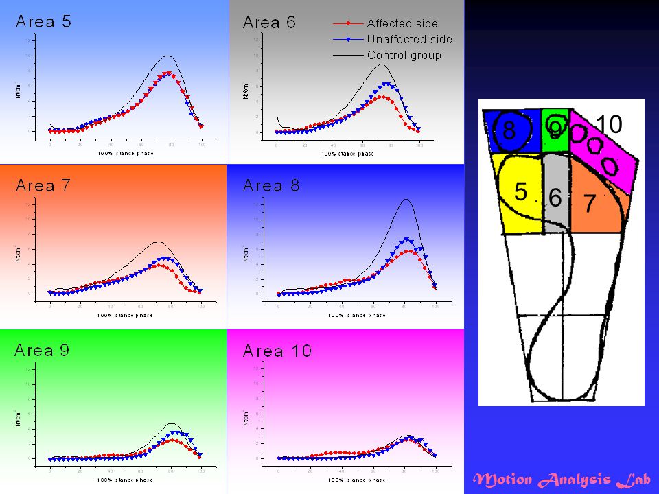
Setting the purpose Choosing the instrumentation (kinematics, kinetic, EMG…..) Data collection Data reduction & analysis Results and interpretation
 Data collection. Data reduction & analysis. Results and interpretation.")
29
Data reduction & analysis
Signal output Signal processing - data smoothing - interpolation - filter: low pass, band pass, high pass filter…... - re-sampling Setting the appropriate parameters

30
Planes of Motion: 1 = Frontal plane 2 = Sagittal plane 3 = Transverse plane

31
Data analysis Calculation - kinematics
Translation and Rotation of different coordinate systems Resolve the joint angles: Step 1: compute b Step 2: compute a Step 3: compute g

32
Data analysis Calculation kinetic(ground reaction force, joint moment)
Inverse dynamics EMG: Rectified Linear envelope Integrated……..

33
Take home message How to choose the appropriate instrument?
- according to the research purpose - understanding the pros and cons How to collect data well? - following the manuscript - setting the appropriate mode What should be noticed in data analysis? - avoid distortion after signal processing - understanding the limitation and problems of different computing method What should be noticed while reading the report? - does the result make sense?

34
TAKE A BREAK

35
Part II : Applications Projects in motion analysis laboratory
Gait analysis Clinical applications Sports medicine

36
Gait analysis Bipedal locomotion, or gait, is a functional task requiring complex interactions and coordination among most of the major joints of the body, particularly of the lower extremity.

37
Anatomical considerations_ hip joint
flexion-extension occurs about a mediolateral axis. adduction-abduction occurs about an anteroposterior axis. internal-external rotation occurs about a longitudinal axis.

38
Anatomical considerations_ knee joint
3 degrees of freedom of angular rotation are also possible during gait. The primary motion is knee flexion-extension. Knee internal-external rotation and adduction-abduction may also occur, but with less consistency and amplitude among healthy individuals owing to soft tissue and bony constraints to these motion.

39
Anatomical considerations_ ankle and foot
Ankle motion is restricted by the morphological constraints of the talocrural joint, which permits only plantarflexion (extension) and dorsiflexion (flexion). In gait analysis as a rigid segment, the foot is required to act as both a semirigid structure and a rigid structure that permits adequate stability
 and dorsiflexion (flexion). In gait analysis as a rigid segment, the foot is required to act as both a semirigid structure and a rigid structure that permits adequate stability.")
40
Gait Cycle Stance phase:60%, including foot flat, midstance, terminal stance, and pre-swing. Swing phase: 40%, including initial swing, midswing, and terminal swing.

41
Time-distance variable
Ranges of normal values for time-distance parameters of adult gait at free walking velocity Stride or cycle time 1.0 to 1.2 sec Stride or cycle length 1.2 to 1.9 m Step length 0.56 to 1.1 m Step width 7.7 to 9.6 cm cadence 90 to 140 step/min velocity 0.9 to 1.8 m/sec

43
Clinical Applications
Musculoskeletal pathology polio, muscle atrophy, amputation, osteoarthritis rheumatoid arthritis, trauma muscle weakness, restricted joint mobility, pain Upper motor neuron pathology cerebral palsy, stroke, brain trauma combine spasticity, sensory disturbance, error in control mechanisms

44
Chair-rise in Patients after Total Knee Arthroplasty
Fong-Chin Su, Kuo-An Lai, Wei-Hsien Hong Clin Biomech 13: , 1998

45
Objectives To understand the biomechanics and compensatory mechanisms of chair-rise in patients after TKA. Functional evaluation of pre-op patients compared to the normal elderly.

46
Subjects * : (yr) (cm) (kg) normal elderly OA patient TKA patient 12
subject N age body height body weight (yr) (cm) (kg) normal elderly OA patient TKA patient 12 14* 12* 159.5 ± 6.9 154.5 ± 5.2 157.0 ± 5.7 61.0 ± 9.90 58.2 ± 10.5 73.3 ± 14.0 60.7±6.67 61.2 ± 7.60 64.8 ± 8.00 * : OA patients ( 10 bilateral, 4 unilateral ) TKA patients ( 8 unilateral, 4 bilateral )
 (cm) (kg) normal elderly. OA patient. TKA patient * 12* ± ± ± ± ± ± ± ± ± * : OA patients ( 10 bilateral, 4 unilateral ) TKA patients ( 8 unilateral, 4 bilateral )")
47
Experiment Setup A/D converter computer disk storage camera interface

48
Marker Set

49
Sit-to-Stand a b c d a - b : flexion momentum phase * 4 chair heights:
b - c : momentum transfer phase c - d : extension phase * 4 chair heights: 115%, 100%, 80%, 65% knee height

50
Duration of the STS

51
COM displacement

52
Vertical velocity of COM

53
Angular changes_ hip joint

54
Angular changes_ knee joint

55
Joint flexion moment_ hip

56
Joint flexion moment_ knee

57
Joint flexion moment_ ankle

58
TKA patient elderly 1.97 sec 1.81 sec seat-off 41% 49% 59% 51%

59
elderly TKA patient Fh Fk Fa

60
Joint Moment COM COM elderly TKA patient

61
Conclusion TKA patient has larger displacement and horizontal velocity of COM compared to the normal elderly. No significant difference in angles of three joints. TKA patients has special pattern in nearly knee full extension. TKA has larger ankle and hip moments. Increased chair height, decreased joint angles and moments.

62
Common Abnormal Kinetic Patterns of the Knee in Gait in Cerebral Palsy
C.J. Lin, L.Y. Guo, F.C. Su, Y.L. Chou Gait & Posture, 11: , 2000

63
Objectives To investigate the detailed kinetic characteristics of each abnormality. 23 children suffering from cerebral palsy with spastic diplegia, were recruited 46 limbs into four groups: jump (n = 7) crouch (n = 8) recurvatum (n = 14) mild (n = 17)
 crouch (n = 8) recurvatum (n = 14) mild (n = 17)")
64
Crouch Gait Results show that crouch gait usually has larger and long-lasting knee extensor moments at stance. This reveals that rectus femoris has relatively high activation.

65
Knee flexor moments are large and long-lasting during stance.
The biceps femoris muscle shows less activation in EMG the soft tissue behind the knee joint provides this flexor moment. This may result in worse recurvatum knee due to overstretch by the external forces. Recurvatum Knee

66
Knee Angle FLEX EXT Recurvatum GAIT CYCLE % 20 40 60 80 100 10 30 50
20 40 60 80 100 10 30 50 70 EXT FLEX Jump Recurvatum Crouch Mild Normal (Degree)
")
67
Knee Joint Moment

68
Gait Analysis After Ankle Arthrodesis
W.L. Wu, F.C. Su, Y.M. Cheng, P.J. Huang, P.J. Chou, Y.L. Chou, C.K. Chou Gait & Posture, 11:54-61, 2000

69
Aims To employ a computerized motion analysis system to identify the effect of ankle arthrodesis on three-dimensional kinematic and kinetic behaviors of the rear and fore foot and muscle activities of the lower extremity during level walking.

70
Subjects Patients: 10 (7 males and 3 females)
with single-side solid arthrodesis of the ankle performed due to trauma, degenerative osteoarthritis or rheumatic arthritis, were recruited for this study. The mean age was 39.6 years old (13 to 64 y/o). The mean duration of follow-up after arthrodesis was 1.7 years (0.5 to 4 years). Controls: 10 normal subjects, mean age 28.8 yrs (20 to 35 y/o)
. The mean duration of follow-up after arthrodesis was 1.7 years (0.5 to 4 years). Controls: 10 normal subjects, mean age 28.8 yrs (20 to 35 y/o)")
71
Markers set & coordinate system
x f z h t y

72
Experiments

73
Spatiotemporal parameters

74
RANGE OF MOTION

75
Hindfoot Motion

76
Forefoot Motion

77
Ground Reaction Force Vertical Force Ankle arthrodesis Normal F3-T3
150 F3-T3 F1-T1 Vertical Force 100 F2-T2 % BW 50 white: no change red: increase green: decrease 10 20 30 40 50 60 70 80 90 100 % Stance Phase

78
Ground Reaction Force Fore-aft force Ankle arthrodesis Normal F5-T5
20 % BW F4-T4 F6-T6 -20 10 20 30 40 50 60 70 80 90 100 white: no change red: increase green: decrease % Stance Phase

79
Ground Reaction Force Medial-lateral force Ankle arthrodesis Normal
10 20 30 40 50 60 70 80 90 100 -10 % BW % Stance Phase F7-T7 F8-T8 F9-T9 white: no change red: increase green: decrease

80
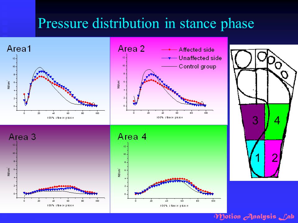
Pressure distribution in stance phase
1 2 3 4
81
5 6 7 8 9 10
82
Conclusion - motion Hindfoot No 2nd rocker
Increased eversion and external rotation throughout whole gait cycle. Forefoot Increased third rocker motion at toe-off. Increased adduction Significant increased eversion at toe-off.

83
Rage of Motion Ankle fusion causes
decrease of sagittal movements of hindfoot. increase of transverse movements of hindfoot. increase of forefoot motion in three planes.

84
Ground Reaction Force Decreased loading rate.
Weak body support in midstance and push-off in preswing. Smaller fore-aft shear force. Greater lateral shear force.

85
Force and Pressure in 10 Masks
Affected side : pressure and force in rearfoot, forefoot and toe areas lack of the force absorption and forward progression pressure and force in two midfoot areas supposing pronated gait

86
Biomechanical Evaluation of New Type Stair Climbing Machine (S770)
")
87
Purpose To investigate the biomechanics of new type stair climbing machine (S770). (1) kinematics: - movement range of hip, knee, and ankle joints (2) kinetics: - foot contact force - joint moment (3) muscle activities
 kinetics: - foot contact force. - joint moment. (3) muscle activities.")
88
Methods_ subject 12 healthy young adults - 2 females, 10 males
- Age: 24.5±1.2 y/o - Body height: 171.1±5.5 cm - Body weight: 64.0±9.6 kg

89
Methods_ instrumentation
6-axis load cell: - embedded in the left foot pedal Surface EMG system: - MA 300 Motion analysis system: - 8 Eagle digital cameras - 15 reflective markers

90
Methods_ EMG Adductor Abductor Hamstring RF,VM,VL
Tibialis anterior and Peroneus longus Gastrocnemius

92
Methods _ data collection
Four conditions: - step to end range, trunk static - step to end range, trunk shift - step at selected range, trunk static - step at selected range, trunk shift Stepping at selected speeds 3 trials/condition, 15 secs/trial * The resistance was consistent

93
Animation_ step to end range
Trunk static Trunk shift

94
Animation_ trunk static
Step to end range Step at selected range

95
Results_ joint angle PF(+) DF(-) Flex(+) Ext(-) Abd(+) Abd(+) Add(-)
Angular displacement change in a cycle.

96
Result_ foot contact force
Three directions of foot contact force in a cycle Blue line (ML) mediolateral direction Green line (AP) anteroposterior direction
 mediolateral direction. Green line (AP) anteroposterior direction.")
97
Results_ foot contact force
Trunk shift > Trunk static Trunk shift End range Trunk shift Selected range Trunk static End range Trunk static Selected range Stair climbing machine (S770) < walking and normal stair climbing ( > 1 BW)
 < walking and normal stair climbing ( > 1 BW)")
98
Results_ joint moment (+) abd
 abd")
99
Results_ joint moment flex(+) flex(+) PF(+) ext(-) ext(-) DF(-)
Internal moment: is the same with muscle contraction. Plot force and moment figure (CHANGE)
")
100
Results _ muscle activities

101
Results_ muscle activities

102
Thanks for your attention ~

Similar presentations