Download presentation
Presentation is loading. Please wait.

1
Respiration: from Physiology to Phonetics
Alain Marchal Laboratoire Parole et Langage CNRS – Aix-en-Provence We first give a brief review of the physiology of respiration and the mechanics of breathing We then indicate how the respiratory cycle is modified during speech production. We revise Ladefoged’s view of the linear organization of muscular activity during phonation and consider the speech specific control of exhalation in the framework of the general theory of coordinated movements. The following issues are discussed: Aerodynamic conditions and constraints, Synergistic action of the inhalatory and expiratory muscles, Correlation between muscula activity , V/C distinction and syllables, The control of subglottal pressure is discussed in the last part with focus on the following points: Stability of Fo and Int despite variations of supraglottal impedance, Subglottal pressure/acoustic parameters, Subglottal pressure/accentuation, Finally, we indicate some of the phonetic consequences of respiratory troubles.

2
F. Rohrer (1925): Treatise on respiratory movements: the basis of Respiratory Physiology. W. Fenn: Extension of this work in the 1940s. Ladefoged et al. (1957): First phonetic studies examining the relationship between respiration and phonation. The way in which respiration is modified to accommodate speech production. Until the seventeenth century, man’s knowledge of respiration was limited to his belief that the prime purpose of breathing was to cool the blood. Not until the twentieth century did any work on ventilatory mechanics develop. In 1925, F. Rohrer published his treatise on respiratory movements, which forms the basis of respiratory physiology. W. Fenn extended this work in the 1940s. Finally, we are indebted to Ladefoged et al. (1957) for the first phonetic studies examining the relationship between respiration and phonation. This was the first clear evocation of the way in which respiration is modified to accommodate speech production.
: First phonetic studies examining the relationship between respiration and phonation. The way in which respiration is modified to accommodate speech production. Until the seventeenth century, man’s knowledge of respiration was limited to his belief that the prime purpose of breathing was to cool the blood. Not until the twentieth century did any work on ventilatory mechanics develop. In 1925, F. Rohrer published his treatise on respiratory movements, which forms the basis of respiratory physiology. W. Fenn extended this work in the 1940s. Finally, we are indebted to Ladefoged et al. (1957) for the first phonetic studies examining the relationship between respiration and phonation. This was the first clear evocation of the way in which respiration is modified to accommodate speech production.")
3
Vital Function of Respiration
To ensure the exchange of gases between air and blood. The respiratory cycle comprises two phases: inhalation and exhalation. Inhalation: Intake of air into the lungs, bringing oxygen to the organism. Exhalation: Emptying the lungs and expelling the carbon dioxide accumulated by the blood. The vital function of respiration is to ensure the exchange of gases between air and blood. The respiratory cycle comprises two phases: inhalation and exhalation. Inhalation allows a certain quantity of air to be stored in the lungs, bringing oxygen to the organism. The function of exhalation is to empty the lungs and expel gaseous waste from the body, in particular the carbon dioxide accumulated by the blood.

4
The Lungs Situated in the rib cage, 2 lungs:
Situated in the rib cage, 2 lungs: - shape of air-filled pyramids, - separated by the mediastinum Divided into two bronchial tubes, subdividing into bronchioles and alveoli. The lungs, situated in the rib cage, have the shape of air-filled pyramids. They are two in number, one on the left, the other on the right, separated by the mediastinum. They resemble two wet sponges, with tree-like ramifications. They are divided into two bronchial tubes which then subdivide into bronchioles and alveoli.

5
The pleural fluid allows the layers to slide over one another;
The two lungs are envelopped in a serous membrane: the pleura. 2 layers: visceral and parietal. The 2 lungs are envelopped in a serous membrane: the pleura. The pleura has 2 layers: - the internal or visceral layer which covers and clings to the lungs - The external or parietal layer which is fixed to the internal wall of the rib cage. The pleura ensures the functional coupling between the chest wall and the lungs. The pleural liquid allows the layers to slide over one another The pleural fluid allows the layers to slide over one another; The pleura ensures the functional coupling between the chest wall and the lungs.

6
Mechanics of breathing
The lungs and thorax have elastic properties: The property of elasticity plays a great role in normal respiration: - elongation during inhalation - return to rest position during exhalation due to the relaxation forces. Elasticity can be estimated using the pleural pressure Pulmonary tissue is elastic and can therefore follow the movements of the rib cage. The lungs and thorax undergo elongation due to muscular activity in the inhalation phase. When this force stops, they resume their natural position and shape; thus reducing the pulmonary volume and forcing out the air previously inhaled. Lung elasticity forces can be estimated by measuring the pleural pressure. The amount of pleural pressure varies according to the stages of the respiratory cycle. The intra-pleural pressure is exerted over the organs contained in the thorax, in particular the heart, the thoracic channel and the oesophagus, where it can be measured more easily.

7
The Respiratory System - The Structural Supports
1– The rib cage 2– The visceral thorax 3- The respiration muscles The rib cage : 12 spinal vertebrae, 12 pairs of ribs, : The sternum. Bounded at the top by the neck and at the bottom by the diaphragm. The structural supports of the respiratory system has three components: the rib cage, the visceral thorax and the respiration muscles. The rib cage is made up of twelve spinal vertebrae, twelve pairs of ribs, and the sternum. It is bounded at the top by the neck and at the bottom by the diaphragm.

8
Rib Cage = Protective shield
The ribs constitute a barrel-shaped protective shield around the thorax: the rib cage. At the back, the head of each rib is joined to the spinal column by sliding joints. At the front, the first seven ribs are attached directly to the sternum by means of costal cartilage. The next three are attached to the lower extremity of the sternum by the cartilage of the seventh rib. The last two ribs (the ‘floating’ ribs) have no anterior attachment; their costal cartilage is embedded in muscle fibres. From Marchal (2007)
 have no anterior attachment; their costal cartilage is embedded in muscle fibres. From Marchal (2007)")
9
- Raising of the ribs: transverse and lateral increase
Head of each rib joined to the spinal column by sliding joints. Displacement of the ribs = enlargement of the cage - Raising of the ribs: transverse and lateral increase - Forward and upward movement of the sternum: increase of the antero-posterior diameter. Movements of the diaphragm: vertical dimension. Because of their shape and mode of attachment, front and back, Raising of the ribs will cause an increase in thoracic volume in two directions: transverse and lateral. The simultaneous forward and upward movement of the sternum results in an increase of the anteroposterior diameter. The vertical dimension can be altered by movements of the diaphragm which lower the abdominal internal organs.

10
Some Principles of Aerodynamics
Boyle’s law: When the dimensions of a container are enlarged, its volume increases; the molecules of air become more spaced out, and air pressure falls. Conversely, when the dimensions are reduced, the volume decreases, the air molecules become compressed, and the pressure increases The pressure of air in the lungs depends on the force exerted on the thoracic walls by the molecules of air inside them. When the dimensions of a container are enlarged, its volume increases, the molecules of air become more spaced out, and air pressure falls. Conversely, when the dimensions are reduced, the volume decreases, the air molecules become compressed, and the pressure increases (Boyle’s law). The pressure of air in the lungs depends on the force exerted by the molecules of air inside them.
. The pressure of air in the lungs depends on the force exerted by the molecules of air inside them.")
11
Some Principles of Aerodynamics
An increase in pulmonary volume > lowering of pressure which results in the drawing in of air from outside. A decrease in pulmonary volume > increase in pressure which pushes the air out. An increase in pulmonary volume provides a lowering of pressure which results in the drawing in of air from outside. Conversely, a decrease in pulmonary volume induces in return an increase in pressure which pushes the air out.

12
How to Measure Air Pressure ?
The U tube manometer measures the height of a column of water when a given pressure is applied to one arm of the tube Electronic transducers for dynamic speech pressure measurements are now used Early studies were conducted using The U tube manometer , measuring the height of a column of water . Not really suited for speech research! Now, there is a wide array of electronic transducers available for recording air pressure.

13
Air Pressures in the Vocal Tract
Pressure is defined as the force per unit area acting perpendicular to a surface Absolute pressure is of little value to the speech scientist or speech therapist Pressures in the respiratory tract (vocal tract) are expressed relative to the atmospheric pressure = gauge or gage pressure Speech pressures are commonly expressed in CmH2o Pressure is defined as the force per unit area acting perpendicular to a surface Absolute pressure is of little value to the speech scientist or speech therapist Pressures in the respiratory tract (vocal tract) are expressed relative to the atmospheric pressure = gauge or gage pressure Speech pressures are commonly expressed in CmH2o
 are expressed relative to the atmospheric pressure = gauge or gage pressure. Speech pressures are commonly expressed in CmH2o. Pressure is defined as the force per unit area acting perpendicular to a surface. Absolute pressure is of little value to the speech scientist or speech therapist. Pressures in the respiratory tract (vocal tract) are expressed relative to the atmospheric pressure = gauge or gage pressure. Speech pressures are commonly expressed in CmH2o.")
14
Airflow Airflow occurs when there is a difference between pressures; Air flows from a region of high pressure to one of low pressure. The bigger the difference, the faster the flow When flow is low and through narrow tubes, it tends to flow in a straight line: Laminar airflow When air flows at higher velocities, flow is disorganized, chaotic and forms eddies: Turbulent airflow Airflow occurs when there is a difference between pressures; Air will flow from a region of high pressure to one of low pressure. The bigger the difference, the faster the flow When flow is low and through narrow tubes, it tends to flow in a straight line: this type of flow is called laminar When air flows at higher velocities, flow is disorganized, chaotic and forms eddies: Turbulent airflow

15
How to Measure Flow ? Spirometer:
Basic equipment for pulmonary function tests Differential pressure transducers for the measurement of flow rates Unsuited for speech The spirometer constitutes the: Basic equipment for pulmonary function tests It uses differential pressure transducers for the measurement of flow rates Unsuited of course for speech

16
How to Measure Flow during a Speech Task ?
Pneumotachograph Measurement of pressure differences across a fine mesh Electronic transducers of various types For speech Airtight mask which fits over the mouth and nose Mouth mask and catheter in nostrils Microphone Body plethysmograph Beware of artifacts: potential perturbation of articulation, air leaks, pressure variation due to jaw movements, resonance in the facemask, … Best solution is to use a body plethysmograph. However, few phonetic studies, with the exception of Proctor & Mead, Ohala and myself.

17
A: The signal from the box represents intrathoracic pressure changes from which airflow can be deducted. (beware of temperature fluctuation in the box in case of prolonged session)
")
19
With air conditioning…

20
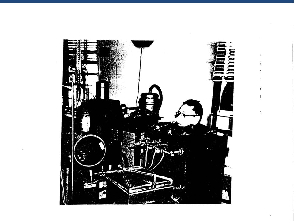
Recording Session with EVA2 in Aix
Multichannel, multisensor recording system developped at Aix lab in the ACCOR and Value European projects: oral – nasal airflow data, air-pressures at different locations in the vocal tract, glottographic data, …., Acoustic speech signal, EMG data……

21
Normal Respiration Inhalation:
Contraction of the external intercostal muscles and of the diaphragm > raising and widening the rib cage > Increase of the pulmonary volume. The intrapulmonary pressure > negative relative to the atmospheric pressure > the lungs fill by aspiration. Air intake: about 1/2 liter Inhalation is the result of raising and widening the rib cage, effected by the contraction of the external intercostal muscles and the flattening of the diaphragm, which presses down on the abdominal viscera. This action, because of the functional coupling between the lungs and the thorax, lowers the intra-pulmonary pressure. The amount of air inhaled is in the region of half a litre.

22
Normal Respiration Exhalation:
Normal exhalation is an entirely passive, involuntary process caused by the elastic recoil of the pulmonary tissue and the ribs. Return to equilibrium. Air out: same volume as intake Exhalation When the inhalation impulse ceases, the lungs deflate and return to the rest position. This constitutes a return to equilibrium. Normal exhalation is an entirely passive process caused by the elastic recoil of the pulmonary tissue and the ribs, from their weight and the pressure exerted by the abdominal organs. The combination of these forces constitutes what is termed: the pressure of relaxation. Air out equals air intake

23
Aerodynamic Data for Normal Respiration
Ratio between inhalation and exhalation is 1:1. Rate is 12 to 18 cycles per minute. Flow: 0,3 -0,5 L/s Volume: 500 cm3 Pulmonary Pressure: 1-3 cm H2O (With forced inhalation and severe muscular effort during exhalation, the rate of flow can increase to more than 50 l/s and intra-pulmonary pressure can go up to 100 cm H2O). The ratio between inhalation and exhalation is 1:1. The typical rate of normal respiration is 12 to 18 cycles per minute. In the adult, mean values are litres per second (l/s) for rate of flow, 500 cm3 for volume, and 1-3 cm H2O for pressure. These values change and increase with work. Thus with forced inhalation and severe muscular effort during exhalation, the rate of flow can increase to more than 50 l/s and intra-pulmonary pressure can go up to 100 cm H2O.
. The ratio between inhalation and exhalation is 1:1. The typical rate of normal respiration is 12 to 18 cycles per minute. In the adult, mean values are litres per second (l/s) for rate of flow, 500 cm3 for volume, and 1-3 cm H2O for pressure. These values change and increase with work. Thus with forced inhalation and severe muscular effort during exhalation, the rate of flow can increase to more than 50 l/s and intra-pulmonary pressure can go up to 100 cm H2O.")
24
Respiration Muscles The 3 dimensions of the rib cage (vertical, transversal and antero-posterior increase during inhalation and decrease during exhalation. muscles of inhalation As mentionned before, the 3 dimensions of the rib cage (vertical, transversal and antero-posterior increase during inhalation and decrease during exhalation. muscles of inhalation

25
External intercostals
INSPIRATORY Principal Diaphragm External intercostals Interchondral part of internal intercostals Accessory Scalenes Pectoralis major Pectoralis minor Sternocleidomastoid In normal respiration, the expansion of the lungs is caused by the contraction of the diaphragm and the external intercostals. In forced respiration, a certain number of supplementary muscles come into play : The major and minor pectoral muscles and the scalene muscles They raise the clavicles, alter the curvature of the ribs and increase still further their elevation.

26
Respiratory Muscles for Inhalation

27
Inhalation Muscle: action of the diaphragm
The diaphragm is the chief inhalation muscle. This muscle is domed at the top and its fibres are attached to the base of the sternum, the lumbar vertebrae and to the inner surfaces of the cartilages of the lower ribs. It separates the thoracic cavity from the abdominal cavity. When the diaphragm contracts, the effect is to flatten the dome and push the abdominal organs down; this enlarges the thoracic cavity in the vertical dimension. The diaphragm: Flattens the dome Pushes the abdominal organs down Enlarges the thoracic cavity in the vertical dimension.

28
Action of the Thoracic Muscles in the Inhalation Phase
The external intercostals run between the ribs and connect the lower edge of each rib vertically and horizontally with the upper edge of the rib immediately below. Their function is to strengthen the thoracic walls so that they do not bulge through the ribs. Their action aims at overcoming the forces of relaxation. Because of their origin and insertion, their contraction makes the ribs rotate outwards and upwards, increasing the anteroposterior dimension (Dickson and Dickson, 1995). After Hardcastle, 1976 The external intercostals: Rotation outwards and upwards: Antero-posterior dimension increase. Supplementary muscles: Major and minor pectoral muscles , Scalene muscles.
. After Hardcastle, The external intercostals: Rotation outwards and upwards: Antero-posterior dimension. increase. Supplementary muscles: Major and minor pectoral muscles , Scalene muscles.")
29
Exhalation Muscles In normal respiration, exhalation is an entirely passive phenomenon due the combination of the forces of relaxation: the lungs deflate and return to their rest position. In forced respiration, supplementary pressure must be exerted on the rib cage to prolong the exhalation phase. This action results from the working of three groups of muscles: the thoracic, the abdominal and the dorsal muscles In normal respiration, exhalation is an entirely passive phenomenon. When the inhalatory effort ceases, the combination of the forces of relaxation is all that is needed to make the lungs deflate and return to their rest position. To prolong the exhalation phase in forced respiration, supplementary pressure must be exerted on the rib cage. This action results from the working of three groups of muscles: The thoracic, the abdominal and the dorsal muscles.

30
Exhalation Muscles The thoracic muscles:
the internal intercostals and the transverse thoracic The abdominal muscles: the transverse abdominal, the internal and external oblique and the rectus abdominis - The dorsal muscles: the great dorsal and the iliocostal. The internal intercostals are the most important of the exhalation muscles. - The thoracic muscles with the internal intercostals, The internal intercostals are the most important of the exhalation muscles the subcostals and the transverse thoracic ; - The abdominal muscles, i.e. the transverse abdominal, the internal oblique, the external oblique and the rectus abdominis ; - The dorsal muscles with the great dorsal and the iliocostal Are supplementary muscles for forced exhalation

31
EXPIRATORY Principal Internal intercostals Accessory Transversus abdominis External obliques Internal obliques Rectus abdominis

32
- The thoracic muscles with the internal intercostals and the transverse thoracic ;
- The abdominal muscles, i.e. the transverse abdominal, the internal oblique, the external oblique and the rectus abdominis ; - The dorsal muscles with the great dorsal and the iliocostal. The internal intercostals are the most important of the exhalation muscles

33
Action of some Thoracic Muscles in the Exhalation Phase
The internal intercostals are the most important of the exhalation muscles. These muscles lie under the external intercostals and their fibres are at right angles to those of the external intercostals. They are situated along a line from lower back to front-back. When they contract, this orientation sets in motion a lowering of the ribs. The contraction of the internal and external obliques, together with the rectus abdominis and the transverse thoracic, helps to reinforce this action and produces compression in the abdomen which makes the diaphragm rise, thus reducing the vertical dimension of the rib cage. After Hardcastle, 1976

34
Pulmonary Capacity and Pulmonary Volume
Pulmonary volume corresponds to the quantity of air that the lungs can contain, whereas Pulmonary capacity refers to their functional limits. Ventilation amplitude, on the other hand, refers to the oxygen requirements of the organism. The total pulmonary volume is obtained after a forced inhalation. It corresponds to the total lung capacity. After a forced exhalation, a certain amount of air always remains in the lungs: this is the residual volume. The difference between the maximal volume and the residual volume corresponds to the vital capacity. The vital capacity is important for determining how long phonation can be maintained whether for singing or speaking. The difference between the inhaled and exhaled volumes in normal respiration is the tidal volume. The expiratory reserve represents the difference between the residual volume and the tidal volume. (see table 1.3).
.")
35
Pulmonary Capacity and Pulmonary Volume
Pulmonary volume = quantity of air that the lungs contain Ventilation amplitude = fn of oxygen need Total pulmonary volume = total lung capacity Residual volume = Air in the lungs after forced exhalation Vital capacity. Quantity of air that can be expired down to the residual volume. The vital capacity is important for determining how long phonation can be maintained whether for singing or speaking Tidal volume. The difference between the inhaled and exhaled volumes in normal respiration Expiratory reserve = difference between the residual volume and the tidal volume. Pulmonary volume corresponds to the quantity of air that the lungs contain, Ventilation amplitude, on the other hand, refers to the oxygen requirements of the organism. The total pulmonary volume is obtained after a forced inhalation. It corresponds to the total lung capacity. After a forced exhalation, a certain amount of air always remains in the lungs: this is the residual volume. The difference between the maximal volume and the residual volume corresponds to the vital capacity. The vital capacity is important for determining how long phonation can be maintained whether for singing or speaking. The difference between the inhaled and exhaled volumes in normal respiration is the tidal volume. The expiratory reserve represents the difference between the residual volume and the tidal volume. ILLUSTRATION Next slide

36
Respiration in Phonation
Normal respiration is automatic Respiration in speech is very finely controlled: to allow for breathing and simultaneously producing a complete utterance without a need for taking a breath at an inappropriate moment. Normal respiration is an automatic aerodynamic phenomenon; respiration in the act of speech is very finely controlled. A sufficient quantity of air must be inhaled to allow for breathing and simultaneously producing a complete utterance without a need for taking a breath at an inappropriate moment. Exhalation must provide an output of air sufficient to maintain stable subglottal pressure for the whole duration of the utterance. The act of speaking can be thought of as a resistance to the flow of exhaled air (Slifka, 2003). Respiration must thus be modified to increase the volume of available air by an increase of inhalation, and by control of exhalation to prolong and regulate the output of air.
. Respiration must thus be modified to increase the volume of available air by an increase of inhalation, and by control of exhalation to prolong and regulate the output of air.")
37
Control of Respiration during Speech
Respiration must thus be modified to increase the volume of available air: - increase of inhalation, - control of exhalation to prolong and regulate the output of air. Exhalation must provide an output of air sufficient to maintain stable subglottal pressure for the whole duration of the utterance. Respiration must thus be modified to increase the volume of available air: - increase of inhalation, - control of exhalation to prolong and regulate the output of air. Exhalation must provide an output of air sufficient to maintain stable subglottal pressure for the whole duration of the utterance.

38
The respiratory cycle during Speech
The respiratory cycle is profoundly altered by speech production The ratio between inhalation and exhalation > 1:4 and up to > 1:10 Inhalation is much faster (via the mouth rather than the nose), to avoid lengthy interruptions. Exhalation > Longer: from 2-3 seconds in resting respiration to seconds, varying according to the length of the utterance. Pulmonary volume: About 1 l.; double that of resting respiration. Half that of vital capacity. The respiratory cycle is profoundly altered by speech production . The ratio between inhalation and exhalation, which is 1:1 at rest, takes on a ratio of 1:4 and can rise as high as 1:10 during speech production. During speech, inhalation is much faster, to avoid lengthy interruptions. To achieve this, respiration occurs largely via the mouth rather than the nose, and there is more use of the diaphragm and internal intercostals. The pulmonary volume required to initiate speech is approximately double that of resting respiration and half that of vital capacity, i.e. about a litre. Exhalation in speech is no longer automatic, but controlled. Its duration lengthens from 2-3 seconds to seconds, varying according to the length of the utterance. The pulmonary volume used is not very different from the functional residual capacity.
, to avoid lengthy interruptions. Exhalation > Longer: from 2-3 seconds in resting respiration to seconds, varying according to the length of the utterance. Pulmonary volume: About 1 l.; double that of resting respiration. Half that of vital capacity. The respiratory cycle is profoundly altered by speech production . The ratio between inhalation and exhalation, which is 1:1 at rest, takes on a ratio of 1:4 and can rise as high as 1:10 during speech production. During speech, inhalation is much faster, to avoid lengthy interruptions. To achieve this, respiration occurs largely via the mouth rather than the nose, and there is more use of the diaphragm and internal intercostals. The pulmonary volume required to initiate speech is approximately double that of resting respiration and half that of vital capacity, i.e. about a litre. Exhalation in speech is no longer automatic, but controlled. Its duration lengthens from 2-3 seconds to seconds, varying according to the length of the utterance. The pulmonary volume used is not very different from the functional residual capacity.")
39
Exhalation is organized in Breath Groups (after Lieberman, 1965)
Spectrogram, Fo, Subglottal pressure, Volume of air in the speaker’s lungs, plotted as a function of time Fig 4.10: unmarked BG Fig 4.18 same sentence: different BG pattern

40
Declination line of Fo from start to end of a breath group
The pitch span as the range of Fo values : baseline and plateau extending during a breath group (after Vaissière, 1983) Declination line of Fo from start to end of a breath group The pitch span as the range of Fo values : baseline and plateau (after Vaissière, 1983)
 Declination line of Fo from start to end of a breath group. The pitch span as the range of Fo values : baseline and plateau (after Vaissière, 1983)")
41
Muscular Control during Exhalation for Speech after Ladefoged (1967)
At the start of exhalation, the inhalation muscles: external intercostals Then : the exhalation muscles Increasingly strong contractions of the internal intercostals compress the rib cage and force out the air remaining in the lungs. Towards the end of exhalation, their action is reinforced by the exhalation accessory muscles According to Ladefoged (1967), exhalation control is essentially provided in the following way: at the start of exhalation, the external intercostals (inhalation m.) continue to be active to slow the lowering of the rib cage, which, because of its weight and the forces of elasticity in the lungs, would otherwise tend to return too quickly to its resting state. The activity of the external intercostals decreases progressively according to pulmonary volume and ceases when the quantity of air necessary for phonation cannot be provided. At this point the exhalation muscles come into play. Increasingly strong contractions of the internal intercostals compress the rib cage and force out the air remaining in the lungs. Towards the end of exhalation their action is reinforced by the accesssory exhalation muscles.
, exhalation control is essentially provided in the following way: at the start of exhalation, the external intercostals (inhalation m.) continue to be active to slow the lowering of the rib cage, which, because of its weight and the forces of elasticity in the lungs, would otherwise tend to return too quickly to its resting state. The activity of the external intercostals decreases progressively according to pulmonary volume and ceases when the quantity of air necessary for phonation cannot be provided. At this point the exhalation muscles come into play. Increasingly strong contractions of the internal intercostals compress the rib cage and force out the air remaining in the lungs. Towards the end of exhalation their action is reinforced by the accesssory exhalation muscles.")
42
Some Neglected Aerodynamic Issues
Transglottal pressure = Subglottal pressure - intraoral pressure Subglottal pressure Level of intensity Subglottal pressure and laryngeal tension Fo Consonantal constrictions and closures modify the impedance of the buccal cavity Consonantal closures change intraoral pressure. For a given laryngeal state, why changes of intraoral pressure do not necessarily result in Fo variations ? How can the absence of continuous variations of intensity be explained ?

43
Recording of some of the Respiratory Muscles
Simultaneous recording of the pulmonary volume, the acoustic signal and EMG of the internal and external intercostals, the diaphragm and the abdominal muscles. Respiration tasks: normal, forced…, apnea List of 30 plurisyllabic words, 40 non sense words, 10 sentences varying in length and syntaxic complexity, and spontaneous speech. 15 repetitions, 2 speakers, standard french Simultaneous recording of: the pulmonary volume, the acoustic signal EMG of the internal and external intercostals, the diaphragm and the abdominal muscles. Respiration tasks: normal, forced…, apnea Change of posture. List of plurisyllabic words, 10 sentences varying in length and syntaxic complexity, and spontaneous speech. 15 repetitions, 2 speakers. Experiment conducted with professors Jammes, Y and Grimmaud, Ch at University Hospital La Timone in Marseille

44
General Theory of Co-ordinated Movement
Hoshiko (1960), Adam and Munro (1973), and Marchal (1988) reconsider the organization of muscular activity during speech. For speech activity, the intercostal muscles and the diaphragm appear to act synergistically during both the inhalation and exhalation phase. The diaphragm has a role up from the start to the end of the exhalation involved in both speech and singing as hypothesized by Sundberg et al., 1999; Lindblom and Sundberg, 2005). This very linear idea of the organisation of muscular activity has been revised by Hoshiko (1960), Adam and Munro (1973), and Marchal (1988) in the light of the general theory of co-ordinated movement. It is therefore in the synergy of muscular activity during speech that Hoshiko (1962, p.118) sees the essential difference between resting respiration and phonatory respiration: “The electromyograms secured from the intercostal muscles suggest that the function of these muscles does not have a strictly one to one relation with the kinds of movements exhibited during vegetative activity. For speech activity, the intercostal muscles appear to act synergistically.” Adam and Munro (1973) reach the same conclusion, as does Marchal (1988, p.9) “One must envisage the existence of a control process which harmonises the different elements of muscular activity, the facilitating or inhibiting muscular functions, or in other words all kinetic impulses.”
, Adam and Munro (1973), and Marchal (1988) reconsider the organization of muscular activity during speech. For speech activity, the intercostal muscles and the diaphragm appear to act synergistically during both the inhalation and exhalation phase. The diaphragm has a role up from the start to the end of the exhalation involved in both speech and singing as hypothesized by Sundberg et al., 1999; Lindblom and Sundberg, 2005). This very linear idea of the organisation of muscular activity has been revised by Hoshiko (1960), Adam and Munro (1973), and Marchal (1988) in the light of the general theory of co-ordinated movement. It is therefore in the synergy of muscular activity during speech that Hoshiko (1962, p.118) sees the essential difference between resting respiration and phonatory respiration: The electromyograms secured from the intercostal muscles suggest that the function of these muscles does not have a strictly one to one relation with the kinds of movements exhibited during vegetative activity. For speech activity, the intercostal muscles appear to act synergistically. Adam and Munro (1973) reach the same conclusion, as does Marchal (1988, p.9) One must envisage the existence of a control process which harmonises the different elements of muscular activity, the facilitating or inhibiting muscular functions, or in other words all kinetic impulses.")
45
Control of respiration as a Co-ordinated Movement
Zinkin (1958): Diaphragm: control of the air supply and of subglottal air pressure. Marchal (1988, p. 6) looks at the asynchronous peaks of activity in the diaphragm and the internal intercostals which he interprets as a response to the need to modify the supply of air according to the impedance of the larynx and the vocal tract.
: Diaphragm: control of the air supply and of subglottal air pressure. Marchal (1988, p. 6) looks at the asynchronous peaks of activity in the diaphragm and the internal intercostals which he interprets as a response to the need to modify the supply of air according to the impedance of the larynx and the vocal tract.")
46
EMG of the respiratory muscles during a speech task

47
Revised Model of the Control of Respiration
Marchal observes asynchronous peaks of activity in the diaphragm and the intercostals during exhalation It appears that the curve of the diaphragm does not return in a linear way during phonation exhalation . The speed of the rise of the diaphragm varies according to the phonetic structure of the utterance. Hypothesis: a response to the need to modify the supply of air according to the impedance of the larynx and the vocal tract. These findings support Zinkin (1958), for whom the control of the phonatory air-supply is due to the controlled behavior of the diaphragm. Marchal (1988, p. 6) looks at the asynchronous peaks of activity in the diaphragm and the internal intercostals . It appears that the curve of the diaphragm does not return to rest in a linear way during phonatory exhalation. The speed of the rise of the diaphragm varies according to the phonetic structure of the utterance, thus helping to enable instant modulation of the intensity of each phoneme He interprets it as a response to the need to modify the supply of air according to the impedance of the larynx and the vocal tract This implies that the diaphragm has a role up to the end of the speech exhalation phase. These findings support Zinkin (1958), for whom the control of the phonatory air supply and regulation of subglottal air pressure is due to the controlled behaviour of the diaphragm. Observed also by Sundberg et al., 1999; Lindblom and Sundberg, 2005 in both speech and singing. These results also partially explain a certain number of contradictions that arose in Stetson’s work; in particular, why variations in intra-oral pressure do not result in changes in subglottal pressure.
, for whom the control of the phonatory air-supply is due to the controlled behavior of the diaphragm. Marchal (1988, p. 6) looks at the asynchronous peaks of activity in the diaphragm and the internal intercostals . It appears that the curve of the diaphragm does not return to rest in a linear way during phonatory exhalation. The speed of the rise of the diaphragm varies according to the phonetic structure of the utterance, thus helping to enable instant modulation of the intensity of each phoneme. He interprets it as a response to the need to modify the supply of air according to the impedance of the larynx and the vocal tract. This implies that the diaphragm has a role up to the end of the speech exhalation phase. These findings support Zinkin (1958), for whom the control of the phonatory air supply and regulation of subglottal air pressure is due to the controlled behaviour of the diaphragm. Observed also by Sundberg et al., 1999; Lindblom and Sundberg, 2005 in both speech and singing. These results also partially explain a certain number of contradictions that arose in Stetson’s work; in particular, why variations in intra-oral pressure do not result in changes in subglottal pressure.")
48
Linguistic Functions Pulmonary Initiation
Egressive airflow: most common process for the production of speech segments Respiratory activity and the syllables Stetson’s (1951) : Syllables initiated by a contraction of the II , interrupted by contraction of the EI > ballistic pulses Syllables delimited by alternating actions in the internal and external intercostals in delimiting syllables. Stetson’s (1951) work described the existence of alternating actions in the II and EI in delimiting syllables. The syllable would be initiated by a contraction of the internal intercostals. This would push out some air which would be interrupted by a contraction of the external intercostals. These opposing actions by the external and internal intercostals would be used to create a series of ballistic pulses corresponding to syllables.
 : Syllables initiated by a contraction of the II , interrupted by contraction of the EI > ballistic pulses. Syllables delimited by alternating actions in the internal and external intercostals in delimiting syllables. Stetson’s (1951) work described the existence of alternating actions in the II and EI in delimiting syllables. The syllable would be initiated by a contraction of the internal intercostals. This would push out some air which would be interrupted by a contraction of the external intercostals. These opposing actions by the external and internal intercostals would be used to create a series of ballistic pulses corresponding to syllables.")
49
Linguistic Functions Ladefoged (1962) disagreed with Stetson’s theory of the syllable. Not supported by experimentally robust data. Lebrun (1966) considers that muscular activity has been more inferred from observation of the ribcage movements than directly measured. Ladefoged (1962) disagreed with this theory of the syllable which was not supported by experimentally robust data. Lebrun (1966) is of the opinion that muscular activity has been more inferred from observation of the ribcage muscles than directly measured.
 considers that muscular activity has been more inferred from observation of the ribcage movements than directly measured. Ladefoged (1962) disagreed with this theory of the syllable which was not supported by experimentally robust data. Lebrun (1966) is of the opinion that muscular activity has been more inferred from observation of the ribcage muscles than directly measured.")
50
Respiration and the Syllable
Difficult to establish an unequivocal relationship between syllables and muscular activity. (relationship: not systematic; differences between activity peaks and numbers of syllables) Marchal (1988) has only been able to make such a connection for slow read speech (as in lists of words and nonsense words) and in syllables accentuated for phrasal emphasis. Difficult to establish an unequivocal relationship between syllables and muscular activity. (relationship: not systematic; differences between activity peaks and numbers of syllables) Marchal (1988) has only been able to make such a connection for slow read speech (as in lists of words and nonsense words) and in syllables accentuated for phrasal emphasis.
 Marchal (1988) has only been able to make such a connection for slow read speech (as in lists of words and nonsense words) and in syllables accentuated for phrasal emphasis. Difficult to establish an unequivocal relationship between syllables and muscular activity. (relationship: not systematic; differences between activity peaks and numbers of syllables) Marchal (1988) has only been able to make such a connection for slow read speech (as in lists of words and nonsense words) and in syllables accentuated for phrasal emphasis.")
51
Respiration and the Syllable
Variation of impedande of the supralaryngeal tract: Hypothesis of an aerodynamic influence by consonantal closure: “in very rare cases, it may be that the chest movement is a continuous, slow “controlled” movement of expiration, and that the syllable is due to the holistic stroke of the consonant Variation of impedande of the supralaryngeal tract: Hypothesis of an aerodynamic influence by consonantal closure: “in very rare cases, it may be that the chest movement is a continuous, slow “controlled” movement of expiration, and that the syllable is due to the holistic stroke of the consonant

52
EMG Data and V/C Distinction
The data often suggests that vowels are marked by a high point in the diaphragm and consonants more by increased activity in the external intercostals. At a normal rate and for open syllables, an almost syllabic division between the secondary patterns of EMG activity can be seen. (diaphragm; EI) the data often suggests that vowels are marked by a high point in the diaphragm and consonants more by increased activity in the external intercostals.
 the data often suggests that vowels are marked by a high point in the diaphragm and. consonants more by increased activity in the external intercostals.")
53
EMG Data and V/C Distinction
Where there are closed syllables or combinations of consonants followed by liquids, a peak in the diaphragm following the consonant can be seen, as if there were a [ә] that is however not visible on the acoustic trace. Should we therefore see an exceptional structure in the vowel-consonant combination (Lenneberg, 1967)? The question is open. At a normal rate and for open syllables, an almost syllabic division between the secondary patterns of EMG activity can be seen. Where there are closed syllables or combinations of consonants followed by liquids, a peak following the consonant can be seen, as if there were a [ә] that is however not visible on the acoustic trace. Should we therefore see an exceptional structure in the vowel-consonant combination (Lenneberg, 1967)? The question is open.
 The question is open. At a normal rate and for open syllables, an almost syllabic division between the secondary patterns of EMG activity can be seen. Where there are closed syllables or combinations of consonants followed by liquids, a peak following the consonant can be seen, as if there were a [ә] that is however not visible on the acoustic trace. Should we therefore see an exceptional structure in the vowel-consonant combination (Lenneberg, 1967) The question is open.")
54
Air Pressures in the Respiratory system and in the Vocal Tract
Intra-pulmonary Pressure = Alveolar Pressure: Pressure in the lungs Pleural Pressure : pressure in the pleural space due to relaxation forces = Oesophageal pressure is a good approximation Subglottal Pressure: Pressure below the vocal folds Supraglottal pressure: Pressure above the vocal folds Intra-pulmonary Pressure = Alveolar Pressure: Pressure in the lungs Pleural Pressure : pressure in the pleural space due to relaxation forces = Oesophageal pressure is a good approximation Subglottal Pressure: Pressure below the vocal folds Supraglottal pressure: Pressure above the vocal folds

55
Air Pressures in the Vocal Tract
Transglottal pressure = Difference between subglottal and supraglottal pressure. Driving force for the vibration of the Vocal Folds Intra-oral pressure: Pressure in the oral cavity During voiceless stop production: Alveolar pressure = Subglottal pressure = Intra-oral pressure

56
The Subglottal Pressure
Variations in subglottal pressure play a central role in speech production. Subglottal pressure corresponds to the intrapulmonary pressure, when the glottis is closed This pressure has to be sufficiently strong to overcome the resistance to airflow presented by the glottis and upper airways. It must also be controlled to ensure both the stability of phonation and a response to the global demands posed by the evolution of prosodic parameters, principally of intensity and Fo. Several methods, direct and indirect, have been used to measure subglottal pressure. Variations in subglottal pressure play a central role in speech production. This pressure has to be sufficiently strong to overcome the resistance to airflow presented by the glottis and upper airways. It must also be controlled to ensure both the stability of phonation and a response to the global demands posed by the evolution of prosodic parameters, principally of intensity and f0. Several methods, direct and indirect, have been used to measure subglottal pressure

57
Measurement of Subglottal Pressure
Direct methods - The catheter Van den Berg (1956) used an open catheter made of polyethylene which was introduced via the nose into the pharynx, then sucked into the glottis with a very strong inbreath. The vocal cord region was slightly anaesthetised by the catheter. Pressure was registered by an optical manometer. This technique is often difficult to speaker to tolerate (nausea can result), and there is a serious risk of disrupting phonation. This technique is therefore seldom used for phonetic studies. Van den Berg (1956) was the first to invent a direct way of measuring subglottal pressure during speech production. He used an open catheter made of polyethylene which was introduced via the nose into the pharynx, then sucked into the glottis with a very strong inbreath. The vocal cord region was slightly anaesthetised by the catheter. Pressure was registered by an optical manometer. This technique is often difficult to speaker to tolerate (nausea can result), and there is a serious risk of disrupting phonation. This technique is therefore seldom used for phonetic studies. - The intratracheal needle Direct recoding of subglottal pressure can also be obtained by using a very fine intratracheal needle. This is inserted into the trachea at a point two rings below the cricoid cartilage. This method has been used in a great number of studies (Lieberman, 1968; Strik and Boyes, 1992, 1995). Its main advantage is that it provides an immediate direct pressure reading. It is however invasive, and because of the risk of infection, recordings require an appropriate medical infrastructure, which makes it cumbersome to use. In practice, it proves hard to convince professional speakers – and, even more so, singers - that the procedure is harmless.
 used an open catheter made of polyethylene which was introduced via the nose into the pharynx, then sucked into the glottis with a very strong inbreath. The vocal cord region was slightly anaesthetised by the catheter. Pressure was registered by an optical manometer. This technique is often difficult to speaker to tolerate (nausea can result), and there is a serious risk of disrupting phonation. This technique is therefore seldom used for phonetic studies. Van den Berg (1956) was the first to invent a direct way of measuring subglottal pressure during speech production. He used an open catheter made of polyethylene which was introduced via the nose into the pharynx, then sucked into the glottis with a very strong inbreath. The vocal cord region was slightly anaesthetised by the catheter. Pressure was registered by an optical manometer. This technique is often difficult to speaker to tolerate (nausea can result), and there is a serious risk of disrupting phonation. This technique is therefore seldom used for phonetic studies. - The intratracheal needle. Direct recoding of subglottal pressure can also be obtained by using a very fine intratracheal needle. This is inserted into the trachea at a point two rings below the cricoid cartilage. This method has been used in a great number of studies (Lieberman, 1968; Strik and Boyes, 1992, 1995). Its main advantage is that it provides an immediate direct pressure reading. It is however invasive, and because of the risk of infection, recordings require an appropriate medical infrastructure, which makes it cumbersome to use. In practice, it proves hard to convince professional speakers – and, even more so, singers - that the procedure is harmless.")
58
Measurement of Subglottal Pressure
- The intratracheal needle Inserted into the trachea at a point two rings below the cricoid cartilage (Lieberman, 1968; Strik and Boyes, 1992, 1995, Giovanni, 2005, ) It provides an immediate direct pressure reading. Recordings require an appropriate medical infrastructure, which makes it cumbersome to use. In practice, it proves hard to convince professional speakers – and, even more so, singers that the procedure is harmless.
 It provides an immediate direct pressure reading. Recordings require an appropriate medical infrastructure, which makes it cumbersome to use. In practice, it proves hard to convince professional speakers – and, even more so, singers that the procedure is harmless.")
59
Direct Subglottal Pressure Recording
Intratracheal needle (CHU, La Timone, 2012)
")
60
- Measurement of oesophageal pressure
Indirect Methods - Measurement of oesophageal pressure A rubber balloon, about 10cm long, 1cm in diameter with a millilitre of air in it, inserted via the nose into the oesophagus by means of a fine catheter, 34cm from the nostrils. The balloon reaches the lower third of the oesophagus, presses against the membrane that is the posterior wall of the trachea. The variations of pressure in the balloon was in some studies seen as directly relating to subglottal pressure. Oesophageal pressure features as a good estimate of subglottal pressure in some studies on isolated vowels and short read phrases (Van de Berg, 1956; Strenger, 1960). These authors used a small rubber balloon, about 10cm long and 1cm in diameter with about a millilitre of air in it, inserted via the nose into the oesophagus by means of a fine catheter for a length of 34cm from the nostrils. The balloon thus reached the lower third of the oesophagus, slightly above the point where the trachea forks. The balloon pressed against the sensitive membrane that is the posterior wall of the trachea. The increase of pressure in the balloon was seen as directly relating [to subglottal pressure]. This method was in fact subject to an important error: it did not take into account of the effect of the forces of relaxation and elasticity which affect the balance of air pressure in the respiratory organs
. These authors used a small rubber balloon, about 10cm long and 1cm in diameter with about a millilitre of air in it, inserted via the nose into the oesophagus by means of a fine catheter for a length of 34cm from the nostrils. The balloon thus reached the lower third of the oesophagus, slightly above the point where the trachea forks. The balloon pressed against the sensitive membrane that is the posterior wall of the trachea. The increase of pressure in the balloon was seen as directly relating [to subglottal pressure]. This method was in fact subject to an important error: it did not take into account of the effect of the forces of relaxation and elasticity which affect the balance of air pressure in the respiratory organs.")
61
Measurement of Oesophageal Pressure
This method was in fact subject to an important error: it did not take into account the effect of the forces of relaxation and elasticity which affect the balance of air pressure in the respiratory organs. Several studies found a difference between oesophageal pressure and directly measured subglottal pressure at the end of the expiratory phase. See for example the comparative studies by Lieberman (1965, 1967).
.")
63
Subglottal Pressure = Poes – Relaxation Pressure
Research into pulmonary physiology shows that intrapleural pressure equates to intrathoracic pressure. Intrapleural pressure = pulmonaty pressure + pressure generated by elastic forces. It has moreover been established that that oesophageal pressure is a good indication of intrapleural pressure. Thus: oesophageal pressure= subglottal pressure + the pressure resulting from the forces of elasticity in the lungs. If measuring oesophageal pressure, it is therefore appropriate to correct the values by referring to pulmonary volume. Research into pulmonary physiology shows that intrapleural pressure equates to intrathoracic pressure. It has moreover been established that that oesophageal pressure is a good indication of intrapleural pressure. This amounts to saying that oesophageal pressure equates to subglottal pressure plus the pressure resulting from the forces of elasticity in the lungs. If measuring oesophageal pressure it is therefore appropriate to keep adjusting the values by referring to pulmonary volume. Only the use of a body plethysmograph allows continuous feedback as to pulmonary volume without interfering with speech. This indirect method of measuring subglottal pressure has the advantage of being not very invasive, but it requires a large array of equipment available only in a hospital setting. This feature surely explains the small number of studies which have used it (Marchal, 1976; Binazzi, et al., 2006).
.")
64
Subglottal Pressure = Poes – Relaxation Pressure
Only the use of a body-plethysmograph gives reliable continuous information about the pulmonary volume without interfering with speech. (Marchal, 1977; Binazzi, et al., 2006). This indirect method of measuring subglottal pressure has the advantage of being not very invasive, but it requires a large array of equipment available only in a hospital setting. This feature surely explains the small number of studies
. This indirect method of measuring subglottal pressure has the advantage of being not very invasive, but it requires a large array of equipment available only in a hospital setting. This feature surely explains the small number of studies.")
65
Body-Plethysmograph

66
Measurement of Intra-oral Air-pressure
Because of the difficulties posed by the direct methods and the oesophageal method of measuring subglottal pressure, some studies have relied on intra-oral pressure. When the vocal tract is completely closed, pressure is equalised in the whole of the vocal tract below the place of closure. This is what happens in the case of a voiceless plosive consonant: in these circumstances, intrapulmonary pressure is the same as intra-oral pressure and equates to subglottal pressure (Kitajima et Fujita, 1990; Hertegard et al.(1995); Giovanni, et al., 2000). The measure of intra-oral pressure is thus necessarily of limited practicality and can rarely be used to study variations of subglottal pressure in continuous speech. Because of the difficulties posed by the direct methods and the oesophageal method of measuring subglottal pressure, several studies have relied on intra-oral pressure. It is indeed the case that when the vocal tract is completely closed, pressure is equalised in the whole of the vocal tract below the place of closure. This is what happens in the case of a voiceless plosive consonant: in these circumstances, intrapulmonary pressure is the same as intra-oral pressure and equates to subglottal pressure (Kitajima et Fujita, 1990; Hertegard et al.(1995); Giovanni, et al., 2000). The measure of intra-oral pressure is thus necessarily of limited practicality and can rarely be used to study variations of subglottal pressure in continuous speech
; Giovanni, et al., 2000). The measure of intra-oral pressure is thus necessarily of limited practicality and can rarely be used to study variations of subglottal pressure in continuous speech. Because of the difficulties posed by the direct methods and the oesophageal method of measuring subglottal pressure, several studies have relied on intra-oral pressure. It is indeed the case that when the vocal tract is completely closed, pressure is equalised in the whole of the vocal tract below the place of closure. This is what happens in the case of a voiceless plosive consonant: in these circumstances, intrapulmonary pressure is the same as intra-oral pressure and equates to subglottal pressure (Kitajima et Fujita, 1990; Hertegard et al.(1995); Giovanni, et al., 2000). The measure of intra-oral pressure is thus necessarily of limited practicality and can rarely be used to study variations of subglottal pressure in continuous speech.")
67
Perk

68
Values of Subglottal Pressure
In resting respiration, the values of subglottal pressure during exhalation approximate 1-3 cm of water. They can rise to 100 cm during violent exhalatory efforts, as in coughing. Phonation initiation requires pressure above 2cm of water and the current values in normal speech are in the region of 2-15cm of water. Similarly, pressure varies according to linguistics needs. Several studies have examined the relationship between sub-glottal pressure, intensity f0 and a range of variations occasioned by the prosodic organisation of the utterance. In resting respiration, the values of subglottal pressure during exhalation approximate 1-3 cm of water. They can rise to 100 cm during violent exhalatory efforts, as in coughing. Phonation initiation requires pressure above 2cm of water and the current values in normal speech are in the region of 2-15cm of water. Similarly, pressure varies according to linguistics needs. Several studies have examined the relationship between sub-glottal pressure, intensity f0 and a range of variations occasioned by the prosodic organisation of the utterance.

69
Subglottal Pressure and Intensity
Muller (1837) used excised larynxes to show the effects of an increase in subglottal pressure on intensity. Van den Berg (1956) measured the relationship between the level of sound, subglottal pressure and the average output of air for the vowel /a/ with different fundamental tones, and with chest voice, head voice and falsetto voice. He confirmed that the behaviour of the glottis as a generator of sound is quadratic rather than linear for the vowel /a/. The studies of Marchal (1979),Ladefoged and McKinney (1963), Isshiki (1964), Strik and Boves (1992), show that there is a very strong relationship between subglottal pressure and intensity. Intensity is practically proportional to the square of the pressure across the whole range of voice registers: INT x SGP. 3.3O7
 used excised larynxes to show the effects of an increase in subglottal pressure on intensity. Van den Berg (1956) measured the relationship between the level of sound, subglottal pressure and the average output of air for the vowel /a/ with different fundamental tones, and with chest voice, head voice and falsetto voice. He confirmed that the behaviour of the glottis as a generator of sound is quadratic rather than linear for the vowel /a/. The studies of Marchal (1979),Ladefoged and McKinney (1963), Isshiki (1964), Strik and Boves (1992), show that there is a very strong relationship between subglottal pressure and intensity. Intensity is practically proportional to the square of the pressure across the whole range of voice registers: INT x SGP. 3.3O7.")
70
Ladefoged and Kinney (1963) also find a relationship between sound pressure, perception of intensity and subglottal pressure. Proportional linear relationship between perceived intensity and subglottal pressure. This result suggests that the subjects who did these tests were particularly aware of physiological effort. The classic experiments by Muller (1837) using excised larynxes constituted the first work to show the effects of an increase in subglottal pressure on intensity. Piquet and Ducroix (1956) made one of the first very fast colour films on the movement of the vocal folds, during the course of a laryngectomy. During this operation, they also diverted air outside the larynx using a canula introduced into the opening that had been made to carry out the operation. As a result of their experiment, for which they have been heavily criticized since, they affirmed that the vocal folds could vibrate in the absence of any current of air. As far as they were concerned, the vocal folds were responsible for variations in intensity. Van den Berg (1956) measured the relationship between the level of sound, subglottal pressure and the average output of air for the vowel /a/ with different fundamental tones, and with chest voice, head voice and falsetto voice. His results allowed him to calculate the power and efficiency of the glottal voice generator. He confirmed that the behaviour of the glottis as a generator of sound is quadratic rather than linear for the vowel /a/. The studies of Ladefoged and McKinney (1963), Isshiki (1964), Strik and Boves (1992) show that there is a very strong relationship between subglottal pressure and intensity. Intensity is practically proportional to the square of the pressure across the whole range of voice registers: INT x SGP. 3.3O7
 using excised larynxes constituted the first work to show the effects of an increase in subglottal pressure on intensity. Piquet and Ducroix (1956) made one of the first very fast colour films on the movement of the vocal folds, during the course of a laryngectomy. During this operation, they also diverted air outside the larynx using a canula introduced into the opening that had been made to carry out the operation. As a result of their experiment, for which they have been heavily criticized since, they affirmed that the vocal folds could vibrate in the absence of any current of air. As far as they were concerned, the vocal folds were responsible for variations in intensity. Van den Berg (1956) measured the relationship between the level of sound, subglottal pressure and the average output of air for the vowel /a/ with different fundamental tones, and with chest voice, head voice and falsetto voice. His results allowed him to calculate the power and efficiency of the glottal voice generator. He confirmed that the behaviour of the glottis as a generator of sound is quadratic rather than linear for the vowel /a/. The studies of Ladefoged and McKinney (1963), Isshiki (1964), Strik and Boves (1992) show that there is a very strong relationship between subglottal pressure and intensity. Intensity is practically proportional to the square of the pressure across the whole range of voice registers: INT x SGP. 3.3O7.")
71
Subglottal Pressure – Intensity- Vowel
Subglottal Pressure is not the only factor to influence vocal intensity. Laryngeal adjustment, the impedance of the vocal tract and radiation also play a part. Marchal and Carton (1980) and Lecuit and Demolin (1998) find distinct regression curves according to the vowels and four levels of Fo. They also show, as does Titze (1989), that even if it is the most important, it is not the only factor to influence vocal intensity. Laryngeal adjustment and the impedance of the vocal tract also play a part. This observation is supported by Marchal and Carton (1980) and by Lecuit and Demolin (1998), who find distinct regression curves according to the vowels and four levels of f0.
 and Lecuit and Demolin (1998) find distinct regression curves according to the vowels and four levels of Fo. They also show, as does Titze (1989), that even if it is the most important, it is not the only factor to influence vocal intensity. Laryngeal adjustment and the impedance of the vocal tract also play a part. This observation is supported by Marchal and Carton (1980) and by Lecuit and Demolin (1998), who find distinct regression curves according to the vowels and four levels of f0.")
72
Subglottal Pressure and Fundamental Frequency
Fo is largely conditioned by transglottal pressure, i.e. the difference between pressure above and pressure below the vocal folds. On average, increase of 5 Hz per cm H2O chest voice =1-3 Hz per cm H2O low chest voice = 2-6 Hz par cm H2O falsetto voice (5-10 Hz par cm H2O) (Titze, 1989). a strong positive relationship between subglottal pressure and f0 is highly likely since the latter is largely conditioned by transglottal pressure, i.e. the difference between pressure above and pressure below the vocal folds. On average, it seems there is an increase of 5 Hz per cm H2O. Even so, laryngeal tension and voice register play a very important part. Fundamental frequency variations therefore seem significantly less important for high chest voice (1-3 Hz par cm H2O) and low chest voice (2-6 Hz par cm H2O) than for falsetto voices (5-10 Hz par cm H2O) (Titze, 1989). Fundamental frequency variation does not depend exclusively on subglottal pressure (Plant and Younger, 2000). A rise in frequency can also result from increased laryngeal tension; when subglottal pressure lowers towards the end of an utterance, f0 can rise, as is particularly apparent in interrogative utterances with rising intonation. Strik and Boves (1992) model the relationship between subglottal pressure and laryngeal adjustments in the control of f0.
 (Titze, 1989). a strong positive relationship between subglottal pressure and f0 is highly likely since the latter is largely conditioned by transglottal pressure, i.e. the difference between pressure above and pressure below the vocal folds. On average, it seems there is an increase of 5 Hz per cm H2O. Even so, laryngeal tension and voice register play a very important part. Fundamental frequency variations therefore seem significantly less important for high chest voice (1-3 Hz par cm H2O) and low chest voice (2-6 Hz par cm H2O) than for falsetto voices (5-10 Hz par cm H2O) (Titze, 1989). Fundamental frequency variation does not depend exclusively on subglottal pressure (Plant and Younger, 2000). A rise in frequency can also result from increased laryngeal tension; when subglottal pressure lowers towards the end of an utterance, f0 can rise, as is particularly apparent in interrogative utterances with rising intonation. Strik and Boves (1992) model the relationship between subglottal pressure and laryngeal adjustments in the control of f0.")
73
Subglottal Pressure and Fundamental Frequency
Fundamental frequency variation also depends from laryngeal tension. when subglottal pressure lowers towards the end of an utterance, Fo can rise, as is particularly apparent in interrogative utterances with rising intonation. Strik and Boves (1992) model the relationship between subglottal pressure and laryngeal adjustments in the control of Fo.
 model the relationship between subglottal pressure and laryngeal adjustments in the control of Fo.")
74
Subglottal Pressure and the Spectrum
Papers by Shutte (1992), Sundberg et al. (1999) and Sjölander and Sundberg (2004) examine the relations between subglottal pressure, the quality of the glottal source and the spectrum. In particular they measured F1 energy in singers and concluded that there was a linear relationship: When subglottal pressure doubled, it produced a rise of 12 dB. Papers by Shutte (1992), Sundberg et al. (1999) and Sjölander and Sundberg (2004) examine the relations between subglottal pressure, the quality of the glottal source and the spectrum. In particular they measured F1 energy in singers and concluded that there was a linear relationship: when subglottal pressure doubled, it produced a rise of 12 dB.
, Sundberg et al. (1999) and Sjölander and Sundberg (2004) examine the relations between subglottal pressure, the quality of the glottal source and the spectrum. In particular they measured F1 energy in singers and concluded that there was a linear relationship: When subglottal pressure doubled, it produced a rise of 12 dB. Papers by Shutte (1992), Sundberg et al. (1999) and Sjölander and Sundberg (2004) examine the relations between subglottal pressure, the quality of the glottal source and the spectrum. In particular they measured F1 energy in singers and concluded that there was a linear relationship: when subglottal pressure doubled, it produced a rise of 12 dB.")
75
Subglottal Pressure and Stress
The notion of expiratory effort Studies have focused on: - the activity of the respiration muscles - the links between variations in: Subglottal pressure and lexical accent ‘emphasis’, phrasal accent. Research has chiefly focused on French and English. The difficulty of identifying stable acoustic correlates for the exponency of intonational accent has led to questions as to the possibility of defining it at a substantive level from the physiological point of view. The notion of exhalatory effort has often been advanced to explain why one segment in the speech-stream should have dynamic value. Studies have focused on the activity of the respiration muscles on the one hand, and on the links between variations in subglottal pressure and the perception of accent on the other hand: lexical accent, accent known as ‘emphasis’, and phrasal accent. Research has chiefly focused on French and English

76
Subglottal Pressure and Lexical Accent
Lieberman (1965): Difference in realisation between “light housekeeper” and “lighthouse keeper”, Accented syllable is marked by a peak in respiratory effort reflected by a peak in subglottal pressure. The example given by Lieberman (1965) of the difference in realisation between “light housekeeper” and “lighthouse keeper”, in which accent shifts from ‘light’ to ‘house’ is good illustration of the view that accented syllables are marked by a peak in respiratory effort reflected by a peak in subglottal pressure
: Difference in realisation between light housekeeper and lighthouse keeper , Accented syllable is marked by a peak in respiratory effort reflected by a peak in subglottal pressure. The example given by Lieberman (1965) of the difference in realisation between light housekeeper and lighthouse keeper , in which accent shifts from ‘light’ to ‘house’ is good illustration of the view that accented syllables are marked by a peak in respiratory effort reflected by a peak in subglottal pressure.")
77
Subglottal Pressure and Lexical Accent (Lieberman, 1965)
")
78
The link between a rapid increase in subglottal pressure and syllable accentuation is also found in French for syllables marked for stylistic effect (Benguerel, 1973; Marchal, 1976). This link between a rapid increase in subglottal pressure and syllable accentuation is also found in French for syllables marked for stylistic effect (Benguerel, 1973; Marchal, 1976).
.")
79
Subglottal Pressure and Emphasis (after Marchal, 1980)
")
80
Subglottal Pressure and Emphasis (after Benguerel, 1973)
")
81
Some credit to the Motor Theory of Speech Perception ?
It would be tempting to see in the link between subglottal pressure variation and the presence of accent a confirmation of the motor theory of perception according to which the listener is aware of the physiological effort of speech production. We think however that variation in subglottal pressure is probably an indicator, but not the only one. Moreover, these same studies find that in French there is an absence of such a link for phrasal accents, which are never associated with any significant variation in subglottal pressure. It would be tempting to see in this link a confirmation of the motor theory of perception according to which the listener is aware of the physiological effort of speech production. We think however that variation in subglottal pressure is probably an indicator, but not the only one. Moreover, these same studies find that in French there is an absence of such a link for phrasal accents, which are never associated with any significant variation in subglottal pressure.

82
Phonetic Consequences of some Respiratory Troubles
Dysarthria: Neurogenic disorder Disturbance in muscular control Possible disruption of all basic motor processes of speech ……………………………. Weak or uncoordinated muscles of breathing Shortness of phrases, prolonged intervals, added pauses, slow rate, loudness decrease Asthma, emphysema: reduction of lung’s capacity Diminution of exhalation volume Shorter breath groups, shorter phrases, loss of intensity, diminution of pitch range

83
Selected References Adam, C, and Munro, R R The Relationship between Internal Intercostal Muscle Activity and Pause Placement in the Connected Utterance of Native and Non-Native Speakers of English. Phonetica 28: Anthony, J. K. F. (1982). Breathing and Speaking. The Modification of Respiration for Speech. Wetherby: British Library. Benguerel, A.P., Corrélat physiologique de l’accent en Français. Phonetica 27: Binazzi, B., Lanini, B., Bianchi, R., Romagnoli, I., Nerini, M., Gigliotti, F., Duranti, R., Milic-Emili, J. & Scano, G. (2006). Breathing Patterns and Kinematics in Normal Subjects in Speech, Singing and Loud Whispering. Acta Physiologica Scandinavica 186(3) Draper, M H, Ladefoged, P, and Whitteridge, D Respiratory Muscles in Speech. Journal of Speech and Hearing Research 2:16-27. Fenn, W O, and Rahn, H eds Handbook of Physiology, Respiration I. Washington: American Physiological Society. Giovanni, A., Heim, C., Demolin, D. & Triglia, J. M. (2000). Estimated Subglottal Pressure in Normal and Dysphonic Subjects. Annals of Oto Rhinol laryngology Hertegard, S., Gauffin, J. & Karlsson, I. (1992). Physiological Correlates of Inverse Filtered Waveforms. Journal of Voice Hixon, T ed Respiratory Function in Speech and Song. London: Taylor & Francis, Ltd.
. Breathing and Speaking. The Modification of Respiration for Speech. Wetherby: British Library. Benguerel, A.P., Corrélat physiologique de l’accent en Français. Phonetica 27: Binazzi, B., Lanini, B., Bianchi, R., Romagnoli, I., Nerini, M., Gigliotti, F., Duranti, R., Milic-Emili, J. & Scano, G. (2006). Breathing Patterns and Kinematics in Normal Subjects in Speech, Singing and Loud Whispering. Acta Physiologica Scandinavica 186(3) Draper, M H, Ladefoged, P, and Whitteridge, D Respiratory Muscles in Speech. Journal of Speech and Hearing Research 2: Fenn, W O, and Rahn, H eds Handbook of Physiology, Respiration I. Washington: American Physiological Society. Giovanni, A., Heim, C., Demolin, D. & Triglia, J. M. (2000). Estimated Subglottal Pressure in Normal and Dysphonic Subjects. Annals of Oto Rhinol laryngology Hertegard, S., Gauffin, J. & Karlsson, I. (1992). Physiological Correlates of Inverse Filtered Waveforms. Journal of Voice Hixon, T ed Respiratory Function in Speech and Song. London: Taylor & Francis, Ltd.")
84
Selected References Hoshiko, M S, and Berger, K W Sequence of Respiratory Muscle Activity during varied Vocal Attack. Speech Monographs 32: Isshiki, N. (1964). Regulatory Mechanism of Voice Intensity Variations. Journal of Speech and Hearing Research Kitajima, K. & Fujita, F. (1990). Estimation of sub-glottal pressure with intra-oral pressure. Acta Otolaryngologica (109) Ladefoged, P., Draper, M. H. & Whitteridge, D. (1957). Respiratory Muscles in Speech. Journal of Speech and Hearing Research Ladefoged, P. (1960). The Regulation of Subglottic Pressure. Folia Phoniatrica Ladefoged, P. (1962). Subglottal Activity during Speech. 4th International Congress of Phonetic Sciences. Mouton, The Hague Ladefoged, P. & Mc Kinney, N. P. (1963). Loudness, Sound Pressure, and Subglottal Pressure in Speech. Journal of the Acoustical Society of America Ladefoged, P. (1967). Three Areas of Experimental Phonetics. London: Oxford University Press Lebrun, Y. (1966). Sur l'activité du diaphragme au cours de la phonation. La Linguistique (2) Lecuit, V. & Demolin, D. (1998). The Relationship between Intensity and Subglottal Pressure with Controlled Pitch. International Congress of Spoken Language Processing. Sydney: Australian Acoustical Society Lieberman, P. (1965). Intonation, Perception and Language. Cambridge: MIT Press
. Regulatory Mechanism of Voice Intensity Variations. Journal of Speech and Hearing Research Kitajima, K. & Fujita, F. (1990). Estimation of sub-glottal pressure with intra-oral pressure. Acta Otolaryngologica (109) Ladefoged, P., Draper, M. H. & Whitteridge, D. (1957). Respiratory Muscles in Speech. Journal of Speech and Hearing Research Ladefoged, P. (1960). The Regulation of Subglottic Pressure. Folia Phoniatrica Ladefoged, P. (1962). Subglottal Activity during Speech. 4th International Congress of Phonetic Sciences. Mouton, The Hague Ladefoged, P. & Mc Kinney, N. P. (1963). Loudness, Sound Pressure, and Subglottal Pressure in Speech. Journal of the Acoustical Society of America Ladefoged, P. (1967). Three Areas of Experimental Phonetics. London: Oxford University Press. Lebrun, Y. (1966). Sur l activité du diaphragme au cours de la phonation. La Linguistique (2) Lecuit, V. & Demolin, D. (1998). The Relationship between Intensity and Subglottal Pressure with Controlled Pitch. International Congress of Spoken Language Processing. Sydney: Australian Acoustical Society Lieberman, P. (1965). Intonation, Perception and Language. Cambridge: MIT Press.")
85
Selected References Lindblom, B. & Sundberg, J. (2005). The Human Voice in Speech and Singing. Berlin: Springer Marchal, A. (1977). Quelques notions de physiologie pulmonaire appliquées à la description de l'accent d'insistance en Français. In Séguinot, A. (ed.), L'accent d'insistance. Montréal: Didier Marchal, A. & Carton, F. (1979). La pression sous-glottique: mesure et relation avec l'intensité et la fréquence fondamentale. In Boë, L. J., Descout, R. & Guérin, B. (eds.), Larynx et Parole. Grenoble: GALF Marchal, A Contrôle de la respiration dans la phonation. Folia Phoniatrica 40:1-11. Marchal, A. (2009). From Speech Physiology to Linguistic Phonetics. Hoboken, NJ: Wiley-ISTE Mc Farland, D H Respiratory Markers of Conversational Interaction. Journal of Speech, Language and Hearing research 44: Mead, J. (1973). Respiration: Pulmonary Mechanics. Annals of Otolaryngology. Mead, J. & Bunn, J. C. (1974). Speech as Breathing. In Wyke, B. (ed.), Ventilatory and Phonatory Control Systems. London: Oxford University Press. Chapter 3. Müller, J. (1837). Von der Stimme und Sprache. Handbuch der Physiologie des Menschen. Koblenz: Holscher Netsell, R. (1969). Subglottal and Intraoral Air Pressure during Intervocalic Contrast of /t/ and /d/. Phonetica
. Quelques notions de physiologie pulmonaire appliquées à la description de l accent d insistance en Français. In Séguinot, A. (ed.), L accent d insistance. Montréal: Didier Marchal, A. & Carton, F. (1979). La pression sous-glottique: mesure et relation avec l intensité et la fréquence fondamentale. In Boë, L. J., Descout, R. & Guérin, B. (eds.), Larynx et Parole. Grenoble: GALF Marchal, A Contrôle de la respiration dans la phonation. Folia Phoniatrica 40:1-11. Marchal, A. (2009). From Speech Physiology to Linguistic Phonetics. Hoboken, NJ: Wiley-ISTE. Mc Farland, D H Respiratory Markers of Conversational Interaction. Journal of Speech, Language and Hearing research 44: Mead, J. (1973). Respiration: Pulmonary Mechanics. Annals of Otolaryngology. Mead, J. & Bunn, J. C. (1974). Speech as Breathing. In Wyke, B. (ed.), Ventilatory and Phonatory Control Systems. London: Oxford University Press. Chapter 3. Müller, J. (1837). Von der Stimme und Sprache. Handbuch der Physiologie des Menschen. Koblenz: Holscher Netsell, R. (1969). Subglottal and Intraoral Air Pressure during Intervocalic Contrast of /t/ and /d/. Phonetica")
86
Selected References Rohrer, F. (1925). Physiologie der Atembewegung. Handbuch der Normalen und Pathologischen Physiologie. Berlin: Springer Sjölander, P. & Sundberg, J. (2004). Spectrum Effects of Subglottal Pressure Variation in Professional Baritone Singers. Journal of the Acoustical Society of America 115(3) Slifka, J Respiratory Constraints on Speech Production: Starting an Utterance. Journal of the Acoustical Society of America 114: Stetson, R. H. (1951). Motor Phonetics: A Study of Speech Movements in Action(2nd ed.). Amsterdam: North Holland. Strick, H. & Boves, L. (1992). Control of fundamental frequency, intensity and voice quality in speech. Journal of Phonetics (20) Sundberg, J., Anderson, M. & Hultqvist, C. (1999). Effects of a Subglottal Pressure Variation on Professional Baritone Singers. Journal of the Acoustical Society of America Titze, I. R. (1989). Regulation of Vocal Power and Efficiency by Subglottal Pressure and Glottal Width. In Fujimura, O. (ed.), Vocal Physiology: Voice Production, Mechanism and Functions. New-York: Raven Press Van den Berg, J. (1956). Direct and Indirect Determination of the Mean Subglottic Pressure. Folia Phoniatrica Yanagihara, N, Koike, Y, and Leden, H V Phonation and Respiration. Folia Phoniatrica 18: Zinkin, N. I. (1958). Les mécanismes de la parole (en Russe). Moscou: Académie des sciences pédagogiques
. Spectrum Effects of Subglottal Pressure Variation in Professional Baritone Singers. Journal of the Acoustical Society of America 115(3) Slifka, J Respiratory Constraints on Speech Production: Starting an Utterance. Journal of the Acoustical Society of America 114: Stetson, R. H. (1951). Motor Phonetics: A Study of Speech Movements in Action(2nd ed.). Amsterdam: North Holland. Strick, H. & Boves, L. (1992). Control of fundamental frequency, intensity and voice quality in speech. Journal of Phonetics (20) Sundberg, J., Anderson, M. & Hultqvist, C. (1999). Effects of a Subglottal Pressure Variation on Professional Baritone Singers. Journal of the Acoustical Society of America Titze, I. R. (1989). Regulation of Vocal Power and Efficiency by Subglottal Pressure and Glottal Width. In Fujimura, O. (ed.), Vocal Physiology: Voice Production, Mechanism and Functions. New-York: Raven Press Van den Berg, J. (1956). Direct and Indirect Determination of the Mean Subglottic Pressure. Folia Phoniatrica Yanagihara, N, Koike, Y, and Leden, H V Phonation and Respiration. Folia Phoniatrica 18: Zinkin, N. I. (1958). Les mécanismes de la parole (en Russe). Moscou: Académie des sciences pédagogiques.")
Similar presentations