Download presentation
Presentation is loading. Please wait.

1
Chapter 3: Meiosis & Development Section 3.4: Prenatal Development

2
What are the steps of prenatal development?
Fertilization Cleavage Implantation Gastrulation Organogenesis Embryo development (week 1-8) Fetus development (week 9-40)
 Fetus development (week 9-40)")
3
What happens during fertilization?
Capacitation- chemicals in female body activate sperm Oocyte secretes chemical to attract sperm

4
What happens during fertilization?
Sperm contact corona radiata- the covering of follicle cell that is protecting secondary oocyte Acrosome on sperm bursts and enzymes begin eating through next layer of egg called zona pellucida Conception occurs when sperm head meets cell membrane of egg The egg cell membrane changes its charge which prevents other sperm from entering cell. Fertilization membrane forms- this will hold cells together when they start dividing.

5
What happens during fertilization?
Sperm loses its tail Egg nuclear membrane degenerates Sperm & egg chromosomes duplicate Sperm & egg chromosomes meet, nucleus begins reforming creates zygote

6
What happens during cleavage?
Cleavage- period of frequent cell division (mitosis) that begins about 24 hours after fertilization Blastomere- 2-4 cells mass Morula- 16 cell stage. Solid ball of cells Blastocyst- cell division continues and cells push against outer edge and form a fluid filled cyst in center of blastocyst A group of cells called the inner cell mass will begin accumulating on one side of the blastocyst- cells are beginning to differentiate This entire process takes about 6-7 days.
 that begins about 24 hours after fertilization. Blastomere- 2-4 cells mass. Morula- 16 cell stage. Solid ball of cells. Blastocyst- cell division continues and cells push against outer edge and form a fluid filled cyst in center of blastocyst. A group of cells called the inner cell mass will begin accumulating on one side of the blastocyst- cells are beginning to differentiate. This entire process takes about 6-7 days.")
7
What happens during implantation?
Day 7, blatocyst impants into uterine lining Outer layer of cells (trophoblast) from blastocyst secrete human chorionic gonadotropin (hCG) hCG prevents menstruation This is what a pregnancy test detects in urine or blood.
 from blastocyst secrete human chorionic gonadotropin (hCG) hCG prevents menstruation. This is what a pregnancy test detects in urine or blood.")
8
What happens during gastrulation?
Amniotic cavity forms between inner cell mass and the trophoblast layer Cells in the blastocyst and inner cell mass will begin to form layers Outer layer of cells forms ectoderm Inner layer of cells forms endoderm Middle layer of cells forms mesoderm Cells in each of these layers will differentiate and have different fates.

9
What are the cell fates of the 3 tissue layers?
Ectoderm Skin Nervous tissue (brain & spinal cord) Endoderm Digestive organs Liver pancreas Mesoderm Muscle Bone Reproductive organs Kidneys Gastrulation ends around day 14
 Endoderm. Digestive organs. Liver. pancreas. Mesoderm. Muscle. Bone. Reproductive organs. Kidneys. Gastrulation ends around day 14.")
10
What supportive structures are forming during the first two weeks?
Chorionic villi- finger-like extensions that extend into uterine wall and come close to mom’s blood stream. Mom and baby’s blood stream NEVER mixes but anything in mom’s blood stream can diffuse into baby’s (and vice versa) Baby sends wastes into mom’s blood stream Yolk sac- makes blood cells Allantois- makes umbilical blood vessels Umbilical cord attaches to center of placenta Placenta- fully formed by 10 weeks; links woman to fetus & secretes hormones to maintain pregnancy & sends nutrients & waste back & forth between mom and baby.
 Baby sends wastes into mom’s blood stream. Yolk sac- makes blood cells. Allantois- makes umbilical blood vessels. Umbilical cord attaches to center of placenta. Placenta- fully formed by 10 weeks; links woman to fetus & secretes hormones to maintain pregnancy & sends nutrients & waste back & forth between mom and baby.")
11
What happens during organogenesis?
The ectoderm, endoderm, and mesoderm begin forming organs Embryo is sensitive to environmental influences such as chemicals and viruses

12
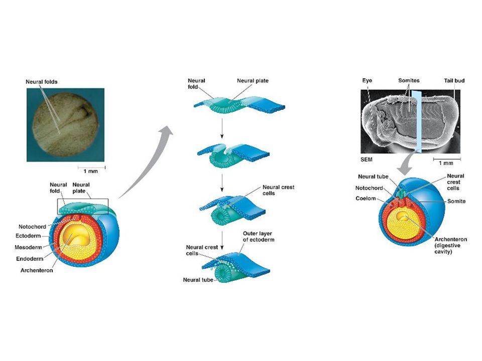
Week 3 Heart begins to forms by day 18
Primitive streak forms along back of embryo. This is a primitive notochord which will become spine Notochord induces cells of ectoderm to fold and form hollow neural tube which eventually becomes brain & spinal cord Neural tube seals around day 20.

14
What is a neural tube defect?
If NT does not seal, Neural Tube Defect (NTD) will form. Ex: Spina bifida Brain or spine is exposed Baby paralyzed from point of exposure down Occurs due to lack of vitamin B or folic acid Pregnant women encouraged to take folic acid supplements/vitamins during pregnancy to prevent NTD NTD can be detected around 15 weeks with alpha fetoprotein (AFP) blood test that detects a protein from fetus’ liver that is leaked at a fast rate If detected and caught early enough can sometimes be corrected with in utero surgery.
 will form. Ex: Spina bifida. Brain or spine is exposed. Baby paralyzed from point of exposure down. Occurs due to lack of vitamin B or folic acid. Pregnant women encouraged to take folic acid supplements/vitamins during pregnancy to prevent NTD. NTD can be detected around 15 weeks with alpha fetoprotein (AFP) blood test that detects a protein from fetus’ liver that is leaked at a fast rate. If detected and caught early enough can sometimes be corrected with in utero surgery.")
15
Week 4 Heart begins beating around day 23
Leg and arm buds begin to form Blood cells form & fill primitive blood vessels Immature lungs and kidneys develop

16
Week 5 & 6 Enlarged head Apoptosis sculpts fingers and toes
Eyes open but no eyelids or irises Gene SRY on y chromosome of a boy will begin forming male hormones that stimulate formation of male organs

17
Week 7 & 8 Skeleton of cartilage is formed
Embryo is about size of a paper clip After 8 weeks it is now called a fetus.

18
Weeks 9-12 Body proportions equal out Bone begins to replace cartilage
Fetus begins coordinating muscle and nerves and begins to move Fetus sucks thumb, kicks Urinates & defecates into amniotic sac Breathes in amniotic fluid Week 12 ends 1ST TRIMESTER

19
2nd Trimester Weeks 13-24 Hair Lanugo- downy hair all over body
Eyelashes Eyebrows Nipples Nails Week 15- gender determined Skin appears wrinkled due to lack of fat & pink with new capillary formation Can feel distinct movement Baby is about 9 inches long

20
3rd Trimester Weeks 25-40 Fetal brain cells grow and form numerous connections to organs Fat forms under skin Digestive & respiratory systems mature Premature babies often have respiratory diseases and have difficulty digesting milk

21
How can genetic disorders or birth defects be detected?
Chorionic villi sampling- cells are removed from chorionic villi and tested for abnormalities in number or shape of chromosomes. Done at 10 weeks Amniocentesis- fluid from amniotic sac is removed by needle and test for abnormalities Done at 14 weeks

22
How do multiples form? Monozygotic multiples- Dizygotic multiples-
Identical twins Fertilized egg splits Twins are genetically identical Share placenta Dizygotic multiples- Fraternal twins Two sperm fertilized two different eggs Twins are genetically different Different placentas

23
What are conjoined twins?
Separation of egg cell begins to occur while organs are developing. Attachment point depends on where split was occurring & what organs were developing at the time. Ex: Brittany & Abigail Hensel

24
Brittany & Abigail Hensel
Dicephalic twins- have two heads Share- liver, bloodstream, all organs below navel, 3 kidneys Separate- neck, head, heart, stomach, gallbladder, lungs, nervous system Parents chose not to separate b/c one child usually dies during surgery

Similar presentations