Download presentation
Presentation is loading. Please wait.

1
L. J. SALOMON, Z. ALFIREVIC, V. BERGHELLA, C. BILARDO, E
L. J. SALOMON, Z. ALFIREVIC, V. BERGHELLA, C. BILARDO, E. HERNANDEZ-ANDRADE, S. L. JOHNSEN, K. KALACHE, K.-Y. LEUNG, G. MALINGER, H. MUNOZ, F. PREFUMO, A. TOI and W. LEE on behalf of the ISUOG Clinical Standards Committee

2
Why ? The International Society of Ultrasound in Obstetrics and Gynecology (ISUOG) is a scientific organization that encourages sound clinical practice, teaching and research for diagnostic imaging in women’s healthcare. Practice Guidelines and Consensus are intended to reflect what is considered by ISUOG to be the best practices at the time at which they were issued. Although ISUOG has made every effort to ensure that guidelines are accurate when issued, neither the Society nor any of its employees or members accepts any liability for the consequences of any inaccurate or misleading data, opinions or statements issued by the CSC. Guidelines are not intended to establish a legal standard of care because interpretation of the evidence that underpins the guidelines may be influenced by individual circumstances and available resources. Approved guidelines can be distributed freely with the permission of ISUOG
 is a scientific organization that encourages sound clinical practice, teaching and research for diagnostic imaging in women’s healthcare. Practice Guidelines and Consensus are intended to reflect what is considered by ISUOG to be the best practices at the time at which they were issued. Although ISUOG has made every effort to ensure that guidelines are accurate when issued, neither the Society nor any of its employees or members accepts any liability for the consequences of any inaccurate or misleading data, opinions or statements issued by the CSC. Guidelines are not intended to establish a legal standard of care because interpretation of the evidence that underpins the guidelines may be influenced by individual circumstances and available resources. Approved guidelines can be distributed freely with the permission of ISUOG")
4
What is the purpose of a mid-trimester fetal ultrasound scan?
Provide accurate diagnostic information for the delivery of optimized antenatal care with the best possible outcomes for mother and fetus. The procedure is used to determine gestational age and to perform fetal measurements for the timely detection of are to detect congenital malformations and multiple pregnancies. Prenatal screening examination includes an evaluation of the following: cardiac activity; fetal number (and chorionicity if multiple pregnancy); fetal age/size; basic fetal anatomy; placental appearance and location. Although many malformations can be identified, it is acknowledged that some may be missed, even with sonographic equipment in the best of hands, or that they may develop later in pregnancy. Before starting the examination, a healthcare practitioner should counsel the woman/couple regarding the potential benefits and limitations of a routine mid-trimester fetal ultrasound scan.
; fetal age/size; basic fetal anatomy; placental appearance and location. Although many malformations can be identified, it is acknowledged that some may be missed, even with sonographic equipment in the best of hands, or that they may develop later in pregnancy. Before starting the examination, a healthcare practitioner should counsel the woman/couple regarding the potential benefits and limitations of a routine mid-trimester fetal ultrasound scan.")
5
Who should perform the scan?
In order to achieve optimal results from routine screening examinations, it is suggested that scans should be performed by individuals who fulfil the following criteria: trained in the use of diagnostic ultrasonography and related safety issues; regularly perform fetal ultrasound scans; participate in continuing medical education activities; have established appropriate referral patterns for suspicious or abnormal findings; routinely undertake quality assurance and control measures. If the examination cannot be performed completely in accordance with adopted guidelines, the scan should be repeated, at least in part, at a later time, or the patient can be referred to another practitioner. This should be done as soon as possible, to minimize unnecessary patient anxiety and unnecessary delay in the potential diagnosis of congenital anomalies or growth disturbances.

6
What equipment? For routine screening, equipment should have at least the following: real time, gray-scale ultrasound capabilities; transabdominal transducers (3–5-MHz range); adjustable acoustic power output controls with output display standards; freeze frame capabilities; electronic calipers; capacity to print/store images; regular maintenance and servicing, important for optimal equipment performance.
; adjustable acoustic power output controls with output display standards; freeze frame capabilities; electronic calipers; capacity to print/store images; regular maintenance and servicing, important for optimal equipment performance.")
7
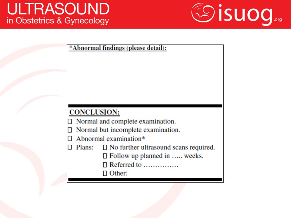
What document? An examination report should be produced as an electronic and/or a paper document, to be sent to the referring care provider in reasonable time. A sample reporting form is available at the end of this guidelines. Images of standard views (stored either electronically or as printed copies) should also be produced and stored. Motion videoclips are recommended for the fetal heart. Local laws should be followed. Many jurisdictions require image storage for a defined period of time.
 should also be produced and stored. Motion videoclips are recommended for the fetal heart. Local laws should be followed. Many jurisdictions require image storage for a defined period of time.")
8
Is prenatal ultrasonography safe?
Prenatal ultrasonography appears to be safe for clinical practice. To date, there has been no independently confirmed study to suggest otherwise. Fetal exposure times should be minimized, using the lowest possible power output needed to obtain diagnostic information, following the ALARA principle (As Low As Reasonably Achievable). More details are available from the ISUOG Safety Statement.
. More details are available from the ISUOG Safety Statement.")
10
Well being? Amniotic fluid assessment: Fetal movement:
Amniotic fluid volume can be estimated subjectively or using sonographic measurements. Subjective estimation is not inferior to the quantitative measurement techniques (e.g. deepest pocket, amniotic fluid index) when performed by experienced examiners. Fetal movement: Normal fetuses typically have a relaxed position and show regular movements. There are no specific movement and temporary absence or reduction of fetal movements during the scan should not be considered as a risk factor. Abnormal positioning or unusually restricted or persistently absent fetal movements may suggest abnormal fetal conditions such as arthrogryposis. The biophysical profile is not considered part of a routine mid-trimester scan.
 when performed by experienced examiners. Fetal movement: Normal fetuses typically have a relaxed position and show regular movements. There are no specific movement and temporary absence or reduction of fetal movements during the scan should not be considered as a risk factor. Abnormal positioning or unusually restricted or persistently absent fetal movements may suggest abnormal fetal conditions such as arthrogryposis. The biophysical profile is not considered part of a routine mid-trimester scan.")
11
Fetal biometry? The following sonographic parameters can be used to estimate gestational age and for fetal size assessment: biparietal diameter (BPD); head circumference (HC); abdominal circumference (AC) or diameter; femur diaphysis length (FDL). Measurements should be performed in a standardized manner on the basis of strict quality criteria. An audit of results can help to ensure accuracy of techniques with regard to specific reference tables. The chosen reference standards should be indicated in the report An image(s) should be taken to document the measurement(s). The application of Doppler techniques is not currently recommended as part of the routine second-trimester ultrasound examination. There is insufficient evidence to support universal use of uterine or umbilical artery Doppler evaluation for the screening of low-risk pregnancies.
; head circumference (HC); abdominal circumference (AC) or diameter; femur diaphysis length (FDL). Measurements should be performed in a standardized manner on the basis of strict quality criteria. An audit of results can help to ensure accuracy of techniques with regard to specific reference tables. The chosen reference standards should be indicated in the report. An image(s) should be taken to document the measurement(s). The application of Doppler techniques is not currently recommended as part of the routine second-trimester ultrasound examination. There is insufficient evidence to support universal use of uterine or umbilical artery Doppler evaluation for the screening of low-risk pregnancies.")
13
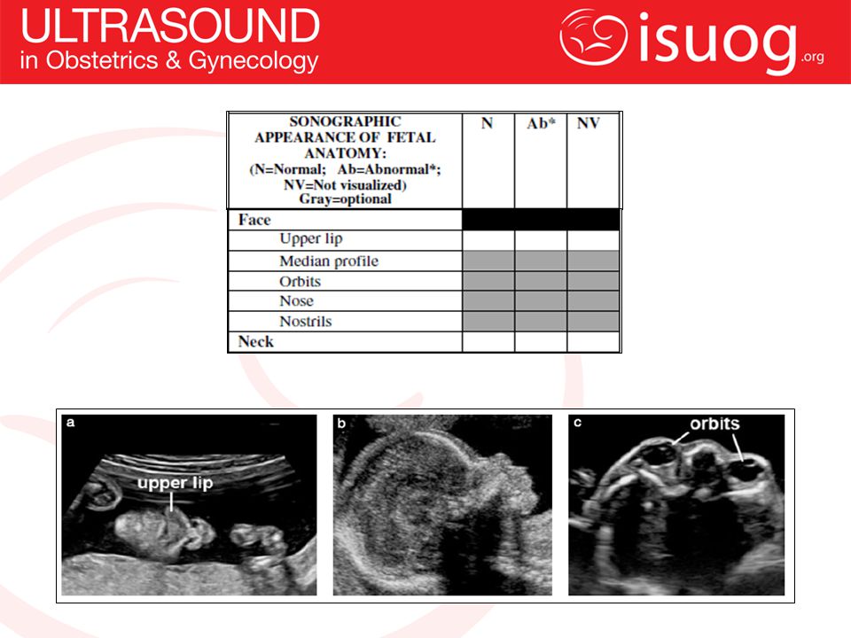
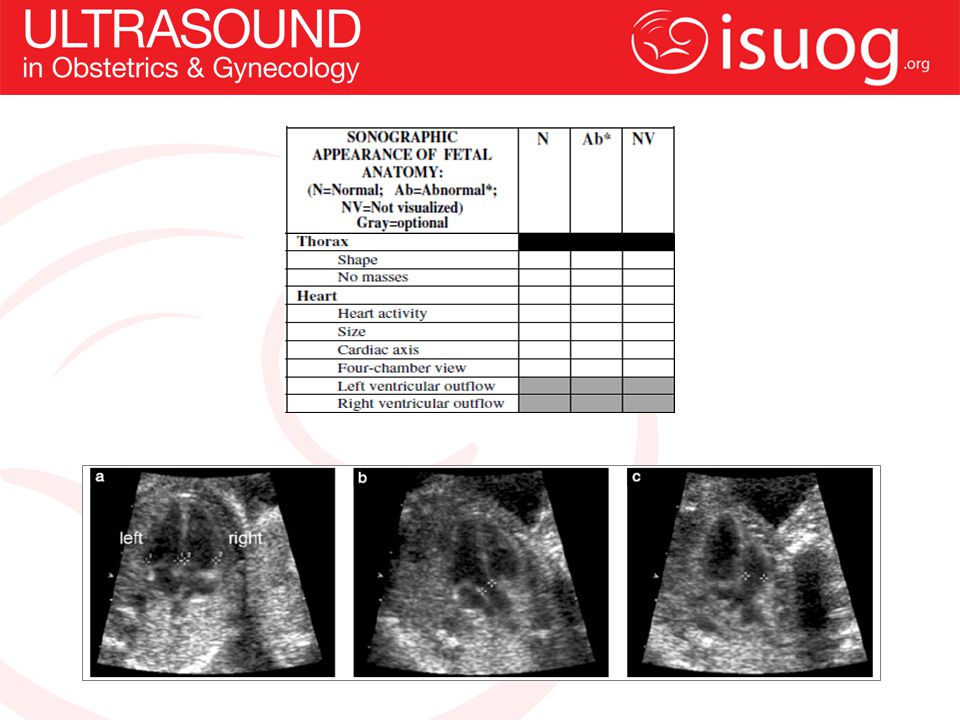
Fetal anatomy? Recommended minimum requirements for a basic fetal anatomical survey during the mid-trimester of pregnancy are summarized in the report form Survey includes: Head: skull, brain.. Face Neck Thorax: chest, heart. Abdomen/urinary tract. Skeletal Limbs Placenta, umbilical cord…

19
Cervix, uterine and adnexa?
Several studies have demonstrated a strong correlation between short cervical length on transvaginal scan and subsequent preterm birth. However, several randomized controlled trials that examined the combination of routine cervical length measurement and subsequent interventions (cerclage, progesterone) failed to demonstrate conclusively any cost-effectiveness of such screening programs. Currently, there is insufficient evidence to recommend routine cervical length measurements at the mid trimester in an unselected population. Uterine fibroids and adnexal masses should be documented if they are likely to interfere with labor.
 failed to demonstrate conclusively any cost-effectiveness of such screening programs. Currently, there is insufficient evidence to recommend routine cervical length measurements at the mid trimester in an unselected population. Uterine fibroids and adnexal masses should be documented if they are likely to interfere with labor.")
21
USE THE FORM!!

22
ISUOG’s website is a great resource for more information: www. isuog
ISUOG’s website is a great resource for more information:

Similar presentations


 Maternal Newborn Nursing Care. Forth Edition. Addison Wesley.>")


