Download presentation
Presentation is loading. Please wait.

1
Radiology Artifacts

2
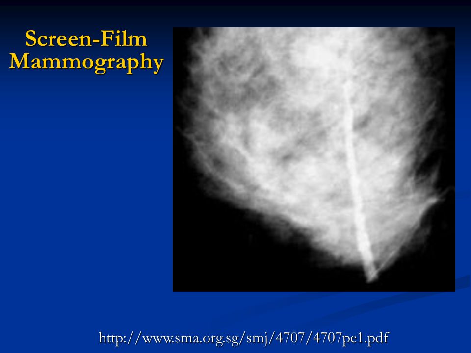
Screen-Film Mammography

3
Screen-Film Mammography
Pressure mark or crimp mark due to excessive pressure on film appears as increased optical density.

4
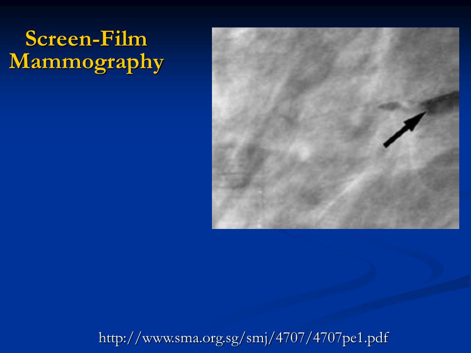
Screen-Film Mammography

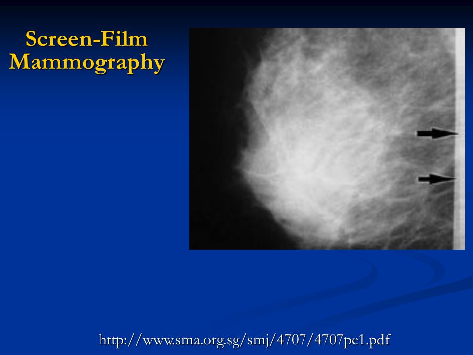
5
Screen-Film Mammography
Artifacts caused by dust and dirt on intensifying screen creating white spots simulating calcifications

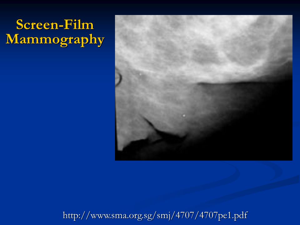
6
Screen-Film Mammography

7
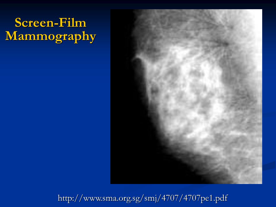
Screen-Film Mammography
Blurring due to motion

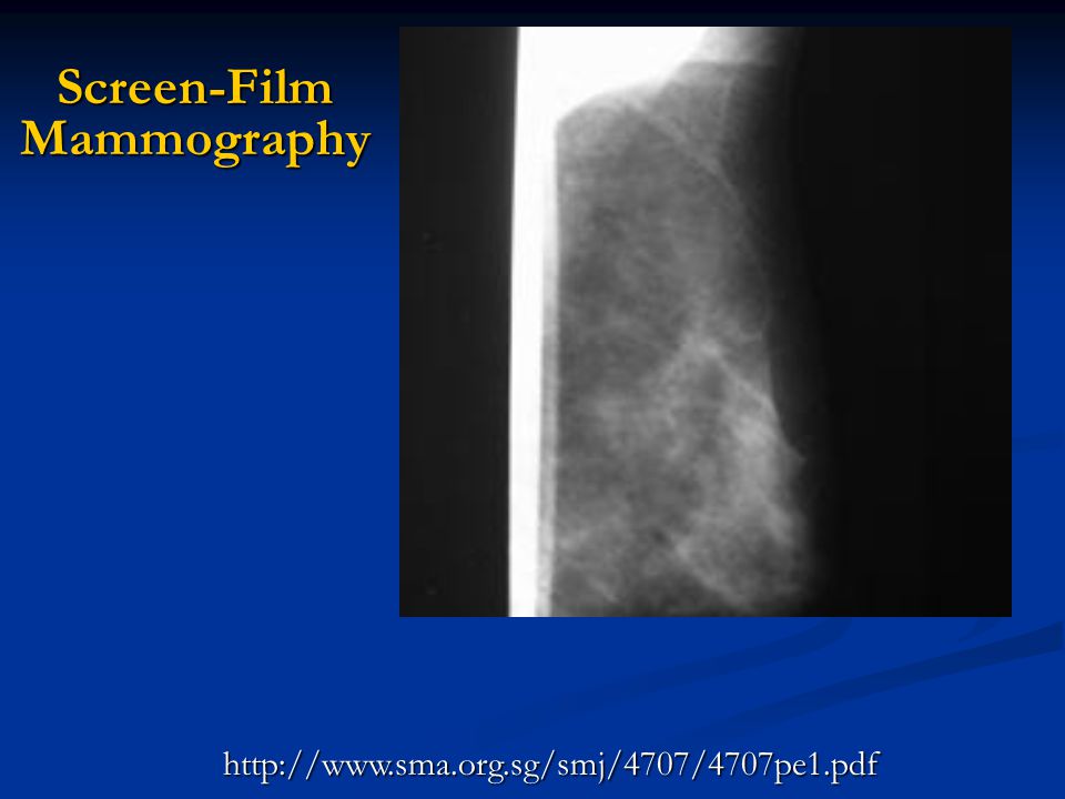
8
Screen-Film Mammography

9
Screen-Film Mammography
Fingerprints

10
Screen-Film Mammography
11
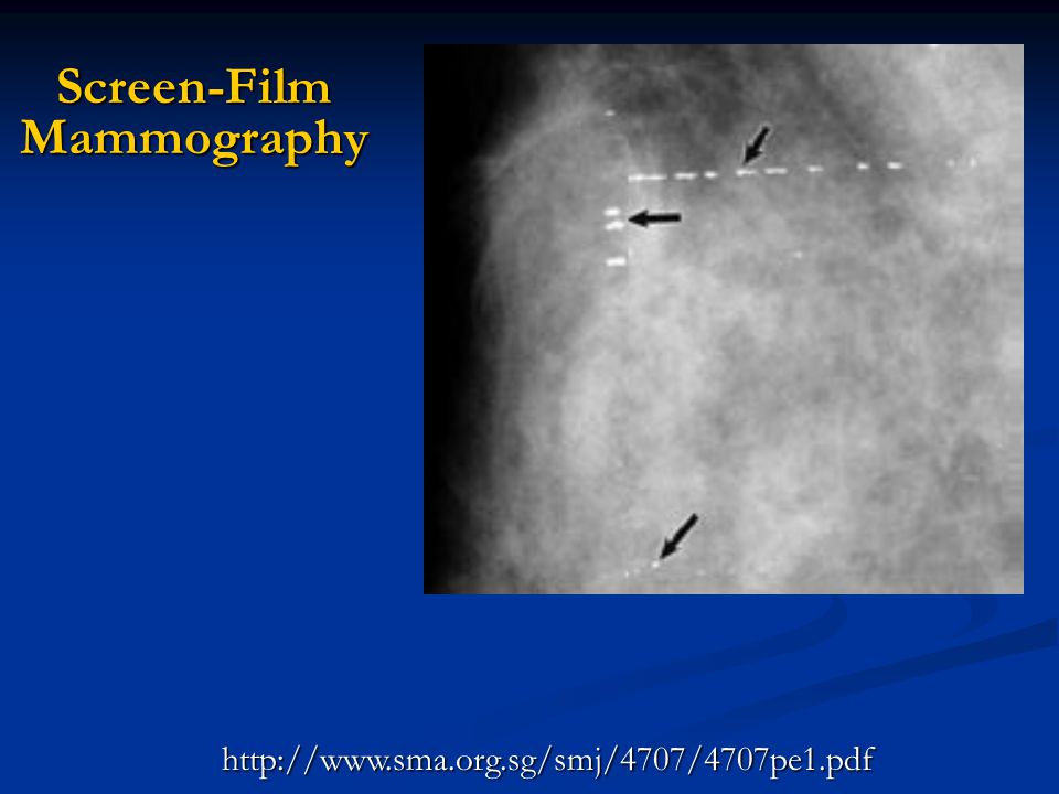
Screen-Film Mammography
Long streak of stain on intensifying screen appearing as minus density

12
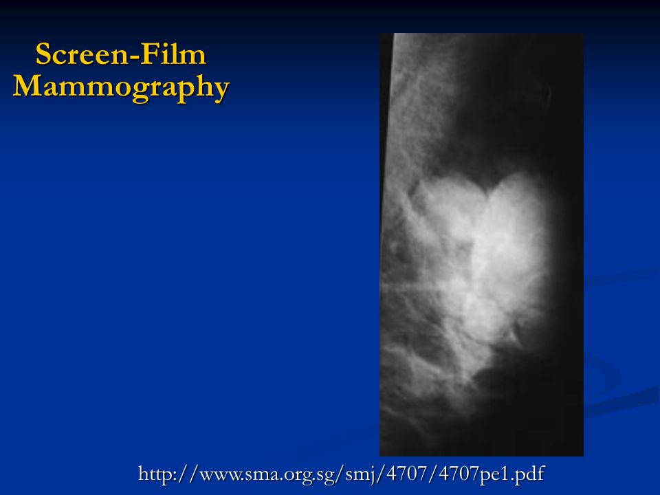
Screen-Film Mammography
Obvious underexposure but not caused by technique or processing

13
Screen-Film Mammography
Two films loaded into same cassette

14
Screen-Film Mammography
15
Screen-Film Mammography
Wet or damp screen causing plus density

16
Screen-Film Mammography
Easy one. Why is ID superimposed on breast?

17
Screen-Film Mammography
Cassette loaded backwards (front-back) into bucky tray.
 into bucky tray.")
18
Screen-Film Mammography
Obvious underexposure but not caused by technique or processing

19
Screen-Film Mammography
Film loaded upside down into cassette

20
Screen-Film Mammography
Easy?

21
Screen-Film Mammography
Cassette loaded upside down into bucky tray.

22
Screen-Film Mammography
23
Screen-Film Mammography
Light leak in cassette Incompletely latched crack

24
Screen-Film Mammography
25
Screen-Film Mammography
Double exposure

26
Screen-Film Mammography
27
Screen-Film Mammography
Compression paddle vertical section (anterior lip)
")
28
Screen-Film Mammography

29
Screen-Film Mammography
Films run through processor too close together (overlapped)
")
30
Screen-Film Mammography
31
Screen-Film Mammography
Static electricity

32
Screen-Film Mammography

33
Screen-Film Mammography
Emulsion pick-off caused by pulling stuck films apart

34
Screen-Film Mammography

35
Screen-Film Mammography
Black arrows point at vertical black lines perpendicular to direction of film travel through processor. Cause: pressure from developer roller or moisture on entrance roller.

36
Screen-Film Mammography
37
Screen-Film Mammography
Dust in darkroom and dirty rollers causing emulsion pick-off.

38
Screen-Film Mammography
39
Screen-Film Mammography
Over-development caused by chemical fog.

40
Two images of same anatomy. What changed?
Radiography Two images of same anatomy. What changed?

41
Radiography No grid 102 cm SID .33R ESE Grid 122 cm SID 2.2 R ESE

42
CR What are white spots?

43
Dust / dirt on phosphor screen

44
Dirt on CR plate. Right image repeated on screen-film

45
L a s e r R o t i n g M B m c d f l P T u b A / D C p h Plate Travel

46
CR What are white lines?

47
Cracks on image plate due to mechanical wear

48
CR

49
Transport problems in CR reader
g M B m c d f l P T u b A / D C p h Plate Travel

50
What are funny dark patterns on ventilator tube metal stabilizer?
CR What are funny dark patterns on ventilator tube metal stabilizer?

51
CR Moire effect because of interference between scan frequency of matrix and spiral

52
CR

53
CR Diagonal collimation caused algorithm to improperly recognize collimated area

54
CR

55
CR Chest incorrectly specified by technologist to CR reader as lumbar spine

56
There’s flow but it isn’t showing up. Why?
Ultrasound There’s flow but it isn’t showing up. Why?

57
Ultrasound Improper setting of velocity scale (set too high). Low flow not displayed

58
What’s that funny reversed flow?
Ultrasound What’s that funny reversed flow?

59
Improper setting of velocity scale (set too low). Aliasing occurs
Ultrasound Improper setting of velocity scale (set too low). Aliasing occurs
. Aliasing occurs.")
60
Low flow in both directions?
Ultrasound Low flow in both directions?

61
Ultrasound Doppler angle is 90o; sample volume has vessel perpendicular to beam. Right image alters Doppler angle.

62
Same anatomy. Why is there spectral broadening on the left image?
Ultrasound Same anatomy. Why is there spectral broadening on the left image?

63
Minimize spectral broadening artifact by using Doppler angles < 60o
Ultrasound 71o Doppler angle 81o Doppler angle Minimize spectral broadening artifact by using Doppler angles < 60o

Similar presentations


 BPKIHS,Dharan.>")


