Download presentation
Presentation is loading. Please wait.

2
Pathogenesis and clinical consequences of endodontic infections
Dag Ørstavik Department of Endodontics Institute of Clinical Dentistry Dental Faculty University of Oslo, Norway Expertise Economics Ethics Education

3
Pathogenesis Greek, pathos + genesis, disease + creation ..the development of morbid conditions or of disease; more specifically the cellular events and reactions and other pathologic mechanisms occurring in the development of disease (medical dictionary) Etiology: Gk, aitia, cause, logos, science = agent(s) initiating pathogenesis
 Etiology: Gk, aitia, cause, logos, science = agent(s) initiating pathogenesis.")
4
Pulpitis and Apical Periododntitis
= manifestations of the body’s response to tooth infection

5
Pulpitis and Apical Periododntitis
= manifestations of the body in response to tooth infection = the disease entities as experienced by the individual

6
Pulpitis and Apical Periododntitis
= manifestations of the body in response to tooth infection = the disease entities as experienced by the individual provide us with diagnostic tools to assess the extent and severity of endodontic infections

7
Changing concepts 1940: Grossman: Endodontic Practice
1998: Ørstavik/Pitt Ford: Essential Endodontology: Prevention and Treatment of Apical Periodontitis 2011: Siqueira: Treatment of Endodontic Infections

8
start -> 1987 -> > Resorptions & trauma necessitate treatment for prevention of apical periodontitis

9
-> 1987 -> > In practice, focus remain on: Prevention and Treatment of Apical Periodontitis. (Tronstad 1988; Ørstavik 1988) We cannot see the infection if it is there; only its manifestation by clinical and radiographic signs and symptoms
 We cannot see the infection if it is there; only its manifestation by clinical and radiographic signs and symptoms.")
10
Pulpitis and apical periodontitis: the primordial dental afflictions
Pulpitis may be a nuisance, but is essentially a harmless step on the way to an oral infection

12
Etiology and pathogenesis
No caries in germ-free animals Traumatic/irritative pulpitis dismissed in the 1970’s: Brännström et al. Kakehashi et al. 1965 Bergenholtz 1974; Katz 1974; Sundqvist 1976 Möller et al 1982

13
Kakehashi et al. 1965 ”You can fill a microbe-free root with anything, as long as it is sterile” Sundqvist 1976 ”If there is no lesion, there is no infection”

14
Radiographic examination Histologic examination
Infectious status of pulp Clinical examination Radiographic examination Histologic examination Non-infected, lacerated pulps 0/26 2*/24 Infected, lacerated pulps 12/52 47/52 10/10 * scattered leukocytes Möller et al 1982

15
Etiology Kakehashi et al. 1965 Bergenholtz 1974; Katz 1974; Sundqvist 1976 Möller et al 1982 The etiology is confirmed by the (almost) universal observation that absence of cultivable bacteria at the time of filling ensures optimal (not 100%) prognosis
 universal observation that absence of cultivable bacteria at the time of filling ensures optimal (not 100%) prognosis.")
16
Etiology Kakehashi et al. 1965 Bergenholtz 1974; Katz 1974; Sundqvist 1976 Möller et al 1982 The etiology is confirmed by the (almost) universal confirmation that absence of cultivable bacteria at the time of filling ensures optimal (not 100%) prognosis The etiology needs confirmation because it is forgotten at the earliest convenience! Necrotic tissue Tissue remnants – smear and debris Overinstrumentation – tissue damage Materials and medicaments (AAE Colleagues for Excellence Winter 2011: ”The major causes of pulpal and periapical diseases are living and nonliving irritants. The latter group includes mechanical, thermal and chemical irritants.”)
 universal confirmation that absence of cultivable bacteria at the time of filling ensures optimal (not 100%) prognosis. The etiology needs confirmation because it is forgotten at the earliest convenience! Necrotic tissue. Tissue remnants – smear and debris. Overinstrumentation – tissue damage. Materials and medicaments. (AAE Colleagues for Excellence Winter 2011: The major causes of pulpal and periapical diseases are living and nonliving irritants. The latter group includes mechanical, thermal and chemical irritants. )")
17
What do we do? and when? Caries removal ”reversible pulpitis”
Pulp capping ”reversible pulpitis” Pulp amputation ”limited[?] pulpitis” Pulp extirpation ”irreversible pulpitis” Treatment of the ”pulp necrosis” necrotic pulp ”apical periodontitis” Apical surgery ”persistent apical periodontitis”

18
What we should do when Caries removal: bacteria in dentin only
Pulp capping: bacteria near but not into pulp Pulp amputation: superficial pulp infection Pulp extirpation: bacteria infecting coronal pulp Canal disinfection: bacteria infesting pulp space Apical surgery: bacteria in inaccessible areas

19
What we should do when The issue is: Where are the baceria?
Caries removal: bacteria in dentin only Pulp capping: bacteria near but not into pulp Pulp amputation: superficial pulp infection Pulp extirpation: bacteria infecting coronal pulp Canal disinfection: bacteria infesting pulp space Apical surgery: bacteria in inaccessible areas Therefore: ”Prevention and Treatment of Apical Periodontitis” or ”Treatment of Endodontic Infections” The issue is: Where are the baceria?

20
Etiology and Pathogenesis
Etiologic agents and situations Microbes from caries Microbes from trauma Microbes from leakage Body responses Dentin reactions Pulpal reactions Vascular events Cellular events Periapical reactions Spread and containment

21
Dentin reactions Microbes from caries; Dentine and pulpal reactions

22
Pulpal reactions Tertiary dentin (reactionary or reparative or irregular secondary dentin) is the outcome of odontoblastic response to irritation occurring mainly during secondary dentinogenesis and is caused by dental abrasion, attrition, cavity preparation, erosion or dental caries (Torneck 1994). Lesot et al. (1993) defines reactionary dentin to be the result of irritation of post-mitotic odontoblasts, whereas reparative dentin is formed by odontoblasts or odontoblast-like cells which differentiate from pulp cells after the cell death of primary odontoblasts (Magloire et al. 1992, Magloire et al. 1996).
 is the outcome of odontoblastic response to irritation occurring mainly during secondary dentinogenesis and is caused by dental abrasion, attrition, cavity preparation, erosion or dental caries (Torneck 1994). Lesot et al. (1993) defines reactionary dentin to be the result of irritation of post-mitotic odontoblasts, whereas reparative dentin is formed by odontoblasts or odontoblast-like cells which differentiate from pulp cells after the cell death of primary odontoblasts (Magloire et al. 1992, Magloire et al. 1996).")
23
Pulpal reactions 0.5 0.25 Minimal Maximal
reactionary dentin formation by odontoblasts reparative dentin (bridge) formation by recruited blast cells Dahl & Ørstavik 2008 based on Murray PE et al. Am J Dent Feb;15(1):41-6.
 formation by recruited blast cells. Dahl & Ørstavik 2008 based on Murray PE et al. Am J Dent Feb;15(1):41-6.")
24
Pulpal reactions Toxic substances, bacterial products, trauma
Lymphocyte recruitement Increase vascular permeability Recruitment of dendritic cells Chemokines; CCs, CXs MMPs, TGF-β1, BMP-2 VEGF Productive: mineralization Odontoblasts and dentin release bioactive substances and chemokines Modified from Dahl & Ørstavik 2008

25
Odontoblasts in Innate Defense Farges et al. 2009
Pulpal reactions Odontoblasts in Innate Defense Farges et al. 2009 Pattern Recognition Receptors (PRRs), including receptors for PAMPs Toll-like Receptors (TLRs) are an important subclass of PRRs. 10 in humans (2009), also odontoblasts Trigger the NF-κB pathway: (nuclear factor kappa-light-chain-enhancer of activated B cells) Proinflammatory cytokines Antimicrobial peptides, incl defensins Maturation of dendritic cells
, including receptors for PAMPs. Toll-like Receptors (TLRs) are an important subclass of PRRs. 10 in humans (2009), also odontoblasts. Trigger the NF-κB pathway: (nuclear factor kappa-light-chain-enhancer of activated B cells) Proinflammatory cytokines. Antimicrobial peptides, incl defensins. Maturation of dendritic cells.")
26
Odontoblasts in Innate Defense Farges et al. 2009
TLR1 to 6 and TLR9 in odontoblasts may recognize PAMPs such as Triacylated lipoproteins – TLR1, TLR2 LTAs, diacylated lipopeptides – TLR2, TLR6 LPS – TLR4 Viralt RNA – TLR3 Flagellin – TLR5 They participate in a balance between induction and suppression of immune responses All genes are expressed in the normal pulp; ie, ready for action! Pulpal reactions

27
DAMPs derived from mitochondria
DAMPs derived from mitochondria. The mitochondrion is a cellular organelle that might function as a source of mito-DAMPs that are released during different modes of cell death (apoptosis, secondary necrosis, or necrosis) and tissue injury. (a) Once these mito-DAMPs are released into the extracellular space, they can stimulate the innate and adaptive immune responses. *Note that there are also extra-mitochondrial sources of ATP. (b) Intact mitochondria derived from cells dying by accidental necrosis after mechanical disruption can induce IL-1β production by macrophages, and attract neutrophils upon i.p. injection [63]. IL-1β production and neutrophil influx are not significantly decreased in P2X7R-deficient mice, which indicates that DAMPs other than ATP might contribute to these responses. Question marks indicate links that are not yet proven. (Krysko et al 2011)
 and tissue injury. (a) Once these mito-DAMPs are released into the extracellular space, they can stimulate the innate and adaptive immune responses. *Note that there are also extra-mitochondrial sources of ATP. (b) Intact mitochondria derived from cells dying by accidental necrosis after mechanical disruption can induce IL-1β production by macrophages, and attract neutrophils upon i.p. injection [63]. IL-1β production and neutrophil influx are not significantly decreased in P2X7R-deficient mice, which indicates that DAMPs other than ATP might contribute to these responses. Question marks indicate links that are not yet proven. (Krysko et al 2011)")
28
NF-κB pathway Pulpal reactions Proinflammatory cytokines
TNF-α, IL-1β, increased after LPS stimulation in pulp Chemokines: regulate attraction and emigration of leukocytes; angiogenesis, apoptosis. Homeostatic chemokines found in pulp including odontoblasts; inflammatory cs only from accumulating inflammatory cells. Antimicrobial peptides Defensins increased in odontoblast-like cells in culture after, eg, LPS stimulation Maturation of dendritic cells The CCL2 chemokine is produced by odontoblasts under caries and may attract dendritic cells to the odontoblast layer and activate them

29
Bacterial effects on dentinogenesis
Pulpal reactions Bacterial effects on dentinogenesis LTA downregulates collagen type1 synthesis (Farges) Dentin sialophosphoprotein gene expression abolished (blocks ->phosphoprotein->hydroxyapatite) TGF-β1 (”the modulator”) downregulated: TGF-β1 attenuates TLR signalling; its absence opens for immune activation by more active TLR signalling TGF-β1 also inhibits responses and activation of T helper and B cells; its absence may thus also increase this side of the immune response.
 Dentin sialophosphoprotein gene expression abolished (blocks ->phosphoprotein->hydroxyapatite) TGF-β1 ( the modulator ) downregulated: TGF-β1 attenuates TLR signalling; its absence opens for immune activation by more active TLR signalling. TGF-β1 also inhibits responses and activation of T helper and B cells; its absence may thus also increase this side of the immune response.")
30
Dentin infection summary
Tubule occlusion and precipitation Odontoblast stimulation -> death -> repair Direct (toxins, LPS) and indirect activation of innate and specific immune responses PAMPs & DAMPs -> TLRs-> TNF-α, IL-1β; chemokines Recruitement of phagocytes for innate response Stimulation/maturation of antigen-presenting cells, notably dendritic cells
 and indirect activation of innate and specific immune responses. PAMPs & DAMPs -> TLRs-> TNF-α, IL-1β; chemokines. Recruitement of phagocytes for innate response. Stimulation/maturation of antigen-presenting cells, notably dendritic cells.")
31
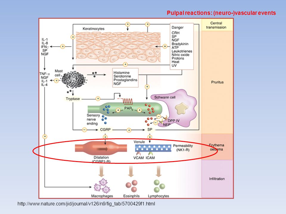
Initial, neurovascular events
Pulpal reactions: (neuro-)vascular events Initial, neurovascular events Activation of nerves Triggers secretion of neuropeptides Increased vasodilation Chemoattractants
vascular events. Initial, neurovascular events. Activation of nerves. Triggers secretion of neuropeptides. Increased vasodilation. Chemoattractants.")
32
Pulpal reactions: (neuro-)vascular events
Heyeraas & Mjør in Ørstavik & Pitt Ford 2008

33
Pulpal reactions: (neuro-)vascular events
34
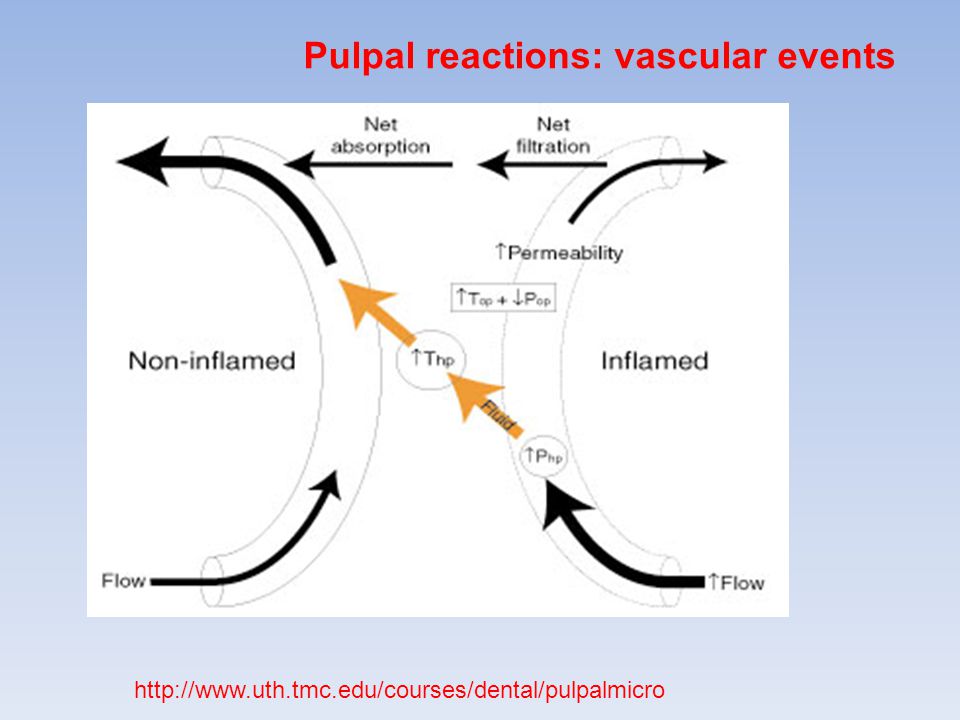
Pulpal reactions: vascular events

35
Pulpal reactions: vascular events
36
reversible-irreversible
Pulpal reactions: cellular events Vital, inflamed: reversible-irreversible microabscesses induced by carious dentin: They could be ”irreversible pulpitis”, but heal anyway: Irreversibility is when the bacteria are inside. Mjør & Tronstad 1972

40
Establishment and progression of the pulpal infection
Pulpal reactions: cellular events Establishment and progression of the pulpal infection Necrosis and infection Little info available on progression through pulp Biofilms and infestation of accessory canals Dental tubule infection Cementum surface colonization Granuloma formation Epithelial stimulation Abscess and sinus tract formation

41
Ørstavik, Essential Endodontology 1998; courtesy of Lambjerg Hansen & Langeland

42
Fig 1. Electron photomicrograph of the apical pulp from a tooth with irreversible pulpitis. Note the swollen, distended endothelial cells (E) with increased rough endoplasmic reticulum (RE) and mitochondria (M). Red blood cells (RBC's) are packed within the lumen. Reference bar represents 2 µm (uranyl acetate, lead citrate; original magnification x9,117). Mendoza et al 1987
 with increased rough endoplasmic reticulum (RE) and mitochondria (M). Red blood cells (RBC s) are packed within the lumen. Reference bar represents 2 µm (uranyl acetate, lead citrate; original magnification x9,117). Mendoza et al")
43
Fig 5. Electron photomicrograph of an experimental pulp exhibiting degradation of perivascular collagen (C). Also noted is a degenerated rnyelinated axon (Ax), degranulation of a myelin sheath (arrow), and a degenerating fibroblast (F). Reference bar represents 2 ~m (uranyl acetate, lead citrate; original magnification x6,860). Mendoza et al 1987 Note: The relationship between bacteria and tissues are poorly characterized histologically
. Mendoza et al Note: The relationship between bacteria and tissues are poorly characterized histologically.")
44
Intracanal biofilms; Riccucci & Siqueira 2010

45
Figure 3. The primary body cells involved in the pathogenesis of apical periodontitis.
The primary body cells involved in the pathogenesis of apical periodontitis. Neutrophils (NG in a) in combat with bacteria (BA) in an exacerbating apical periodontitis. Lymphocytes (LY in b) are the major components of chronic apical periodontitis, but their subpopulations cannot be identified on a structural basis. Plasma cells (PL in c) form a significant component of chronic asymptomatic lesions. Note the highly developed rough endoplasmic reticulum of the cytoplasm and the localized condensation of heterochromatin subjacent to the nuclear membrane, which gives the typical ‘cartwheel’ appearance in light microscopy. Macrophages (MA in d) are voluminous cells with elongated or U-forming nuclei and cytoplasm with rough endoplasmic reticulum. Magnifications: (a,b,c,d) 3900x. (From Nair, 1998b.) The primary body cells involved in the pathogenesis of apical periodontitis. Neutrophils (NG in a) in combat with bacteria (BA) in an exacerbating apical periodontitis. Lymphocytes (LY in b) are the major components of chronic apical periodontitis, but their subpopulations cannot be identified on a structural basis. Plasma cells (PL in c) form a significant component of chronic asymptomatic lesions. Note the highly developed rough endoplasmic reticulum of the cytoplasm and the localized condensation of heterochromatin subjacent to the nuclear membrane, which gives the typical ‘cartwheel’ appearance in light microscopy. Macrophages (MA in d) are voluminous cells with elongated or U-forming nuclei and cytoplasm with rough endoplasmic reticulum. Magnifications: (a,b,c,d) 3900x. (From Nair, 1998b.) Nair P CROBM 2004;15: Copyright © by International & American Associations for Dental Research
 in combat with bacteria (BA) in an exacerbating apical periodontitis. Lymphocytes (LY in b) are the major components of chronic apical periodontitis, but their subpopulations cannot be identified on a structural basis. Plasma cells (PL in c) form a significant component of chronic asymptomatic lesions. Note the highly developed rough endoplasmic reticulum of the cytoplasm and the localized condensation of heterochromatin subjacent to the nuclear membrane, which gives the typical ‘cartwheel’ appearance in light microscopy. Macrophages (MA in d) are voluminous cells with elongated or U-forming nuclei and cytoplasm with rough endoplasmic reticulum. Magnifications: (a,b,c,d) 3900x. (From Nair, 1998b.) The primary body cells involved in the pathogenesis of apical periodontitis. Neutrophils (NG in a) in combat with bacteria (BA) in an exacerbating apical periodontitis. Lymphocytes (LY in b) are the major components of chronic apical periodontitis, but their subpopulations cannot be identified on a structural basis. Plasma cells (PL in c) form a significant component of chronic asymptomatic lesions. Note the highly developed rough endoplasmic reticulum of the cytoplasm and the localized condensation of heterochromatin subjacent to the nuclear membrane, which gives the typical ‘cartwheel’ appearance in light microscopy. Macrophages (MA in d) are voluminous cells with elongated or U-forming nuclei and cytoplasm with rough endoplasmic reticulum. Magnifications: (a,b,c,d) 3900x. (From Nair, 1998b.) Nair P CROBM 2004;15: Copyright © by International & American Associations for Dental Research.")
46
Intracanal biofilms; Riccucci & Siqueira 2010 [b]..A biofilm is present covering the root canal walls, and a dense bacterial aggregate is evidenced more coronally. Empty spaces are shrinkage artefacts (original magnification ×100). (c) Higher magnification of the area demarcated by the rectangle in b. Bacterial filamentous forms prevail, and the extracellular component is abundant at this level (original magnification ×400). (d) Higher magnification of the bacterial aggregate indicated by the arrow in b. Different morphotypes are present. Note the concentration of polymorphonuclear neutrophils in contact with the biofilm surface (original magnification ×400).
. (c) Higher magnification of the area demarcated by the rectangle in b. Bacterial filamentous forms prevail, and the extracellular component is abundant at this level (original magnification ×400). (d) Higher magnification of the bacterial aggregate indicated by the arrow in b. Different morphotypes are present. Note the concentration of polymorphonuclear neutrophils in contact with the biofilm surface (original magnification ×400).")
47
Riccucci & Siqueira 2010: Maxillary lateral incisor with a large periapical radiolucency (>5 mm) (inset). (a and b) The section taken at the transition between the apical and the middle third showing a bacterial biofilm covering the dentinal walls. The dentinal tubules subjacent to the biofilm are heavily invaded and colonized to varying depths (Taylor's modified Brown and Brenn, original magnification ×100 and ×400).
 The section taken at the transition between the apical and the middle third showing a bacterial biofilm covering the dentinal walls. The dentinal tubules subjacent to the biofilm are heavily invaded and colonized to varying depths (Taylor s modified Brown and Brenn, original magnification ×100 and ×400)..")
48
varying depths: Valderhaug 1974

49
Langeland and, particularly, Tronstad have shown extensive colonization of necrotic cementum

50
Enterococci in dentinal tubules Love & Jenkinson 2002
Streptococcal invasion of dentinal tubules (upper diagram) and co-invasion with P. gingivalis (lower diagram). Streptococcal cells (•) adhere to unmineralized collagen type I ( ) via antigen I/II polypeptide adhesin (▴). Growth of streptococci in the presence of collagen peptides leads to up-regulation of antigen I/II production (▴), long-chaining of cells, and colonization along the length of the tubule. In the lower diagram, P. gingivalis cells (•) and S. gordonii cells both adhere to collagen (1), but P. gingivalis is unable to penetrate the tubules further in monoculture. The presence of S. gordonii (2) provides an additional binding substrate for P. gingivalis and promotes intratubular colonization by P. gingivalis. Up-regulation of streptococcal antigen I/II adhesin production (3) provides additional binding sites for P. gingivalis. These bacteria remain in association with the streptococci (4), and the dentinal tubules become invaded by a mixed bacterial population. Haapasalo & Ørstavik 1987: Enterococci in dentinal tubules Love & Jenkinson 2002
 and co-invasion with P. gingivalis (lower diagram). Streptococcal cells (•) adhere to unmineralized collagen type I ( ) via antigen I/II polypeptide adhesin (▴). Growth of streptococci in the presence of collagen peptides leads to up-regulation of antigen I/II production (▴), long-chaining of cells, and colonization along the length of the tubule. In the lower diagram, P. gingivalis cells (•) and S. gordonii cells both adhere to collagen (1), but P. gingivalis is unable to penetrate the tubules further in monoculture. The presence of S. gordonii (2) provides an additional binding substrate for P. gingivalis and promotes intratubular colonization by P. gingivalis. Up-regulation of streptococcal antigen I/II adhesin production (3) provides additional binding sites for P. gingivalis. These bacteria remain in association with the streptococci (4), and the dentinal tubules become invaded by a mixed bacterial population. Haapasalo & Ørstavik 1987: Enterococci in dentinal tubules. Love & Jenkinson")
51
An endodontic disease model related to virulence factors of E. faecalis. The virulence factors of the bacterium inside the dentinal tubules and the root canal are released to the periradicular area, where they elicit leukocyte attraction or stimulate leukocytes to produce inflammatory mediators or lytic enzymes. Some of the bacteria may translocate to the periradicular lesion as well. The injurious virulence factors and leukocyte products are shown in the zone between the interrupted lines. In a magnified window, the adhesion of the bacterium to diverse elements of the dentin is depicted. Bacterial products fighting other bacteria are also included. Note that names in black boxes are the products of the bacterium. Abbreviations: Adh, surface adhesins; AS, aggregation substance; Bact, bacteriocins; BS, binding substance; CP, collagen peptides; Cyl, cytolysin; Ef, Enterococcus faecalis; Elas, elastase; Gel, gelatinase; Hya, hyaluronidase; H2O2, hydrogen peroxide; IFN-γ, gamma interferon; IL, interleukin; LE, lysosomal enzymes; LTA, lipoteichoic acid; NO, nitric oxide; O2.−, superoxide anion; PGE2, prostaglandin E2; SP, sex pheromones; and TNF, tumor necrosis factor. O2.−: The dot denotes the presence of an unpaired electron, and the superscript denotes the negative charge. Kayaoglu & Ørstavik 2004

52
In a magnified window, the adhesion of the bacterium to diverse elements of the dentin is depicted.
Ef, Enterococcus faecalis; Adh, surface adhesins; AS, aggregation substance; LTA, lipoteichoic acid Adhesion causes resistance; so does plaque/biofilm formation Kayaoglu & Ørstavik 2004

53
Granuloma formation: Asymtomatic chronic apical periodontitis is also referred to as solid dental or periapical granuloma. Histopathologically the lesion consists of a granulomatous tissue with infiltrate cells, fibroblasts and a well-developed fibrous capsule. (Nair 2008) Riccucci & Siqueira 2010
 Riccucci & Siqueira")
54
Although the reported prevalence of cysts among apical periodontitis lesions varies from 6 to 55% .., investigations based on meticulous serial sectioning and strict histopathological criteria show that the actual prevalence of the cysts may be well below 20%.... During the first phase the dormant epithelial cell-rests are believed to proliferate, probably under the influence of growth factors that are released by various cells residing in the lesion. [integrin expression for cell adherence in an established lining] Nair 2008
![Although the reported prevalence of cysts among apical periodontitis lesions varies from 6 to 55% .., investigations based on meticulous serial sectioning and strict histopathological criteria show that the actual prevalence of the cysts may be well below 20%.... During the first phase the dormant epithelial cell-rests are believed to proliferate, probably under the influence of growth factors that are released by various cells residing in the lesion. [integrin expression for cell adherence in an established lining]](http://slideplayer.com/slide/3898202/13/images/54/Although+the+reported+prevalence+of+cysts+among+apical+periodontitis+lesions+varies+from+6+to+55%25+..%2C+investigations+based+on+meticulous+serial+sectioning+and+strict+histopathological+criteria+show+that+the+actual+prevalence+of+the+cysts+may+be+well+below+20%25....+During+the+first+phase+the+dormant+epithelial+cell-rests+are+believed+to+proliferate%2C+probably+under+the+influence+of+growth+factors+that+are+released+by+various+cells+residing+in+the+lesion.+%5Bintegrin+expression+for+cell+adherence+in+an+established+lining%5D.jpg "Nair")
55
Summary of pathogenesis
Microbial ingress into dentin and pulp canal system essential A multitude of bioreactions primarily geared at killing and containing microbes Dentinal sclerosis Reactionary and reparative dentine formation Neural and vascular responses for localization of inflammation and evasion of strangulation Secondary immune responses for containment of pathogens Establishment of battlefield by granuloma formation after necrosis and tubule infection Activation of epithelium for walling off the lesion into a cyst Now for Clinical Manifestations

56
The National Museum, Reykjavik, Iceland 1998
Historical ”A skull of a woman from a heathen grave at Hólaskógi in Thjórsárdal. It is probable that a dental infection in the upper jaw was the cause of her death. ” The National Museum, Reykjavik, Iceland 1998

57
Toothaches and death. Pulpitis & apical periodontitis Clarke JH.
J Hist Dent Mar;47(1):11-3. Toothaches and death. Clarke JH. Department of Behavioral Sciences, Oregon Health Sciences University, Portland, USA. Abstract Deaths from dental abscesses today are so rare, that it is difficult to fathom that only 200 years ago, this was a leading cause of death. When the London (England) Bills of Mortality began listing the causes of death in the early 1600's, "teeth" were continually listed as the fifth or sixth leading cause of death. (This does not include the category of "Teething" which was probably erroneously blamed for many children's deaths.) As we examine several historic factors of this period, it is apparent that the number of deaths attributed to "teeth" in the seventeenth and eighteenth centuries was probably fairly accurate, and it was not antibiotics, nor the discovery of asepsis, that brought about the dramatic reduction in these dental mortalities, but two much earlier dental innovations…. “anatomic forceps” Cyrus Fay 1826, “anesthesia” Horace Wells 1844
:11-3. Toothaches and death. Clarke JH. Department of Behavioral Sciences, Oregon Health Sciences University, Portland, USA. Abstract. Deaths from dental abscesses today are so rare, that it is difficult to fathom that only 200 years ago, this was a leading cause of death. When the London (England) Bills of Mortality began listing the causes of death in the early 1600 s, teeth were continually listed as the fifth or sixth leading cause of death. (This does not include the category of Teething which was probably erroneously blamed for many children s deaths.) As we examine several historic factors of this period, it is apparent that the number of deaths attributed to teeth in the seventeenth and eighteenth centuries was probably fairly accurate, and it was not antibiotics, nor the discovery of asepsis, that brought about the dramatic reduction in these dental mortalities, but two much earlier dental innovations…. anatomic forceps Cyrus Fay 1826, anesthesia Horace Wells")
58
"London's Bill of Mortality (December 1664-December 1665) [Official Document]," in Children and Youth in History, Item #159, (accessed February 8, 2011). Annotated by Lynda Payne
![London s Bill of Mortality (December 1664-December 1665) [Official Document], in Children and Youth in History, Item #159, (accessed February 8, 2011).](http://slideplayer.com/slide/3898202/13/images/58/London+s+Bill+of+Mortality+%28December+1664-December+1665%29+%5BOfficial+Document%5D%2C+in+Children+and+Youth+in+History%2C+Item+%23159%2C+++%28accessed+February+8%2C+2011%29..jpg "Annotated by Lynda Payne.")
60
Toothache today

61
Pulpitis Toothache in US children. children Lewis C, Stout J. Abstract
Arch Pediatr Adolesc Med Nov;164(11): Toothache in US children. Lewis C, Stout J. Department of Pediatrics, Division of General Pediatrics and the Child Health Institute, University of Washington School of Medicine, Seattle, WA 98195, USA. Abstract OBJECTIVES: To describe the prevalence of and risk factors for recent toothache among US children and to estimate frequency of contact between children with toothache and their pediatric primary care providers (PPCP). DESIGN: Cross-sectional analysis of nationally representative data. SETTING: The 2007 National Survey of Children's Health. PARTICIPANTS: Population-based sample of parents/guardians of children aged 1 through 17 years from 50 states and the District of Columbia. OUTCOME MEASURE: Parent-reported toothache in the last 6 months. RESULTS: A total of 10.7% of US children and 14% of children aged 6 to 12 years experienced toothache in the last 6 months. Poor and low-income minority children and those with special needs were significantly more likely to have had a toothache on multivariable analysis. Most children with toothache in the last 6 months had their own physician (88.9%) and had a preventive medical visit in the last year (88.1%), pointing to opportunities for PPCP to identify and intervene with children who have untreated dental decay and toothache. CONCLUSIONS: Toothache is not the universal experience it was before the advent of modern dentistry. Nevertheless, a substantial number of US children recently had a toothache, with noteworthy variability between states. There are opportunities for PPCP to address oral health prevention, assess for dental decay and toothache, and treat complications. We propose toothache as a potential quality indicator reflecting disparities in oral health for a population. Pulpitis
: Toothache in US children. Lewis C, Stout J. Department of Pediatrics, Division of General Pediatrics and the Child Health Institute, University of Washington School of Medicine, Seattle, WA 98195, USA. Abstract. OBJECTIVES: To describe the prevalence of and risk factors for recent toothache among US children and to estimate frequency of contact between children with toothache and their pediatric primary care providers (PPCP). DESIGN: Cross-sectional analysis of nationally representative data. SETTING: The 2007 National Survey of Children s Health. PARTICIPANTS: Population-based sample of parents/guardians of children aged 1 through 17 years from 50 states and the District of Columbia. OUTCOME MEASURE: Parent-reported toothache in the last 6 months. RESULTS: A total of 10.7% of US children and 14% of children aged 6 to 12 years experienced toothache in the last 6 months. Poor and low-income minority children and those with special needs were significantly more likely to have had a toothache on multivariable analysis. Most children with toothache in the last 6 months had their own physician (88.9%) and had a preventive medical visit in the last year (88.1%), pointing to opportunities for PPCP to identify and intervene with children who have untreated dental decay and toothache. CONCLUSIONS: Toothache is not the universal experience it was before the advent of modern dentistry. Nevertheless, a substantial number of US children recently had a toothache, with noteworthy variability between states. There are opportunities for PPCP to address oral health prevention, assess for dental decay and toothache, and treat complications. We propose toothache as a potential quality indicator reflecting disparities in oral health for a population. Pulpitis.")
62
children Arch Pediatr Adolesc Med Nov;164(11): Toothache in US children. Lewis C, Stout J. Department of Pediatrics, Division of General Pediatrics and the Child Health Institute, University of Washington School of Medicine, Seattle, WA 98195, USA. Abstract OBJECTIVES: To describe the prevalence of and risk factors for recent toothache among US children and to estimate frequency of contact between children with toothache and their pediatric primary care providers (PPCP). DESIGN: Cross-sectional analysis of nationally representative data. SETTING: The 2007 National Survey of Children's Health. PARTICIPANTS: Population-based sample of parents/guardians of children aged 1 through 17 years from 50 states and the District of Columbia. OUTCOME MEASURE: Parent-reported toothache in the last 6 months. RESULTS: A total of 10.7% of US children and 14% of children aged 6 to 12 years experienced toothache in the last 6 months. Poor and low-income minority children and those with special needs were significantly more likely to have had a toothache on multivariable analysis. Most children with toothache in the last 6 months had their own physician (88.9%) and had a preventive medical visit in the last year (88.1%), pointing to opportunities for PPCP to identify and intervene with children who have untreated dental decay and toothache. CONCLUSIONS: Toothache is not the universal experience it was before the advent of modern dentistry. Nevertheless, a substantial number of US children recently had a toothache, with noteworthy variability between states. There are opportunities for PPCP to address oral health prevention, assess for dental decay and toothache, and treat complications. We propose toothache as a potential quality indicator reflecting disparities in oral health for a population. A total of 10.7% of US children and 14% of children aged 6 to 12 years experienced toothache in the last 6 months. Pulpitis
. DESIGN: Cross-sectional analysis of nationally representative data. SETTING: The 2007 National Survey of Children s Health. PARTICIPANTS: Population-based sample of parents/guardians of children aged 1 through 17 years from 50 states and the District of Columbia. OUTCOME MEASURE: Parent-reported toothache in the last 6 months. RESULTS: A total of 10.7% of US children and 14% of children aged 6 to 12 years experienced toothache in the last 6 months. Poor and low-income minority children and those with special needs were significantly more likely to have had a toothache on multivariable analysis. Most children with toothache in the last 6 months had their own physician (88.9%) and had a preventive medical visit in the last year (88.1%), pointing to opportunities for PPCP to identify and intervene with children who have untreated dental decay and toothache. CONCLUSIONS: Toothache is not the universal experience it was before the advent of modern dentistry. Nevertheless, a substantial number of US children recently had a toothache, with noteworthy variability between states. There are opportunities for PPCP to address oral health prevention, assess for dental decay and toothache, and treat complications. We propose toothache as a potential quality indicator reflecting disparities in oral health for a population. A total of 10.7% of US children and 14% of children aged 6 to 12 years experienced toothache in the last 6 months. Pulpitis.")
63
children Oral Health Prev Dent. 2008;6(4): Prevalence, intensity and impact of dental pain in 5-year-old preschool children. Moura-Leite FR, Ramos-Jorge ML, Bonanato K, Paiva SM, Vale MP, Pordeus IA. Federal University of Minas Gerais, Belo Horizonte, Brazil. Abstract PURPOSE: The aim of this study was to assess the impact of clinical oral health conditions, and the prevalence, intensity and the impact of dental pain on daily living among 5-year-old preschool children. MATERIALS AND METHODS: A cross-sectional survey was carried out on a sample of 578 children attending preschools in Belo Horizonte, Brazil. Data were collected by means of a pretested questionnaire given to the parents and a visual analogue scale of faces applied to the children. The children underwent dental examinations. RESULTS: According to the parents' reports, the lifetime prevalence of dental pain was 25.0% (95% confidence interval, 95% CI = 21.4 to 28.6), and dental pain caused crying in 16.8% (95% CI = 13.6 to 19.9) of the children; 10.7% (95% CI = 8.1 to 13.3) of children had dental pain in the 2 months prior to the dental examination. Among this group of children with dental pain, 59.3% experienced a negative impact as a result of pain. The following clinical conditions had mostly caused dental pain in the 2 months prior to the dental examination: root remnants, fistula and pulp caries. This recent pain resulted in a visit to the clinician in 13.6% of the children. CONCLUSIONS: Prevalence, intensity and the impact of dental pain in 5-year-old children were high in Belo Horizonte, Brazil. Dental pain assessed in the present study was associated with avoidable pathological factors. However, only few children were treated professionally for the dental pain they were experiencing. Public policies should be developed and implemented to promote fair, comprehensive treatment for the population. Pulpitis
, and dental pain caused crying in 16.8% (95% CI = 13.6 to 19.9) of the children; 10.7% (95% CI = 8.1 to 13.3) of children had dental pain in the 2 months prior to the dental examination. Among this group of children with dental pain, 59.3% experienced a negative impact as a result of pain. The following clinical conditions had mostly caused dental pain in the 2 months prior to the dental examination: root remnants, fistula and pulp caries. This recent pain resulted in a visit to the clinician in 13.6% of the children. CONCLUSIONS: Prevalence, intensity and the impact of dental pain in 5-year-old children were high in Belo Horizonte, Brazil. Dental pain assessed in the present study was associated with avoidable pathological factors. However, only few children were treated professionally for the dental pain they were experiencing. Public policies should be developed and implemented to promote fair, comprehensive treatment for the population. Pulpitis.")
64
children Oral Health Prev Dent. 2008;6(4): Prevalence, intensity and impact of dental pain in 5-year-old preschool children. Moura-Leite FR, Ramos-Jorge ML, Bonanato K, Paiva SM, Vale MP, Pordeus IA. Federal University of Minas Gerais, Belo Horizonte, Brazil. Abstract PURPOSE: The aim of this study was to assess the impact of clinical oral health conditions, and the prevalence, intensity and the impact of dental pain on daily living among 5-year-old preschool children. MATERIALS AND METHODS: A cross-sectional survey was carried out on a sample of 578 children attending preschools in Belo Horizonte, Brazil. Data were collected by means of a pretested questionnaire given to the parents and a visual analogue scale of faces applied to the children. The children underwent dental examinations. RESULTS: According to the parents' reports, the lifetime prevalence of dental pain was 25.0% (95% confidence interval, 95% CI = 21.4 to 28.6), and dental pain caused crying in 16.8% (95% CI = 13.6 to 19.9) of the children; 10.7% (95% CI = 8.1 to 13.3) of children had dental pain in the 2 months prior to the dental examination. Among this group of children with dental pain, 59.3% experienced a negative impact as a result of pain. The following clinical conditions had mostly caused dental pain in the 2 months prior to the dental examination: root remnants, fistula and pulp caries. This recent pain resulted in a visit to the clinician in 13.6% of the children. CONCLUSIONS: Prevalence, intensity and the impact of dental pain in 5-year-old children were high in Belo Horizonte, Brazil. Dental pain assessed in the present study was associated with avoidable pathological factors. However, only few children were treated professionally for the dental pain they were experiencing. Public policies should be developed and implemented to promote fair, comprehensive treatment for the population. 5-year-olds: lifetime prevalence of dental pain was 25.0% …dental pain caused crying in 16.8% ..of the children; 10.7% …of children had dental pain in the 2 months prior to the dental examination. Pulpitis
, and dental pain caused crying in 16.8% (95% CI = 13.6 to 19.9) of the children; 10.7% (95% CI = 8.1 to 13.3) of children had dental pain in the 2 months prior to the dental examination. Among this group of children with dental pain, 59.3% experienced a negative impact as a result of pain. The following clinical conditions had mostly caused dental pain in the 2 months prior to the dental examination: root remnants, fistula and pulp caries. This recent pain resulted in a visit to the clinician in 13.6% of the children. CONCLUSIONS: Prevalence, intensity and the impact of dental pain in 5-year-old children were high in Belo Horizonte, Brazil. Dental pain assessed in the present study was associated with avoidable pathological factors. However, only few children were treated professionally for the dental pain they were experiencing. Public policies should be developed and implemented to promote fair, comprehensive treatment for the population. 5-year-olds: lifetime prevalence of dental pain was 25.0% …dental pain caused crying in 16.8% ..of the children; 10.7% …of children had dental pain in the 2 months prior to the dental examination. Pulpitis.")
65
adolescents BMC Oral Health Aug 13;10:20. Contextual and individual assessment of dental pain period prevalence in adolescents: a multilevel approach. Peres MA, Peres KG, Frias AC, Antunes JL. Oral Epidemiology and Public Health Dentistry, Post-graduate Program in Public Health, Department of Public Health, Universidade Federal de University of Santa Catarina, Florianópolis, Brazil. Abstract BACKGROUND: Despite evidence that health and disease occur in social contexts, the vast majority of studies addressing dental pain exclusively assessed information gathered at individual level. OBJECTIVES: To assess the association between dental pain and contextual and individual characteristics in Brazilian adolescents. In addition, we aimed to test whether contextual Human Development Index is independently associated with dental pain after adjusting for individual level variables of socio-demographics and dental characteristics. METHODS: The study used data from an oral health survey carried out in São Paulo, Brazil, which included dental pain, dental exams, individual socioeconomic and demographic conditions, and Human Development Index at area level of 4, year-old and 1, year-old schoolchildren. The Poisson multilevel analysis was performed. RESULTS: Dental pain was found among 25.6% (95%CI = ) of the adolescents and was 33% less prevalent among those living in more developed areas of the city than among those living in less developed areas. Girls, blacks, those whose parents earn low income and have low schooling, those studying at public schools, and those with dental treatment needs presented higher dental-pain prevalence than their counterparts. Area HDI remained associated with dental pain after adjusting for individual level variables of socio demographic and dental characteristics. CONCLUSIONS: Girls, students whose parents have low schooling, those with low per capita income, those classified as having black skin color and those with dental treatment needs had higher dental pain prevalence than their counterparts. Students from areas with low Human Development Index had higher prevalence of dental pain than those from the more developed areas regardless of individual characteristics. Pulpitis
 of the adolescents and was 33% less prevalent among those living in more developed areas of the city than among those living in less developed areas. Girls, blacks, those whose parents earn low income and have low schooling, those studying at public schools, and those with dental treatment needs presented higher dental-pain prevalence than their counterparts. Area HDI remained associated with dental pain after adjusting for individual level variables of socio demographic and dental characteristics. CONCLUSIONS: Girls, students whose parents have low schooling, those with low per capita income, those classified as having black skin color and those with dental treatment needs had higher dental pain prevalence than their counterparts. Students from areas with low Human Development Index had higher prevalence of dental pain than those from the more developed areas regardless of individual characteristics. Pulpitis.")
66
adolescents BMC Oral Health Aug 13;10:20. Contextual and individual assessment of dental pain period prevalence in adolescents: a multilevel approach. Peres MA, Peres KG, Frias AC, Antunes JL. Oral Epidemiology and Public Health Dentistry, Post-graduate Program in Public Health, Department of Public Health, Universidade Federal de University of Santa Catarina, Florianópolis, Brazil. Abstract BACKGROUND: Despite evidence that health and disease occur in social contexts, the vast majority of studies addressing dental pain exclusively assessed information gathered at individual level. OBJECTIVES: To assess the association between dental pain and contextual and individual characteristics in Brazilian adolescents. In addition, we aimed to test whether contextual Human Development Index is independently associated with dental pain after adjusting for individual level variables of socio-demographics and dental characteristics. METHODS: The study used data from an oral health survey carried out in São Paulo, Brazil, which included dental pain, dental exams, individual socioeconomic and demographic conditions, and Human Development Index at area level of 4, year-old and 1, year-old schoolchildren. The Poisson multilevel analysis was performed. RESULTS: Dental pain was found among 25.6% (95%CI = ) of the adolescents and was 33% less prevalent among those living in more developed areas of the city than among those living in less developed areas. Girls, blacks, those whose parents earn low income and have low schooling, those studying at public schools, and those with dental treatment needs presented higher dental-pain prevalence than their counterparts. Area HDI remained associated with dental pain after adjusting for individual level variables of socio demographic and dental characteristics. CONCLUSIONS: Girls, students whose parents have low schooling, those with low per capita income, those classified as having black skin color and those with dental treatment needs had higher dental pain prevalence than their counterparts. Students from areas with low Human Development Index had higher prevalence of dental pain than those from the more developed areas regardless of individual characteristics. Dental pain was found among 25.6% …of the adolescents and was …less prevalent among those living in more developed areas of the city than among those living in less developed areas. Pulpitis
 of the adolescents and was 33% less prevalent among those living in more developed areas of the city than among those living in less developed areas. Girls, blacks, those whose parents earn low income and have low schooling, those studying at public schools, and those with dental treatment needs presented higher dental-pain prevalence than their counterparts. Area HDI remained associated with dental pain after adjusting for individual level variables of socio demographic and dental characteristics. CONCLUSIONS: Girls, students whose parents have low schooling, those with low per capita income, those classified as having black skin color and those with dental treatment needs had higher dental pain prevalence than their counterparts. Students from areas with low Human Development Index had higher prevalence of dental pain than those from the more developed areas regardless of individual characteristics. Dental pain was found among 25.6% …of the adolescents and was …less prevalent among those living in more developed areas of the city than among those living in less developed areas. Pulpitis.")
67
adults Impact of oral disease on quality of life in the US and Australian populations. Sanders AE, Slade GD, Lim S, Reisine ST. Community Dent Oral Epidemiol Apr;37(2): Epub 2009 Jan 17. …..prevalence was quantified as the proportion of adults who reported experiencing one or more impacts fairly often or very often within the past year.
: Epub 2009 Jan 17. …..prevalence was quantified as the proportion of adults who reported experiencing one or more impacts fairly often or very often within the past year.")
68
adults J Orofac Pain Fall;22(4): Chronic orofacial pain in southern Chinese people: experience, associated disability, and help-seeking response. Leung WS, McMillan AS, Wong MC. University of Hong Kong. Abstract AIMS: To investigate chronic orofacial pain experience, psychosocial impact, and help-seeking response in adult Chinese people in Hong Kong. METHODS: A cross-sectional population-based telephone interview survey identified 1352 randomly selected people aged > or =18 years. Standard questions were asked about current or episodic and prior (> or = 6 months) experience of 7 orofacial pain symptoms. … RESULTS: Current or episodic symptoms of orofacial pain were reported by 57.0% of respondents, and 13.2% of this group reported symptoms that had lasted for a 6 months (chronic subgroup). …. CONCLUSION: The prevalence of current/episodic orofacial pain was relatively high, whereas chronic orofacial pain was much less common. Although the intensity of chronic orofacial pain was significant, associated psychosocial disability was low, as was the level of perceived need for treatment. ….
 experience of 7 orofacial pain symptoms. … RESULTS: Current or episodic symptoms of orofacial pain were reported by 57.0% of respondents, and 13.2% of this group reported symptoms that had lasted for a 6 months (chronic subgroup). …. CONCLUSION: The prevalence of current/episodic orofacial pain was relatively high, whereas chronic orofacial pain was much less common. Although the intensity of chronic orofacial pain was significant, associated psychosocial disability was low, as was the level of perceived need for treatment. ….")
69
adults J Orofac Pain Fall;22(4): Chronic orofacial pain in southern Chinese people: experience, associated disability, and help-seeking response. Leung WS, McMillan AS, Wong MC. University of Hong Kong. Abstract AIMS: To investigate chronic orofacial pain experience, psychosocial impact, and help-seeking response in adult Chinese people in Hong Kong. METHODS: A cross-sectional population-based telephone interview survey identified 1352 randomly selected people aged > or =18 years. Standard questions were asked about current or episodic and prior (> or = 6 months) experience of 7 orofacial pain symptoms. … RESULTS: Current or episodic symptoms of orofacial pain were reported by 57.0% of respondents, and 13.2% of this group reported symptoms that had lasted for a 6 months (chronic subgroup). …. CONCLUSION: The prevalence of current/episodic orofacial pain was relatively high, whereas chronic orofacial pain was much less common. Although the intensity of chronic orofacial pain was significant, associated psychosocial disability was low, as was the level of perceived need for treatment. …. Current or episodic symptoms of orofacial pain were reported by 57.0% of respondents, and 13.2% of this group reported symptoms that had lasted for a 6 months (chronic subgroup).
 experience of 7 orofacial pain symptoms. … RESULTS: Current or episodic symptoms of orofacial pain were reported by 57.0% of respondents, and 13.2% of this group reported symptoms that had lasted for a 6 months (chronic subgroup). …. CONCLUSION: The prevalence of current/episodic orofacial pain was relatively high, whereas chronic orofacial pain was much less common. Although the intensity of chronic orofacial pain was significant, associated psychosocial disability was low, as was the level of perceived need for treatment. …. Current or episodic symptoms of orofacial pain were reported by 57.0% of respondents, and 13.2% of this group reported symptoms that had lasted for a 6 months (chronic subgroup).")
70
adults Br Dent J Dec 20;205(12):659-64; discussion 648. Epub 2008 Dec 5. A retrospective investigation of the clinical management of patients attending an out of hours dental clinic in Merseyside under the new NHS dental contract. Tulip DE, Palmer NO. Manchester University, Manchester, UK. Abstract AIM: To investigate the clinical management of patients attending for emergency dental treatment. DESIGN: A retrospective analysis of clinical record cards. METHOD: Information was collected from patient record cards concerning the patient's reason for attendance and their management at an emergency dental clinic in South Sefton, Liverpool. RESULTS: Over a nine month period, 1,718 patients attended the clinic; 1,472 record cards were analysed. Over 80% of the patients attending the out of hours (OOH) clinic had pain associated with a localised dental infection or dental abscess. Where a diagnosis was recorded, only 67% of patients received appropriate treatment. Over 50% of patients received antibiotics alone with no other definitive treatment provided. The principal antibiotic prescribed for both adult and child patients was amoxicillin. CONCLUSION: The current study has highlighted that GDPs working within the OOH services are not adhering to current clinical and best practice guidelines with respect to patient examination, diagnosis, management, in particular the correct prescribing of antibiotics for dental infections, and clinical record keeping.
 clinic had pain associated with a localised dental infection or dental abscess. Where a diagnosis was recorded, only 67% of patients received appropriate treatment. Over 50% of patients received antibiotics alone with no other definitive treatment provided. The principal antibiotic prescribed for both adult and child patients was amoxicillin. CONCLUSION: The current study has highlighted that GDPs working within the OOH services are not adhering to current clinical and best practice guidelines with respect to patient examination, diagnosis, management, in particular the correct prescribing of antibiotics for dental infections, and clinical record keeping.")
71
adults Br Dent J Dec 20;205(12):659-64; discussion 648. Epub 2008 Dec 5. A retrospective investigation of the clinical management of patients attending an out of hours dental clinic in Merseyside under the new NHS dental contract. Tulip DE, Palmer NO. Manchester University, Manchester, UK. Abstract AIM: To investigate the clinical management of patients attending for emergency dental treatment. DESIGN: A retrospective analysis of clinical record cards. METHOD: Information was collected from patient record cards concerning the patient's reason for attendance and their management at an emergency dental clinic in South Sefton, Liverpool. RESULTS: Over a nine month period, 1,718 patients attended the clinic; 1,472 record cards were analysed. Over 80% of the patients attending the out of hours (OOH) clinic had pain associated with a localised dental infection or dental abscess. Where a diagnosis was recorded, only 67% of patients received appropriate treatment. Over 50% of patients received antibiotics alone with no other definitive treatment provided. The principal antibiotic prescribed for both adult and child patients was amoxicillin. CONCLUSION: The current study has highlighted that GDPs working within the OOH services are not adhering to current clinical and best practice guidelines with respect to patient examination, diagnosis, management, in particular the correct prescribing of antibiotics for dental infections, and clinical record keeping. Over 80% of the patients attending the out of hours (OOH) clinic had pain associated with a localised dental infection or dental abscess.
 clinic had pain associated with a localised dental infection or dental abscess. Where a diagnosis was recorded, only 67% of patients received appropriate treatment. Over 50% of patients received antibiotics alone with no other definitive treatment provided. The principal antibiotic prescribed for both adult and child patients was amoxicillin. CONCLUSION: The current study has highlighted that GDPs working within the OOH services are not adhering to current clinical and best practice guidelines with respect to patient examination, diagnosis, management, in particular the correct prescribing of antibiotics for dental infections, and clinical record keeping. Over 80% of the patients attending the out of hours (OOH) clinic had pain associated with a localised dental infection or dental abscess.")
72
adults The most common clinical diagnosis made was caries [254/1,062] % ..cavities caused through decay or lost restorations 17.6% acute periapical periodontitis % and fractured teeth % [pulpal pain ~ pulpitis: %] These proportions were higher in children than in adults. A significant proportion of patients also presented with periodontal infections (6.0%), dental abscesses (6.2%), dry sockets (alveolar osteitis) (4.5%), pericoronitis (4.3%) and retained roots (3.7%).
![adults The most common clinical diagnosis made was. caries [254/1,062] 23.9% ..cavities caused through decay or lost restorations 17.6%](http://slideplayer.com/slide/3898202/13/images/72/adults+The+most+common+clinical+diagnosis+made+was.+caries+%5B254%2F1%2C062%5D+23.9%25+..cavities+caused+through+decay+or+lost+restorations+17.6%25.jpg "acute periapical periodontitis 12.1% and fractured teeth 8.0% [pulpal pain ~ pulpitis: 61.6%] These proportions were higher in children than in adults. A significant proportion of patients also presented with periodontal infections (6.0%), dental abscesses (6.2%), dry sockets (alveolar osteitis) (4.5%), pericoronitis (4.3%) and retained roots (3.7%).")
73
adults Community Dent Health Sep;25(3):143-7. The presenting complaints of low income adults for emergency dental care: an analysis of 35,000 episodes in Victoria, Australia. McGuire S, Hoogeveen J, Bacchia P, Johnstone P, Khew C, Lee B, Marchant H, Morris K, Riley C, Smith K, Kruger E, Tennant M. Dental Health Services, Victoria. Abstract OBJECTIVE: This study examined the mix of presenting problems faced by a large diverse dental service treating low-income Australian adults and provides a basis for communities to understand and manage demand for dental services. DESIGN: A retrospective analysis in a state-wide multi-centre dental health service. Data for all patients (in all public adult dental clinics in the state of Victoria during May-Aug 2005) who used the emergency services in a 12 week period were recorded and analysed. A triage question tree was developed and embedded into a neural network based computer triage tool. RESULTS: Approximately 52% of low income adults presenting for emergency treatment required treatment on the day of triage. The main problem was with natural teeth (89.6%). Of those with natural teeth problems, 41.3% had pain disturbing their sleep patterns and 14.7% had experienced a swelling. Metropolitan patients accessed the services 2.3 times more than rural patients. CONCLUSION: These data clearly highlight that there is significant opportunity to reduce nearly 48% of on-day demand for emergency dental care through the application of appropriately clinical based triage.
 who used the emergency services in a 12 week period were recorded and analysed. A triage question tree was developed and embedded into a neural network based computer triage tool. RESULTS: Approximately 52% of low income adults presenting for emergency treatment required treatment on the day of triage. The main problem was with natural teeth (89.6%). Of those with natural teeth problems, 41.3% had pain disturbing their sleep patterns and 14.7% had experienced a swelling. Metropolitan patients accessed the services 2.3 times more than rural patients. CONCLUSION: These data clearly highlight that there is significant opportunity to reduce nearly 48% of on-day demand for emergency dental care through the application of appropriately clinical based triage.")
74
adults Community Dent Health Sep;25(3):143-7. The presenting complaints of low income adults for emergency dental care: an analysis of 35,000 episodes in Victoria, Australia. McGuire S, Hoogeveen J, Bacchia P, Johnstone P, Khew C, Lee B, Marchant H, Morris K, Riley C, Smith K, Kruger E, Tennant M. Dental Health Services, Victoria. Abstract OBJECTIVE: This study examined the mix of presenting problems faced by a large diverse dental service treating low-income Australian adults and provides a basis for communities to understand and manage demand for dental services. DESIGN: A retrospective analysis in a state-wide multi-centre dental health service. Data for all patients (in all public adult dental clinics in the state of Victoria during May-Aug 2005) who used the emergency services in a 12 week period were recorded and analysed. A triage question tree was developed and embedded into a neural network based computer triage tool. RESULTS: Approximately 52% of low income adults presenting for emergency treatment required treatment on the day of triage. The main problem was with natural teeth (89.6%). Of those with natural teeth problems, 41.3% had pain disturbing their sleep patterns and 14.7% had experienced a swelling. Metropolitan patients accessed the services 2.3 times more than rural patients. CONCLUSION: These data clearly highlight that there is significant opportunity to reduce nearly 48% of on-day demand for emergency dental care through the application of appropriately clinical based triage. The main problem was with natural teeth (89.6%). Of those with natural teeth problems, 41.3% had pain disturbing their sleep patterns and 14.7% had experienced a swelling.
 who used the emergency services in a 12 week period were recorded and analysed. A triage question tree was developed and embedded into a neural network based computer triage tool. RESULTS: Approximately 52% of low income adults presenting for emergency treatment required treatment on the day of triage. The main problem was with natural teeth (89.6%). Of those with natural teeth problems, 41.3% had pain disturbing their sleep patterns and 14.7% had experienced a swelling. Metropolitan patients accessed the services 2.3 times more than rural patients. CONCLUSION: These data clearly highlight that there is significant opportunity to reduce nearly 48% of on-day demand for emergency dental care through the application of appropriately clinical based triage. The main problem was with natural teeth (89.6%). Of those with natural teeth problems, 41.3% had pain disturbing their sleep patterns and 14.7% had experienced a swelling.")
75
Pulp, periapex and pain Affects children down to 5 years old
Pain summary Pulp, periapex and pain Affects children down to 5 years old Incongruous data that are difficult to relate to dental diagnoses, but some VERY vague estimates: 25% in any age group, children, adolescents and adults, may have experienced some dental pain over the past year 75% of emergency visits may be related to pulpal and periapical conditions What does pain indicate? Largely neglected in basic science Inflammation? Hyperemia? Infection? Indirect evidence favors microbial activity as a prerequisite for persistant pain

76
Clinical, objective manifestations
Productive, protective Pulp polyp Pulpitis Pain, necrosis Reorganising, protective (Acute) apical periodontitis: granuloma, cyst Swelling, pain, radiographs Evading, defensive Dangerous and life-threatening: osteomyelitis, fasceitis Systemic symptoms
 apical periodontitis: granuloma, cyst. Swelling, pain, radiographs. Evading, defensive. Dangerous and life-threatening: osteomyelitis, fasceitis. Systemic symptoms.")
77
Clinical manifestations
Productive, protective

78
Pulpal exposure: new knowledge, new materials, consequences for treatment?
Bogen et al 2008

79
J Am Dent Assoc. 2008 Mar;139(3):305-15; quiz 305-15.
Direct pulp capping with mineral trioxide aggregate: an observational study. Bogen G, Kim JS, Bakland LK.

80
The counterpoint: Eur J Oral Sci Jun;118(3):290-7. Treatment of deep caries lesions in adults: randomized clinical trials comparing stepwise vs. direct complete excavation, and direct pulp capping vs. partial pulpotomy. Bjørndal L, Reit C, Bruun G, Markvart M, Kjaeldgaard M, Näsman P, Thordrup M, Dige I, Nyvad B, Fransson H, Lager A, Ericson D, Petersson K, Olsson J, Santimano EM, Wennström A, Winkel P, Gluud C.

81
Eur J Oral Sci Jun;118(3):290-7. Treatment of deep caries lesions in adults: randomized clinical trials comparing stepwise vs. direct complete excavation, and direct pulp capping vs. partial pulpotomy. Bjørndal L, Reit C, Bruun G, Markvart M, Kjaeldgaard M, Näsman P, Thordrup M, Dige I, Nyvad B, Fransson H, Lager A, Ericson D, Petersson K, Olsson J, Santimano EM, Wennström A, Winkel P, Gluud C. NO DIFFERENCE BETWEEN COMPLETE OR STEPWISE EXCAVATION

82
Eur J Oral Sci Jun;118(3):290-7. Treatment of deep caries lesions in adults: randomized clinical trials comparing stepwise vs. direct complete excavation, and direct pulp capping vs. partial pulpotomy. Bjørndal L, Reit C, Bruun G, Markvart M, Kjaeldgaard M, Näsman P, Thordrup M, Dige I, Nyvad B, Fransson H, Lager A, Ericson D, Petersson K, Olsson J, Santimano EM, Wennström A, Winkel P, Gluud C. 65-70% ACUTE FAILURES AFTER PULP EXPOSURE – CAP OR PARTIAL PULPOTOMY!!!!! Incidentally – a study very high up on the evidence ladder

83
Pulpitis What if they had done the root filling at once?
Doubtless Likely Probable Negative development Bones & Martinsen, Master’s Thesis 2011

84
Instead of waiting for the pain and lesion to develop?
Apical periodontitis Doubtless Probable No worse Negative development Bones et al, Master’s Thesis 2011

85
Reorganising, protective
Granuloma/cyst formation: Little pain and containment of infection; ie, effective body response Further to the granuloma later

86
Remeber the Bill of Mortality:
Remeber the Bill of Mortality: Evading, defensive: dangerous and life-threatening Galteland et al. 2009: Necrotizing fasciitis from endodontic treatment Adielsson et al. 2000: Dental diagnosis: ’periapical destruction tooth no. 17’

87
Asymptomatic disease: epidemiology
Prevalence of chronic apical periodontitis %, selected countries, mean age years Eriksen et al., 2002 Subgroup root-filled teeth: 37 % of these have AP

88
Pathogenesis and epidemiology
What does it mean that 40 % of root filled teeth have radiographic evidence of apical periodontitis? Consequences for treatment and prophylaxis?

89
Epidemiology What are the consequences of these findings?
AAE Jackson Hole seminar in 2000 Background: Institutions : 95 % success vital cases; 80+% success treating apical periodontitis Field studies: 60-70% success Why this discrepancy? What is dragging the success rate down? Implications for preoperative diagnosis?

90
If the students’ performance was true in practice, we would have an overall successrate of 87%!
Ørstavik: Jackson Hole Lecture 2000

91
Keeping our heads cool, it is almost unavoidable not to concludethat we have perhaps 50% success treating AP in prfactice Ørstavik: Jackson Hole Lecture 2000

92
Is this likely or even possible?
Ørstavik in Harty’s Endodontic Practice 2010

93
Int Endod J May;39(5): Consequences of and strategies to deal with residual post-treatment root canal infection. Wu MK, Dummer PM, Wesselink PR. A review and commentary

94
[Conclusions first:] “Logically it would appear from the above review that a post-treatment residual infection of the root canal is likely to be present following the use of contemporary treatment procedures. Furthermore, it would seem impossible to eliminate predictably bacteria from the most apical portion of the root canal and the apical ramifications, certainly not without extruding infected debris and/or materials into the apical tissues.”
![[Conclusions first:]](http://slideplayer.com/slide/3898202/13/images/94/%5BConclusions+first%3A%5D.jpg "Logically it would appear from the above review that a post-treatment residual infection of the root canal is likely to be present following the use of contemporary treatment procedures. Furthermore, it would seem impossible to eliminate predictably bacteria from the most apical portion of the root canal and the apical ramifications, certainly not without extruding infected debris and/or materials into the apical tissues.")
95
[Conclusions first:] “Logically it would appear from the above review that a post-treatment residual infection of the root canal is likely to be present following the use of contemporary treatment procedures. Furthermore, it would seem impossible to eliminate predictably bacteria from the most apical portion of the root canal and the apical ramifications, certainly not without extruding infected debris and/or materials into the apical tissues.” [Old knowledge – new discussion]
![[Conclusions first:]](http://slideplayer.com/slide/3898202/13/images/95/%5BConclusions+first%3A%5D.jpg "Logically it would appear from the above review that a post-treatment residual infection of the root canal is likely to be present following the use of contemporary treatment procedures. Furthermore, it would seem impossible to eliminate predictably bacteria from the most apical portion of the root canal and the apical ramifications, certainly not without extruding infected debris and/or materials into the apical tissues. [Old knowledge – new discussion]")
96
“From the above review, it must be concluded that bacteria are likely to remain in dentinal tubules following intra-canal shaping, irrigation and medication procedures.” Kayaoglu G et al., J Endod Mar;37(3): (ART & TM are Propolis extracts)
: (ART & TM are Propolis extracts)")
97
“From the above it may be concluded that there is insufficient evidence to support the assumption that residual bacteria are entombed in the canal system in vivo by placement of a root filling.” ? Saleh et al 2004

98
Eledeniz et al. Discontinuous adaptation of sealer to dentin and guttapercha

99
“From the above, it must be concluded that conventional radiographic techniques lack sufficient sensitivity to serve as a reliable means for diagnosing post-treatment health. Therefore, the absence of a radiolucency does not prove that residual bacteria have been entombed in the canal system by placement of a root filling and thus rendered harmless…. From the above it is concluded that histologically, post-treatment apical periodontitis is likely to be present in more than half of root filled human teeth.” [?] From Ketterl 1965
![From the above, it must be concluded that conventional radiographic techniques lack sufficient sensitivity to serve as a reliable means for diagnosing post-treatment health. Therefore, the absence of a radiolucency does not prove that residual bacteria have been entombed in the canal system by placement of a root filling and thus rendered harmless…. From the above it is concluded that histologically, post-treatment apical periodontitis is likely to be present in more than half of root filled human teeth. [ ]](http://slideplayer.com/slide/3898202/13/images/99/From+the+above%2C+it+must+be+concluded+that+conventional+radiographic+techniques+lack+sufficient+sensitivity+to+serve+as+a+reliable+means+for+diagnosing+post-treatment+health.+Therefore%2C+the+absence+of+a+radiolucency+does+not+prove+that+residual+bacteria+have+been+entombed+in+the+canal+system+by+placement+of+a+root+filling+and+thus+rendered+harmless%E2%80%A6.+From+the+above+it+is+concluded+that+histologically%2C+post-treatment+apical+periodontitis+is+likely+to+be+present+in+more+than+half+of+root+filled+human+teeth.+%5B+%5D.jpg "From Ketterl")
100
Cone beam CT The potential applications of cone beam computed tomography in the management of endodontic problems (Patel & al. 2007) Increased sensitivity = better disease detection BUT Periapical radiographs are still current best or adequate practice From Larheim & Ørstavik 2008
 Increased sensitivity = better disease detection. BUT. Periapical radiographs are still current best or adequate practice. From Larheim & Ørstavik")
101
Int Endod J. 2009 Jun;42(6):507-15. Epub 2009 Mar 2.
Detection of periapical bone defects in human jaws using cone beam computed tomography and intraoral radiography. Patel S, Dawood A, Mannocci F, Wilson R, Pitt Ford T. Endodontic Postgraduate Unit, King's College London Dental Institute, London, UK. Abstract AIM: To compare the diagnostic accuracy of intraoral digital periapical radiography with that of cone beam computed tomography (CBCT) for the detection of artificial periapical bone defects in dry human jaws. METHODOLOGY: Small and large artificial periapical lesions were prepared in the periapical region of the distal root of six molar teeth in human mandibles. Scans and radiographs were taken with a charged couple device (CCD) digital radiography system and a CBCT scanner before and after each periapical lesion had been created. Sensitivity, specificity, positive predictive values, negative predictive values and Receiver Operator Characteristic (ROC) curves as well as the reproducibility of each technique were determined. RESULTS: The overall sensitivity was and 1.0 for intraoral radiography and CBCT respectively, i.e. these techniques correctly identified periapical lesions in 24.8% and 100% of cases, respectively. Both imaging techniques had specificity values of 1.0. The ROC Az values were and for intraoral radiography and CBCT, respectively. CONCLUSIONS: With intraoral radiography, external factors (i.e. anatomical noise and poor irradiation geometry), which are not in the clinician's control, hinder the detection of periapical lesions. CBCT removes these external factors. In addition, it allows the clinician to select the most relevant views of the area of interest resulting in improved detection of the presence and absence of artificial periapical lesions.
 for the detection of artificial periapical bone defects in dry human jaws. METHODOLOGY: Small and large artificial periapical lesions were prepared in the periapical region of the distal root of six molar teeth in human mandibles. Scans and radiographs were taken with a charged couple device (CCD) digital radiography system and a CBCT scanner before and after each periapical lesion had been created. Sensitivity, specificity, positive predictive values, negative predictive values and Receiver Operator Characteristic (ROC) curves as well as the reproducibility of each technique were determined. RESULTS: The overall sensitivity was and 1.0 for intraoral radiography and CBCT respectively, i.e. these techniques correctly identified periapical lesions in 24.8% and 100% of cases, respectively. Both imaging techniques had specificity values of 1.0. The ROC Az values were and for intraoral radiography and CBCT, respectively. CONCLUSIONS: With intraoral radiography, external factors (i.e. anatomical noise and poor irradiation geometry), which are not in the clinician s control, hinder the detection of periapical lesions. CBCT removes these external factors. In addition, it allows the clinician to select the most relevant views of the area of interest resulting in improved detection of the presence and absence of artificial periapical lesions.")
102
“From the above, it is concluded that long-standing inflammation may have systemic effects and influence general health. Thus, it is necessary to develop procedures that reduce root infection to a level that eliminates periapical inflammation.” Caplan et al., 2006, 2009 Galteland et al. 2009 Adielsson et al. 2000 dissemination association Among ARIC [Atherosclerosis Risk in Communities] study participants with 25 or more teeth, those with a greater self-reported history of ET were more likely to have CHD than were those reporting no history of ET. More accurate epidemiologic quantification of endodontic infection and inflammation is required before definitive conclusions can be made about potential relationships between endodontic disease and CHD. Caplan et al., 2006, 2009

103
Back to Wu et al: - agree on all accounts: Prevention is always better than cure: early intervention – complete asepsis! Consequences for pulp capping and uncertain, temporary measures

104
Wu MK, Shemesh H, Wesselink PR.
Int Endod J Aug;42(8): Limitations of previously published systematic reviews evaluating the outcome of endodontic treatment. Wu MK, Shemesh H, Wesselink PR. Department of Endodontology, Academic Centre of Dentistry Amsterdam (ACTA), University of Amsterdam and VU University, Amsterdam, The Netherlands.
: Limitations of previously published systematic reviews evaluating the outcome of endodontic treatment. Wu MK, Shemesh H, Wesselink PR. Department of Endodontology, Academic Centre of Dentistry Amsterdam (ACTA), University of Amsterdam and VU University, Amsterdam, The Netherlands.")
105
Int Endod J. 2009 Aug;42(8):656-66. Epub 2009 Jun 22.
Limitations of previously published systematic reviews evaluating the outcome of endodontic treatment. Wu MK, Shemesh H, Wesselink PR. Department of Endodontology, Academic Centre of Dentistry Amsterdam (ACTA), University of Amsterdam and VU University, Amsterdam, The Netherlands. Abstract ….However, a high percentage of cases confirmed as healthy by radiographs revealed apical periodontitis on cone beam computed tomography (CBCT) and by histology. In teeth, where reduced size of the existing radiolucency was diagnosed by radiographs and considered to represent periapical healing, enlargement of the lesion was frequently confirmed by CBCT. In clinical studies, two additional factors may have further contributed to the overestimation of successful outcomes after root canal treatment: (i) extractions and re-treatments were rarely recorded as failures; and (ii) the recall rate was often lower than 50%. The periapical index (PAI), frequently used for determination of success, was based on radiographic and histological findings in the periapical region of maxillary incisors. The validity of using PAI for all tooth positions might be questionable, as the thickness of the cortical bone and the position of the root tip in relation with the cortex vary with tooth position. In conclusion, the serious limitations of longitudinal clinical studies restrict the correct interpretation of root canal treatment outcomes. Systematic reviews reporting the success rates of root canal treatment without referring to these limitations may mislead readers. The outcomes of root canal treatment should be re-evaluated in long-term longitudinal studies using CBCT and stricter evaluation criteria.
, University of Amsterdam and VU University, Amsterdam, The Netherlands. Abstract. ….However, a high percentage of cases confirmed as healthy by radiographs revealed apical periodontitis on cone beam computed tomography (CBCT) and by histology. In teeth, where reduced size of the existing radiolucency was diagnosed by radiographs and considered to represent periapical healing, enlargement of the lesion was frequently confirmed by CBCT. In clinical studies, two additional factors may have further contributed to the overestimation of successful outcomes after root canal treatment: (i) extractions and re-treatments were rarely recorded as failures; and (ii) the recall rate was often lower than 50%. The periapical index (PAI), frequently used for determination of success, was based on radiographic and histological findings in the periapical region of maxillary incisors. The validity of using PAI for all tooth positions might be questionable, as the thickness of the cortical bone and the position of the root tip in relation with the cortex vary with tooth position. In conclusion, the serious limitations of longitudinal clinical studies restrict the correct interpretation of root canal treatment outcomes. Systematic reviews reporting the success rates of root canal treatment without referring to these limitations may mislead readers. The outcomes of root canal treatment should be re-evaluated in long-term longitudinal studies using CBCT and stricter evaluation criteria.")
107
Int Endod J Mar;44(3): Periapical bone defects of root filled teeth with persistent lesions evaluated by cone-beam computed tomography. Yoshioka T, Kikuchi I, Adorno CG, Suda H. Aim To evaluate and categorize the bone defects of root filled teeth with persistent periapical lesions by cone-beam computed tomography (CBCT). Methodology Slice images of 532 teeth with persistent periapical lesions were obtained by CBCT in 427 patients and were examined by two endodontists. The periapical lesions were categorized into five types according to the characteristics of the bone defect based on CBCT images.
. Methodology Slice images of 532 teeth with persistent periapical lesions were obtained by CBCT in 427 patients and were examined by two endodontists. The periapical lesions were categorized into five types according to the characteristics of the bone defect based on CBCT images.")
108
Figure 4. Type IV: A ‘through and through’ bone defect was observed around the apex of the maxillary left lateral incisor (*). (a) Periapical radiograph. (b) Labio-palatal slice image of the CBCT (3DX). (c) Horizontal slice image of the CBCT (3DX). La: labial side. P: palatal side.
. (a) Periapical radiograph. (b) Labio-palatal slice image of the CBCT (3DX). (c) Horizontal slice image of the CBCT (3DX). La: labial side. P: palatal side..")
110
Lesion type Bone defect Percentage Type I Cancellous 22 Type II Buccal/labial 67 Type III Palatal/lingual 4 Type IV Through-and-through 7

111
Int Endod J Mar;44(3): Periapical bone defects of root filled teeth with persistent lesions evaluated by cone-beam computed tomography. Yoshioka T, Kikuchi I, Adorno CG, Suda H. Abstract Conclusion CBCT accurately identified the type of periapical bone defect in persistent lesions. Because 10% of the teeth had apical root protrusions, which could not be identified by periapical radiography, the diagnostic information obtained by CBCT was an essential component of the treatment planning process. [in addition, the cases were all diagnosed prior to CBCT; ie, even more cases will not show in periapical radiographs when they do not present with clinical symptoms] ?Another 25% failures after the introduction of CBCT?

112
What do we do? Caries removal ”reversible pulpitis”
Pulp capping ”reversible pulpitis” Pulp amputation ”limited pulpitis” Pulp extirpation ”irreversible pulpitis” Canal disinfection ”pulp necrosis” ”apical periodontitis” Root apex amputation ”persistent apical periodontitis”

113
What should we be doing? Caries removal: bacteria in dentin only
Pulp capping: bacteria to but not into pulp Pulp amputation: superficial pulp infection Pulp extirpation: bacteria infecting coronal pulp Canal disinfection: bacteria infesting pulp space Apical surgery: bacteria in inaccessible areas ”Prevention and Treatment of Apical Periodontitis” or ”Treatment of Endodontic Infections”

114
How well do we do? We may be more successful than feared doing endo on teeth with vital pulps; but less successful than hoped when treating teeth with apical periodontitis

115
In conclusion: We should optimize and maintain high-quality procedures for treatment of teeth without apical pulp infection; and be judicious and restrictive in case selection for conservative treatment and retreatment of apical periodontitis

116
It may be a while before we get there!
Thank you for your attention!

Similar presentations


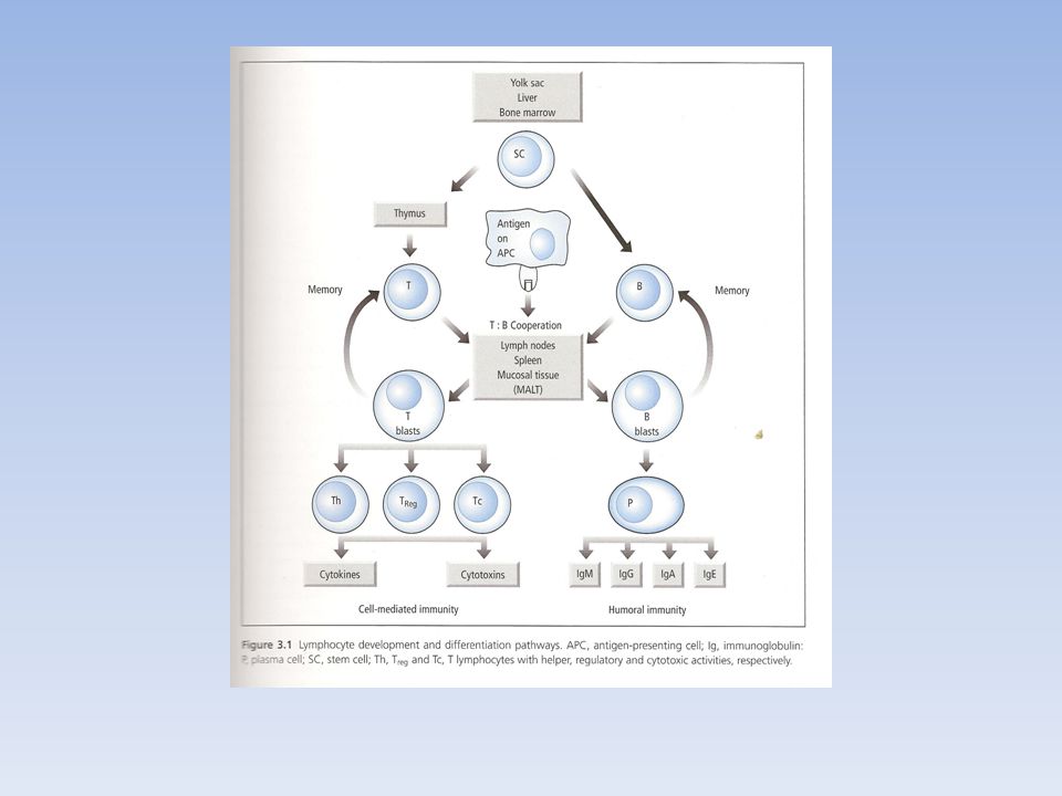

 BIOS 486A/586A>")


 Impacts personal decisions about our lifestyles Affects government, public health agency and.>")


 Lecture 18 Bacterial Pathogenesis (Based on other textbooks such as Madigan’s)>")

>")


