Download presentation
Presentation is loading. Please wait.

1
Recognition and Management of Vascular Injuries
Reza Ghavamian MD Professor and Interim Chairman Department of Urology Montefiore Medical Center Albert Einstein College of Medicine

2
Laparoscopic Complications
Colombo & Gill et al: Single institution analysis 2007: 1867 procedures intraoperative 3.5% Postoperative 8.9% Mortality 0.4% Associated with more complications: lap cystectomy, partial nephrectomy Length of surgery >4hrs Serum Cr> 1.5mg/dl Hemorrhage most common complication intraop and postop Complications decrease with surgeon experience Colombo JR et al: J Urol 178: , 2007.

3
WHY COMPLICATIONS? Experience: 4 fold if > 100 cases
Complexity: 9 fold if more complex Patient risk: As ASA increases so does risk of complications. (Fahlenkamp, D. et al.: J. Urol. 162: 765, 1999 – 2,407 cases) (Parsons, J. et al.: Urology: 63: 27, 2004 – 894 cases)
 (Parsons, J. et al.: Urology: 63: 27, 2004 – 894 cases)")
4
A PLEA FOR CONFORMITY IN REPORTING COMPLICATIONS
Clavien System: I: Any deviation for a normal postoperative course without need for any intervention or medication II: Need for medications, blood transfusion, or parenteral nutrition IIIa: Intervention – without general anesthesia III b: Intervention – with general anesthesia IVa: Life threatening, Single organ dysfunction IVb: Multiple organ dysfunction V: Death (I, II, and IIIa are largely minor whereas IIIb and IV would be considered major complications) (Dindo,D., Clavien, P. et al.: Ann. Surg. 240: 205, 2004)
 (Dindo,D., Clavien, P. et al.: Ann. Surg. 240: 205, 2004)")
5
Entry Pneumoperitoneum Intraoperative Postoperative a. Early b. Late
COMPLICATIONS Entry Pneumoperitoneum Intraoperative Postoperative a. Early b. Late

6
Access Related Complications
Michael Stifelman M.D.

7
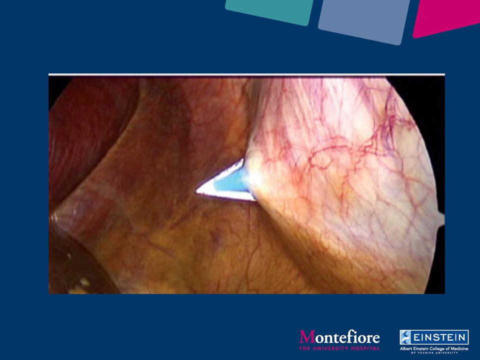
ENTRY: 1. Initial access 2. Trocars

9
ENTRY A good beginning is essential:
“More than one half of the complications related to laparoscopy are related to the entry technique.” Incidence: 0.3 – 1.0% (Magrina, J. F.: Clin. Ob. and Gyn. 45: 469, 2002) (meta-analysis: 1,549,360 laparoscopic cases)
 (meta-analysis: 1,549,360 laparoscopic cases)")
10
ENTRY INJURIES Veress or Open? Veress Open
Vascular injury: 0.08% 0.0%* Bowel injury: 0.08% 0.05% Gas embolism: % 0.0% Death: % 0.0% (n= 12,444) (n= 489,335) *p < .05; (Bonjer, H: Br. J. Surg. 84: 599, 1997) (N.B.: other prospective studies showed no difference!)
 (n= 489,335) *p < .05; (Bonjer, H: Br. J. Surg. 84: 599, 1997) (N.B.: other prospective studies showed no difference!)")
11
Access Related Complications (0.03 – 1%)
Extraperitoneal insertion Vascular injury Abdominal wall vessels Retroperitoneal vessels Mesenteric vessels Visceral injury Stomach, bowel, liver, spleen, bladder

12
Options for Gaining Intraperitoneal Entry:
Closed puncture technique- Veress needle (highest injury rate) FOR THE NOVICE!! Hassan Technique Hand-Assist access first Insert additional trocars with hand in abdomen
 FOR THE NOVICE!! Hassan Technique. Hand-Assist access first. Insert additional trocars with hand in abdomen.")
13
Strategies to avoid access-related complications:
Use Hassan technique or make hand-assist device incision Use visual introducing trocars when using Veress Always verify Veress needle position Saline drop test Move cm insufflation pressure

14
VERESS NEEDLE The operator should feel or sense the needle passing through two distinct planes. The needle is advanced and withdrawn several times. If this is done easily and without obstruction, the tip is in proper position.

15
TRANSPERITONEAL STANDARD ENTRY
Veress needle: Test needle prior to placement. Aspirate, irrigate, aspirate (then irrigate)…drop test and advancement test. Needle rotation. “If in doubt, pull it out.” (High pressure and low flow, remove needle.) Tip: Increase abdominal pressure to 25 mm Hg for initial trocar placement.
…drop test and advancement test. Needle rotation. If in doubt, pull it out. (High pressure and low flow, remove needle.) Tip: Increase abdominal pressure to 25 mm Hg for initial trocar placement.")
16
TRANSPERITONEAL STANDARD ENTRY
Open cannula: Place in an unscarred area of the abdomen. Finger to palpate underside of peritoneum 360 degrees, to insure absence of adherent bowel, etc. Use the balloon trocar – reduces any leak or subcutaneous emphysema Cite work of McKernan…safer and quicker…

17
WHERE’S THE BEST PLACE? Entry sites: 5! Umbilical (Danger – IVC/Aorta)
Right (Palmer’s point) or Left MCL subcostal (Danger – Liver or Liver/spleen) Right or Left side AAL – 2 fingerbreadths above the iliac crest (Danger – colon) (Don’t hesitate to go left when you are operating right!) (McDonald, D., et al.: SLEPT 15: 325, 2005)
 or Left MCL subcostal. (Danger – Liver or Liver/spleen) Right or Left side AAL – 2 fingerbreadths above the iliac crest. (Danger – colon) (Don’t hesitate to go left when you are operating right!) (McDonald, D., et al.: SLEPT 15: 325, 2005)")
18
INTRAOPERATIVE COMPLICATIONS
The BIG 3: 1. Cardiac arrest 2. Vascular 3. Bowel The others: Spleen, Liver, Pancreas, Bladder, Ureter, Diaphragm, Instrumentation, Oliguria

19
Intra-abdominal Vascular Injury:
Ensure skin incision wide enough If Veress aspirate Consider visual obturator If bleeding suspected Leave veress/trocar in place Place accessory ports Beware of hematoma obscuring injury

20
Intraoperative Vascular Injuries

21
VASCULAR INJURY Overview: Incidence: 0.5 – 2.8% Conversion: 50%
Mortality: 9-17% Mechanism: 1. Veress needle: 38% 2. Trocar: % 3. Intraoperative: 17% (Hashizume, M.: Japan. Surg. Endosc. 11: 1198, 1997; Chapron, C. M., J. Am. Coll. Surg. 185: 461, 1997; Mintz, M. :J. Reprod. Med. 18: 269, 1997; Yuzpe, A.: J. Reprod. Med. 35: 485, 1990; Magrina, J. : Clin. Obstet. and Gyn , 2002; Parsons, J. et al.: Urology: 63: 27, 2004)
")
22
PROBLEM: INTRAOPERATIVE HEMORRHAGE
Prevention: 5.5-6 cm. off the midline to avoid the epigastric vessels* “In order to operate fast, it is necessary to go slow.” G. Vallancien Think twice … cut once. Liberal use of energy devices (harmonic, Ligasure) Blunt ports Abdominal inspection at 5 mm Hg: look for “rivulets – red swirls” Port removal under vision at 5 mm Hg *(Hashizume, M.: Japan. Surg. Endosc. 11: 1198, 1997)
 Blunt ports. Abdominal inspection at 5 mm Hg: look for rivulets – red swirls Port removal under vision at 5 mm Hg. *(Hashizume, M.: Japan. Surg. Endosc. 11: 1198, 1997)")
23
TROCAR INJURY: ABDOMINAL WALL
The most common site is from the inferior and superior epigastric vessels. The overall incidence is 0.5% Key point: Lateral ports should be at least cm. off the midline to avoid the epigastric vessels. (Hashizume, M.: Japan. Surg. Endosc. 11: 1198, 1997)
")
24
Intraoperative Vascular Injuries
Risk 2-3% Can occur due to the proximity of the operation to the great vessels in the upper tract Proximity to the iliac vessels in the pelvis Be prepared (extra suction, open basic laparotomy tray) Prompt recognition key Cut only what you see Gentle handling of instruments Control your assistant Always orient yourself
 Prompt recognition key. Cut only what you see. Gentle handling of instruments. Control your assistant. Always orient yourself.")
25
Intraoperative Vascular Injuries
Steps: Transient increase in abdominal pressure to mmHg and maintain pneumoperitoneum Direct pressure with gauze (rolled 4x4) or rolled surgicel and suction irrigator If under control assess extra trocars Obtain optimal exposure, assess what is bleeding, isolate site If possible avoid clips or hem-o-locks Judicious use of : Lapra-Ty, Ligasure, laparoscopic Statinsky, surgical glues Free hand suturing best!! (just like open)
 or rolled surgicel and suction irrigator. If under control assess extra trocars. Obtain optimal exposure, assess what is bleeding, isolate site. If possible avoid clips or hem-o-locks. Judicious use of : Lapra-Ty, Ligasure, laparoscopic Statinsky, surgical glues. Free hand suturing best!! (just like open)")
26
Intraoperative Vascular Injuries
Low treshold to open Transfuse as necessary Have vascular and abdominal tray available There is no shame in conversion! Exposure Pressure, pack, transfuse needed Obtain vascular consult if necessary

27
PROBLEM: INTRAOPERATIVE HEMORRHAGE
Management: Raise pneumoperitoneum pressure to 25 mm Hg Tamponade (rolled 4 x 4 / Satinsky) Hydrate - transfuse Identify what is bleeding! Small - electrosurgery or harmonic +/- fibrin glue / gelfoam / Floseal Large – get blood / call Vascular surgery /suture (EndoStitch/LaparoTy clip/free hand) +/- fibrin glue / gelfoam / Floseal
 Hydrate - transfuse. Identify what is bleeding! Small - electrosurgery or harmonic +/- fibrin glue / gelfoam / Floseal. Large – get blood / call Vascular surgery /suture (EndoStitch/LaparoTy clip/free hand) +/- fibrin glue / gelfoam / Floseal.")
28
WHEN AND HOW TO CONVERT:
1. Tamponade site of bleeding. 2. Open set and blood in the room 3. Second suction unit set up 4. Call out for vascular surgery 5(a). Convert to hand-assist or 5(b). Open: swing endoscope up to underside of abdomen and incise on endoscope; rapidly pack site of bleeding
. Convert to hand-assist. or. 5(b). Open: swing endoscope up to underside of abdomen and incise on endoscope; rapidly pack site of bleeding.")
29
HEMOSTASIS FloSEAL: Collagen derived granules and topical thrombin.
Indications: capillary to arterial bleeding – works on actively bleeding tissues. Package to patient: 2 min. (Baxter BioScience)
")
30
INTRAOPERATIVE COMPLICATIONS: INSTRUMENTATION
Device Malfunction: Stapler Mayhem : FDA databases: Manufacturer and User Facility Device Experience + Alternative Summary Reporting database Mortalities: Injuries: 2,180 Malfunction: ,804 (Brown, S. and Woo, E.: J. Am Col. Surg. 199: 375, 2004)
")
31
Movies

32
HEMORRHAGE TRAY Contents: Laparoty clip applier Set of LaparoTy clip
2 needle holders Endostitch 4-0 Vicryl Klein bulldogs + Klein applicator Satinsky Surgicel Bolsters 4-0 silk on CV needle Endostitch LaparoTy clip

33
Take Home Message: Major vascular injury is a rare but serious complication that occurs in 0.11% to 2% of cases, most frequently involving the aorta and common iliac vessels Campbell’s Urology, 2002 Major vascular injury will present with sudden hypotension/ tachycardia and with rapid accumulation of blood in the abdominal cavity, a mesenteric hematoma, or a expanding retroperitoneal hematoma If bleeding is confined to the retroperitoneum, there may be very little blood intraperitoneally or none at all (thus presenting as an expanding retroperitoneal hematoma) Usal et al, Surgical Endoscopy, 1998
 Usal et al, Surgical Endoscopy,")
34
Take Home Message: Distance from the skin to the great vessels is only a few centimeters, especially in thin pts in a relaxed anesthetic state Nordesgaard et al, Am J Surg, 1995 When performing laparoscopy, must be aware of the potential for injury to major vascular structures and constantly be prepared to rapidly identify and treat this potentially life-threatening complication, with rapid location and control of site of injury and consideration of prompt exploratory laparotomy Geers and Holden, Am Surg, 1996

35
PROBLEM: POSTOPERATIVE HEMORRHAGE
Presentation: Two forms: a. Acute: Sudden vascular collapse (hypotension (70s) /tachy) abd.distention b. Gradual: Mild hypotension (90s) with tachycardia 2. Persistent pulse / pain (Bhayani, S., Kavoussi, L., et al.: J. Urol. 175: 2137, 2006) Diagnostic studies: 1. Hct./Hgb a. Acute: > 10 point drop in hct. from immediate postop b. Gradual: > 5 point drop in hct. – / need for 5 unit transfusion within initial hrs. 2. CT scan: (only for gradual group) Treatment: Exploration (lap. vs. open) check port site/op. site
 /tachy) abd.distention. b. Gradual: Mild hypotension (90s) with tachycardia. 2. Persistent pulse / pain. (Bhayani, S., Kavoussi, L., et al.: J. Urol. 175: 2137, 2006) Diagnostic studies: 1. Hct./Hgb. a. Acute: > 10 point drop in hct. from immediate postop. b. Gradual: > 5 point drop in hct. – / need for 5 unit transfusion within initial hrs. 2. CT scan: (only for gradual group) Treatment: Exploration (lap. vs. open) check port site/op. site.")
36
PROBLEM: POSTOPERATIVE HEMORRHAGE
Results: “Acute” Incidence: 0.4% (4 out of 1,123 laparoscopic renal cases) Approach: 3 open and 1 laparoscopic exploration - < 10 hrs. postop Cause: 3 adrenal and one renal artery. Hospital stay: 8 days Results: “Gradual” Incidence: 0.5% (5 out of 1,123 laparoscopic renal cases) Approach: 1 open and 4 laparoscopic exploration – hrs postop Cause: No source seen – diffuse oozing. Hospital stay: 12 days (Bhayani, S., Kavoussi, L., et al.: J. Urol. 175: 2137, 2006)
 Approach: 3 open and 1 laparoscopic exploration - < 10 hrs. postop. Cause: 3 adrenal and one renal artery. Hospital stay: 8 days. Results: Gradual Incidence: 0.5% (5 out of 1,123 laparoscopic renal cases) Approach: 1 open and 4 laparoscopic exploration – hrs postop. Cause: No source seen – diffuse oozing. Hospital stay: 12 days. (Bhayani, S., Kavoussi, L., et al.: J. Urol. 175: 2137, 2006)")
37
PROBLEM: POSTOPERATIVE HEMORRHAGE
Upper retroperitoneal procedures: Incidence: 0.4% (3.4% nephrectomy 5.4% adrenalectomy 9.9% partial nephrectomy) Units transfused: 56% (1-2) 38% (3-6) 6% (11 and 12) % explored: 12% (2 acute / 2 delayed*) Risk factors: Age and ASA classfication Intraoperative injury to spleen or liver Hosp. stay: 2.7 days *(patient restarted coumadin – bled on postop day 4 – PTT > 100) (Rosevear, H., Roberts, W., Wolf, J. et al.: J. Urol. 176: , 2006)
 Units transfused: 56% (1-2) 38% (3-6) 6% (11 and 12) % explored: 12% (2 acute / 2 delayed*) Risk factors: Age and ASA classfication. Intraoperative injury to spleen. or liver. Hosp. stay: 2.7 days. *(patient restarted coumadin – bled on postop day 4 – PTT > 100) (Rosevear, H., Roberts, W., Wolf, J. et al.: J. Urol. 176: , 2006)")
38
Postoperative Vascular Injuries
Hct decreases by 7-10 points (due to oligiuria and excess ressucitation) Warning signs: Postoperative pain Abdominal distension and discomfort Nausea Tachycardia Continued fall in Hct Treat with open or lap re-exploration depending on stability Assess further with CT scan if stable
 Warning signs: Postoperative pain. Abdominal distension and discomfort. Nausea. Tachycardia. Continued fall in Hct. Treat with open or lap re-exploration depending on stability. Assess further with CT scan if stable.")
Similar presentations

 Mark K. Dodson, M.D. Professor Department of OB/Gyn Division of Gynecologic Oncology University of Utah.>")


 Fellowship in Andrology (U of Ottawa) Fellowship in EndoUrology and Laparoscopy (McMaster.>")


