Download presentation
Presentation is loading. Please wait.

1
Restrictive lung diseases
Acute and Chronic

2
Restrictive lung diseases (Diffuse Interstitial Lung Disease)
A heterogeneous group of disorders characterized predominantly by diffuse and usually chronic involvement of the pulmonary connective tissue Test Obstructive Restrictive FEV1 ↓↓ ↓ VC slight↓ or N FEV1/ VC N or ↑

3
Restrictive Lung Diseases
Definition: Reduced lung compliance More pressure needed to expand lungs Lungs are stiff Pulmonary function test: Low FEV1, Low FVC (the ratio FEV1/FVC is normal)
")
4
Fibrosis Stiff lung Which level?
principally the most peripheral and delicate interstitium in the alveolar walls Fibrosis Stiff lung This result in reduced expansion of lung with reduction in total lung capacity

5
Restrictive lung diseases
Characterized by reduced compliance of the lung. Caused by: 2. parenchymal disease: Prominent changes in the interstitium (interstitial lung disease) 1. Chest wall abnormalities Kyphoscoliosis sever obesity Guilian Barrre’ syndrome Guilian Barrre’ syndrome is an acute inflammatory demyelinating polyneuropathy , an autoimmune disease affecting the peripheral nervous system, usually triggered by an acute infectious process. exhibits as an ascending paralysis noted by weakness in the legs that spreads to the upper limbs With prompt treatment by plasmapheresis or intravenous immunoglobulins, the majority of patients will regain full functional capacity.
 1. Chest wall abnormalities. Kyphoscoliosis. sever obesity. Guilian Barrre’ syndrome. Guilian Barrre’ syndrome is an acute inflammatory demyelinating polyneuropathy , an autoimmune disease affecting the peripheral nervous system, usually triggered by an acute infectious process. exhibits as an ascending paralysis noted by weakness in the legs that spreads to the upper limbs. With prompt treatment by plasmapheresis or intravenous immunoglobulins, the majority of patients will regain full functional capacity.")
6
Restrictive lung diseases (Interstitial lung disease)
Important signs and symptoms: - Dyspnea. - Hypoxia (caused by damage to the alveolar epithelium and interstitial vasculature produces abnormalities in the ventilation-perfusion ratio) - With progressive severe hypoxia, respiratory failure and cor pulmonale. - Chest radiographs show diffuse infiltration by small nodules, irregular lines, or "ground-glass shadows
 - With progressive severe hypoxia, respiratory failure and cor pulmonale. - Chest radiographs show diffuse infiltration by small nodules, irregular lines, or ground-glass shadows.")
7
Restrictive lung diseases (Interstitial lung disease)
Chronic chronic inflammation &fibrosis e.g. Pneumoconiosis Idiopathic pulmonary fibrosis Sarcoidosis Hypersensitivity pneumonitis Immune mediated: SLE Chemical related: berylliosis Acute edema in acute respiratory distress syndrome e.g. Adult respiratory distress syndrome Neonatal respiratory distress syndrome

8
Chronic restrictive lung disease (Chronic interstitial lung disease)
Are a heterogeneous group with little uniformity regarding terminology and classification.

9
Chronic restrictive lung disease
Major Categories of Chronic Interstitial Lung Disease (CILD) -Fibrosing: Pneumoconiosis Usual interstitial pneumonia (idiopathic pulmonary fibrosis) Cryptogenic organizing pneumonia Associated with collagen vascular diseases Drug and Radiation Reactions -Granulomatous: Sarcoidosis Hypersensitivity pneumonitis. Wegener’s granulomatosis -Eosinophilic granuloma -Smoking related: Desquamative interstitial pneumonia Respiratory bronchiolitis-associated interstitial lung disease
 -Fibrosing: Pneumoconiosis. Usual interstitial pneumonia (idiopathic pulmonary fibrosis) Cryptogenic organizing pneumonia. Associated with collagen vascular diseases. Drug and Radiation Reactions. -Granulomatous: Sarcoidosis. Hypersensitivity pneumonitis. Wegener’s granulomatosis. -Eosinophilic granuloma. -Smoking related: Desquamative interstitial pneumonia. Respiratory bronchiolitis-associated interstitial lung disease.")
10
Chronic Interstitial Lung disease CILD
Many entities are of unknown cause and pathogenesis Similar clinical signs, symptoms, radiographic alterations and pathophysiologic changes. Patient have reduced forced vital capacity, however the FEV1/ FVC is normal Account for about 15% of non-infectious lung diseases.

11
Idiopathic pulmonary fibrosis Hypersensitivity pneumonitis
CILD Idiopathic pulmonary fibrosis Hypersensitivity pneumonitis Smoking related Pneumoconiosis

12
Idiopathic pulmonary fibrosis (cryptogenic fibrosing alveolitis) (Usual interstitial pneumonia) UIP
A clinicopathologic syndrome with characteristic radiologic, pathologic and clinical features. Radiology: Bilateral lung nodular infiltratre Histology: diffuse interstitial fibrosis and inflammation. Clinical features: Dyspnea, advanced cases result in sever hypoxemia and cyanosis. Males are more affected than female 2/3 of pt. older than 60 years

13
Pathogenesis Some form of alveolar wall injury result in interstitial edema and alveolitis. Type I pneumocyte is more susceptible to injury. Type II pneumocyte hyperplasia (regenerate). Fibroblast proliferation with progressive fibrosis of intra-alveolar exudate and interalveolar septa. IgG deposits are seen in alveolar wall. FGF, TGF-, PDGF IL-8 leukotriens
. Fibroblast proliferation with progressive fibrosis of intra-alveolar exudate and interalveolar septa. IgG deposits are seen in alveolar wall. FGF, TGF-, PDGF. IL-8 leukotriens.")
14
Morphology of IPF Gross -The lungs are firm.
-Pulmonary edema. The morphologic changes vary according to the stage of the disease. Early cases: -Intraalveolar exudate. -Hyaline membranes. -Infiltration of the alveolar septa with mononuclear cells. -Hyperplasia of type II pneumocytes -

15
Morphology of IPF Advancing disease:
-Organization of the intraalveolar exudates by fibrous tissue. -Thickening of the alveolar septa owing to fibrosis and variable amounts of inflammation. -Alternating areas of fibrosis and normal tissue. - Geographic variation - Temporal variation In the end, the lung consists of spaces lined by cuboidal or columnar epithelium separated by inflammatory fibrous tissue (honeycomb lung).
.")
16
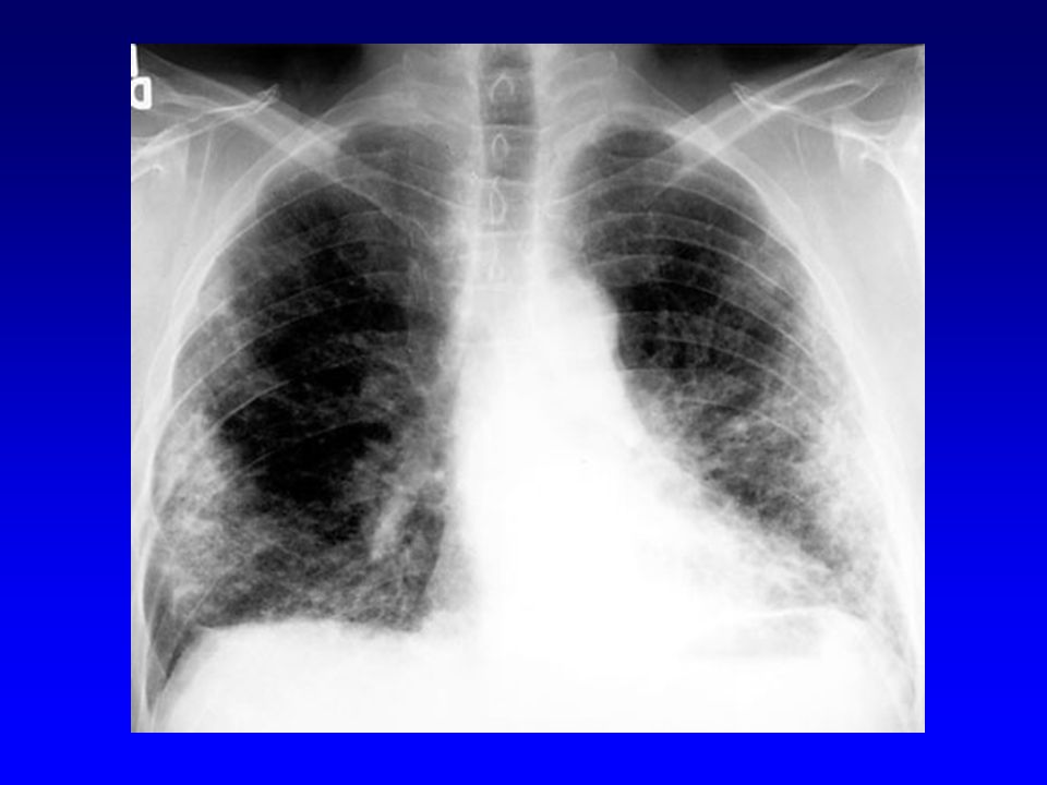
honeycomb lung

21
Prognosis of IPF Gradual onset of dyspnea with respiratory difficulty.
Hypoxemia and cyanosis. Cor pulmonale and cardiac failure may result. The progression in individual cases is unpredictable. The median survival is about 2 to 4 years.

22
Pneumoconiosis Non-neoplastic lung reaction to inhalation of mineral dusts. Most common dusts are coal dust, silica, asbestos and beryllium.

23
Pneumoconiosis Occupational Lung Diseases
Dr. Atif Ali 2010

24
Pneumoconiosis Pathogenesis
The development of pneumoconiosis is dependent on: - The amount of dust retained in the lung and airways. a. Concentration of the dust in the ambient air. b. Duration of the exposure. c. Effectiveness of the clearance mechanisms. - The size (1-5) shape. - Their solubility and physiochemical activity. - The possible additional effects of other irritants, tobacco smoking. The particles are impacted at alveolar duct macrophage, accumulate inflammatory response fibrosis.
 shape. - Their solubility and physiochemical activity. - The possible additional effects of other irritants, tobacco. smoking. The particles are impacted at alveolar duct macrophage, accumulate inflammatory response fibrosis.")
25
Coal worker’s pneumoconiosis (CWP)
Pneumoconioses Coal worker’s pneumoconiosis (CWP) Silicosis Asbestosis
 Silicosis. Asbestosis.")
26
Coal worker’s pneumoconiosis (CWP)
Occurs in coal workers after many years of underground mine work. Two forms: - The simple form: - Focal aggregations of coal dust-laden macrophages (coal macules, 1 to 2 mm) mainly in upper lobes. - Patients have slight cough and blackish sputum. - emphysema ( smoking related). - The complicated form: With heavier pulmonary burdens of coal dust, fibrous scarring appears (complicated CWP) also callled progressive massive fibrosis (PMF).
 mainly in upper lobes. - Patients have slight cough and blackish sputum. - emphysema ( smoking related). - The complicated form: With heavier pulmonary burdens of coal dust, fibrous scarring appears (complicated CWP) also callled progressive massive fibrosis (PMF).")
27
Coal worker pneumoconiosis
Morphology: Complicated CWP: -Black scars exceed 2 cm in diameter some times up to 10 cm -It consists of dense collagen and carbon pigments. -Cor pulmonale. -Miners who have rheumatoid arthritis and PMF are called Caplan’s syndrome.

28
Coal miner with progressive massive fibrosis (unstained)
")
29
Coal worker pneumoconiosis
Clinical course: CWP is usually benign disease with little symptom Minor cases progress to PMF No increased risk to bronchogenic carcinoma

30
Coal worker’s pneumoconiosis (CWP)
Pneumoconioses Coal worker’s pneumoconiosis (CWP) Silicosis Asbestosis
 Silicosis. Asbestosis.")
31
Silicosis Long exposure to silica particles.
Nodular densely fibrosing pneumoconiosis. Encountered in a diversity of industries: mining of gold, tin, copper and coal, sandblasting, metal grinding, ceramic manufacturing, drilling and tunneling. Pathogenesis: Crystalline silica is highly fibrogenic. Scattered lymphocytes and macrophages are drawn rapidly with fibrosis. Some particles are transported to lymph nodes.

32
Morphology of Silicosis
Tiny collagenous nodules that enlarge forming stony-hard large fibrous scars usually in the upper lobes. The lung parenchyma between the scars may be compressed or emphysematous. Calcifications may appear (eggshell calcification) . Similar collagenous nodules within the lymph nodes. Fibrous pleural plaques may develop.
 . Similar collagenous nodules within the lymph nodes. Fibrous pleural plaques may develop.")
33
Morphology of Silicosis
Micro: -Hyalinized collagen fiber surround an amorphous center (fibrous nodules). - Scarring progress to PMF. -Scarring extending and encroching the pulmonary arteries. -Cor pulmonale.
. - Scarring progress to PMF. -Scarring extending and encroching the pulmonary arteries. -Cor pulmonale.")
34
Clinical features of Silicosis
depend on form of silicosis

35
Forms of Silicosis Acute silicosis: Chronic silicosis:
results from exposure to high dose of silica. Fluid in alveoli. Pt. Have rapid onset of tachypnea, cough and repiratory failure. Chronic silicosis: Inhalation of silica for long time with fibrotic nodules ( present in upper lobe of lung & in subpleural spaces Complicated silicosis: Progression of chronic silicosis with PMF with chronic hypoxia Other pulmonary disease: Increased susceptibility to TB Caplan syndrome ( uncommon) Lung cancer
 Lung cancer.")
36
Silica and lung cancer The relationship between silica and lung cancer has been a contentious issue, but in 1997, based on evidence from several epidemiologic studies, the International Agency for Research on Cancer concluded that crystalline silica from occupational sources is carcinogenic in humans. However, this subject continues to be controversial.

37
Coal worker’s pneumoconiosis (CWP)
Pneumoconioses Coal worker’s pneumoconiosis (CWP) Silicosis Asbestosis
 Silicosis. Asbestosis.")
38
Asbestosis Asbestos is a family of crystalline hydrated silicates with a fibrous geometry. Two forms: Serpentine chrysotile (flexible fiber), more common Amphibole (straight and stiff fiber), more pathogenic Both forms are fibrogenic.
, more common. Amphibole (straight and stiff fiber), more pathogenic. Both forms are fibrogenic.")
39
Asbestosis Inhalation of asbestos leads to: - Asbestos pneumoconiosis.
- Pleural effusion. - Pleural adhesions. - Parietal pleural fibrocalcific plaques. - Increased incidence of mesothelioma, bronchogenic carcinoma, laryngeal cancer. These consequences occurs decades after exposure has ended. An increased incidence of asbestos-related cancers in family members of asbestos workers has alerted the general public to the potential hazards of asbestos in the environment.

40
Morphology Of Asbestosis diffuse pulmonary interstitial fibrosis
scarring containing asbestos bodies (ferrougenous bodies).
.")
41
Asbestosis bodies (from human lung)
")
42
Association of asbestos bodies with fibrosis (asbestosis)
")
43
Pleural plaque in asbestos
Parietal pleura over dome of diaphragm

44
Asbestosis In asbestosis, pt. develop progressively worsened dyspnea with cough and sputum progressing to cor pulmonale and death. Both bronchogenic carcinoma and mesothelioma develop in workers exposed to asbestos. The risk of bronchogenic carcinoma is fivefold and for mesothelioma is 1000 fold greater. The risk of bronchogenic carcinoma in 50 X increased in smoking asbestos workers (but not that of mesothelioma)
")
46
Malignant Mesothelioma
Rare cancer of mesothelial cells Arise from parietal or visceral pleura Can arise from peritoneum and pericardium 50% of pt. have history of exposure to asbestos at work It also appeared in relatives of asbestos worker or in people living near asbestos factory The latent period between exposure and malignant mesothelioma is long (25 to 40 years) Nearly all cases are related to amphibole asbestos These minerals cannot be removed from the lung and the risk for malignant mesothelioma is life long Simian virus 40 (SV40) T antigen is found in 60 to 80% of malignant mesothelioma Characteristic E/M finding : numerous long microvilli on cell surface
 Nearly all cases are related to amphibole asbestos. These minerals cannot be removed from the lung and the risk for malignant mesothelioma is life long. Simian virus 40 (SV40) T antigen is found in 60 to 80% of malignant mesothelioma. Characteristic E/M finding : numerous long microvilli on cell surface.")
47
Idiopathic pulmonary fibrosis Hypersensitivity pneumonitis
CILD Idiopathic pulmonary fibrosis Usual interstitial pneumonia Pneumoconiosis Coal worker’s pneumoconiosis (CWP) Silicosis Asbestosis Hypersensitivity pneumonitis Desquamative interstitial pneumonia
 Silicosis. Asbestosis. Hypersensitivity pneumonitis. Desquamative interstitial pneumonia.")
Similar presentations


 Infections (pneumonia, airways disease)>")


 r Thin elastin-rich connective component containing.>")