Download presentation
Presentation is loading. Please wait.

1
Chap. 19– The Heart (Cardiology)
")
2
Chap. 19 (Heart) Study Guide
Critically read Chapter 19 pp before 19.5 “Blood Flow” section Comprehend Terminology (those in bold) Study-- Figure questions, Think About It questions, and Before You Go On (section-ending) questions Do Testing Your Recall— 1, 2, 4-8, 11-19 Do True or False– 1-4, 7, 9-10 Do Testing Your Comprehension-- #1 2 2
 Study-- Figure questions, Think About It questions, and Before You Go On (section-ending) questions. Do Testing Your Recall— 1, 2, 4-8, Do True or False– 1-4, 7, Do Testing Your Comprehension-- #")
3
What are you going to do with your heart?
“The best and most beautiful things in the world cannot be seen or even touched—they must be felt with the heart.” --Hellen Keller “Happiness comes only when we push our brains and hearts to the farthest reaches of which we are capable.” --Leo C. Rosten

4
I. Overview of cardiovascular system

5
§ Introduction The circulatory system—
Three component: the pump, the passageway, and the transport medium What are they respectively? The pump-- The passageway-- The transport medium--

6
§ Two circuits in the cardiovascular system (1)
Pulmonary circulation— Function? The route? R. ventricle Pulmonary arteries Lungs Pulmonary veins L. atrium Figure 19.1

7
Pulmonary circulation Right side of heart Lungs Pulmonary capillaries
veins Pulmonary arteries = O2-poor blood = O2-rich blood Right side of heart

8
§ Two circuits in the cardiovascular system (2)
Systemic circulation— Functions? The route? (Students work on it.) Starts which chamber of the heart? Major vessels? Destinations? Two major veins? Ends at which chamber of the heart? Fig. 19.1
 Starts which chamber of the heart Major vessels Destinations Two major veins Ends at which chamber of the heart Fig")
9
Systemic circulation Left side of heart Systemic veins Systemic
= O2-poor blood = O2-rich blood Systemic veins Systemic arteries Systemic capillaries Organ systems

10
II. Gross anatomy of the heart

11
§ Shape and size of the heart
Base – broad superior portion Apex - inferior end (a blunt point) 3.5 in. wide at base, 5 in. from base to apex, and 2.5 in. anterior to posterior weighs 10 oz (300 gram; size of your fist) Fig. 19.2
 3.5 in. wide at base, 5 in. from base to apex, and 2.5 in. anterior to posterior. weighs 10 oz (300 gram; size of your fist) Fig")
12
Aorta Superior vena cava Base of heart Apex of heart Diaphragm
Next Topic Diaphragm

13
§ Heart Position (1)
")
14
§ Heart Position (2)
")
15
§ Heart Position (3)
")
16
§ Pericardial sac (pericardium) -1
Def. the double-walled, membranous covering that encloses the heart Function– Friction free Peircardial fluid— Figure x Cardiac Disorders here (Table 19.3): Pericarditis– inflammation here Pericardial effusion– fluid in pericardial cavity Cardiac tamponade– accumulation of fluid here
: Pericarditis– inflammation here. Pericardial effusion– fluid in pericardial cavity. Cardiac tamponade– accumulation of fluid here.")
17
Figure x Pericardial cavity Heart

18
§ Pericardial sac (pericardium) - 2
Parietal pericardium– 2 SUBLAYERS A-outer, tough/fibrous layer (CT) + B-deep thin serous layer– turns inward forms #2 below Visceral pericardium (a.k.a. epicardium of heart wall) INNER, thin, smooth, moist serous layer covers heart surface Pericardial cavity: between above filled with ____________________ Fig. 19.3
 + B-deep thin serous layer– turns inward forms #2 below. Visceral pericardium (a.k.a. epicardium of heart wall) INNER, thin, smooth, moist serous layer. covers heart surface. Pericardial cavity: between above. filled with ____________________. Fig")
19
Functions? 3 1 2

20
§ Heart Wall (from outermost layer)
Epicardium (a.k.a. visceral pericardium) serous membrane covers heart Myocardium thick muscular layer fibrous skeleton - network of collagenous and elastic fibers (special section for this one) Endocardium - smooth inner lining What type of epi.? Simple _________ epi. Continuous with endothelium cells . . . Fig. 19.3
 serous membrane covers heart. Myocardium. thick muscular layer. fibrous skeleton - network of collagenous and elastic fibers (special section for this one) Endocardium - smooth inner lining. What type of epi. Simple _________ epi. Continuous with endothelium cells Fig")
21
2 3 1

22
§ Fibrous skeleton of the heart (1)
What is it? Four CT rings fuse with . . . Structure details– Four fibrous rings, surrounds the 4 valves; in sheets of tissue that interconnect these rings Location? In the walls between … Figure 19.8

23
Fibrous skeleton including fibrous rings
(Rear) Fibrous skeleton including fibrous rings Right AV valve Left AV valve Aortic valve Pulmonary semilunar valve Ventricular myocardium (Front)
 Fibrous skeleton including fibrous rings. Right AV valve. Left AV valve. Aortic valve. Pulmonary semilunar. valve. Ventricular. myocardium. (Front)")
24
§ Fibrous skeleton of the heart (2)
Functions– 1. Structure support-- firm base of the heart valves and openings of great vessels 2. It anchors the cardiac muscle 3. An electrical insulator: Separate the atria from the ventricles and direct A.P. to specific pathways

25
Checkpoint Questions Does most of the heart lie to the right or left of the median plane? Name, in order, the three layers of the heart wall beginning with the outermost layer.

26
§ Heart Chambers 4 chambers— 3 sulci (grooves)— on the surface
A. right and left ATRIA auricles? Ear-like structures . . . B. right and left VENTRICLES 3 sulci (grooves)— on the surface Largely fat and coronary blood vessels A. Atrioventricular (coronary) sulcus- B+C. Anterior and posterior interventricular sulci Figure 19.5 a+b
— on the surface. Largely fat and coronary blood vessels. A. Atrioventricular (coronary) sulcus- B+C. Anterior and posterior interventricular sulci. Figure 19.5 a+b.")
27
Coronary sulcus? Anterior view

28
Posterior view

29
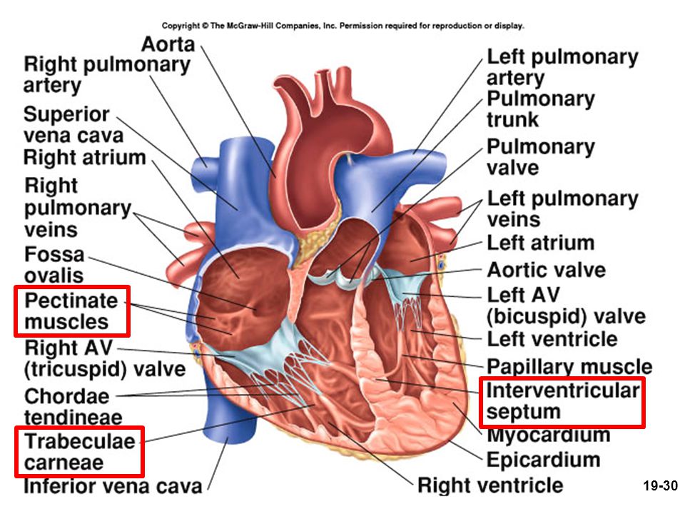
§ Heart Chambers – Internal (Fig. 19.7)
Interatrial septum wall that separates atria Pectinate muscles internal ridges of myocardium in right atrium and both auricles; absorber) Interventricular septum wall that separates ventricles Trabeculae carneae internal ridges in both ventricles absorber) Wave/sound absorber
 Interventricular septum. wall that separates ventricles. Trabeculae carneae. internal ridges in both ventricles absorber) Wave/sound absorber.")
31
Checkpoint Questions Which heart chamber has the thickest walls? What is the significance of this structural difference? Do the atrial pectinate muscles more nearly resemble the ventricular papillary muscles or the trabeculae carneae?

32
blood from the atria to ventricles . . .
§ Heart valves (1) Two atrioventricular (AV) valves— A. Right AV valve– also called the tricuspid valve B. Left AV valve– also called . . . Function-- blood from the atria to ventricles . . . Figure 19.8 (a,b)
 Two atrioventricular (AV) valves— A. Right AV valve– also called the tricuspid valve. B. Left AV valve– also called Function-- blood from the atria to ventricles Figure 19.8 (a,b)")
33
Superior views of these valves – next slide
Aorta Pulmonary artery Superior vena cava Pulmonary valve Pulmonary veins Pulmonary veins Left atrium Left AV valve Right atrium Aortic valve Chordae tendineae Right AV valve Papillary muscle Left ventricle Right ventricle Interventricular septum Inferior vena cava Superior views of these valves – next slide

34
Right AV valve Left AV valve
Aortic/pulmonary valve

35
Heart valves (2) Chordae tendineae— Structure–
Fibrous cords anchor the cusps to the ventricle walls via papillary muscles Function– Prevent valves from being _________ Figure 19.7, 19.8

36
Right atrium Chordae tendineae Right AV valve Direction of backflow of blood Septum Right ventricle Papillary muscle

37
Right AV valve seen from within the right ventricle

38
§ Heart valves (3) Semilunar valves include: One ______ valve and one __________ valve Where are they located respectively? Major arteries leave the ventricles How to prevent them from everting? Anatomical structure— leakproof “seam” Function– (of all valves) Ensure unidirectional flow of blood Figure 19.7 and Fig. Z
 Ensure unidirectional flow of blood. Figure 19.7 and Fig. Z.")
39
Next slide Aorta Pulmonary artery Pulmonary valve Aortic valve
Superior vena cava Pulmonary valve Pulmonary veins Pulmonary veins Left atrium Left AV valve Right atrium Aortic valve Chordae tendineae Right AV valve Papillary muscle Left ventricle Right ventricle Interventricular septum Inferior vena cava

40
(Pulmonary trunk or Aorta) (Right or Left Ventricle)
Direction of backflow of blood Leakproof “seam” Aortic valve (Right or Left Ventricle)
")
41
§ Valve Mechanics (Fig. 19.9, 19.19)
Ventricles filling & isovolumetric contraction AV valves open (semilunar valves close); blood flows from atria to ventricles (v. fillings) AV valves open/closed (circle one)—S1 ventricle pressure continues to rise Momentarily before ventricle ejection Ventricles ejection & isovolumetric relaxation semilunar valves open (AV valves close); ventricle ejection; ventricle pressure drops semilunar valves open/closed (circle one)—S2 Isovolumetric relaxation
; blood flows from atria to ventricles (v. fillings) AV valves open/closed (circle one)—S1. ventricle pressure continues to rise. Momentarily before ventricle ejection. Ventricles ejection & isovolumetric relaxation. semilunar valves open (AV valves close); ventricle ejection; ventricle pressure drops. semilunar valves open/closed (circle one)—S2. Isovolumetric relaxation.")
42
Operation of Atrioventricular Valves

43
Operation of Semilunar Valves

44
Before You Go On (p. 730) Reminder: Remember to go over each question of Before You Go On in the text. P. 730– Trace the flow of blood through the heart, naming each chamber, valve, and the great vessels in order (from the superior vena cava to the aorta). Do it yourself. Fig is a great figure to help you with this. \
. Do it yourself. Fig is a great figure to help you with this. \")
45
Fig. 19.9 Pathway of blood flow through the heart
Figure 19.10 Fig Pathway of blood flow through the heart

46
III. The Coronary Circulation

47
§ Coronary arteries Right C.A. Left C.A.

48
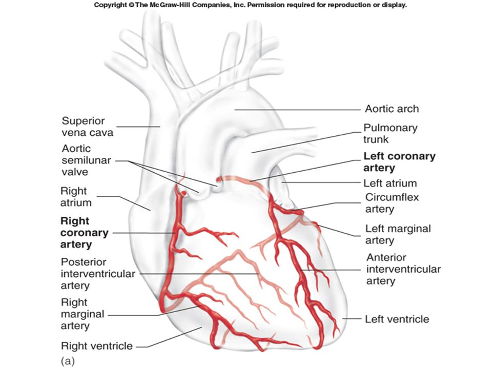
§ Coronary Arterial Supply
1. Left coronary artery (LCA)– 2 branches 1A--anterior interventricular branch supplies blood to interventricular septum and anterior walls of both ventricles 1B--circumflex branch (Fig a+b) passes around left side of heart in coronary sulcus, supplies left atrium and posterior wall of left ventricle; it gives off a left marginal branch (1C) 2. Right coronary artery (RCA)– 2 branches 2A--right marginal branch supplies lateral side of R atrium and ventricle 2B--posterior interventricular branch supplies posterior walls of ventricles
– 2 branches. 1A--anterior interventricular branch. supplies blood to interventricular septum and anterior walls of both ventricles. 1B--circumflex branch (Fig a+b) passes around left side of heart in coronary sulcus, supplies left atrium and posterior wall of left ventricle; it gives off a left marginal branch (1C) 2. Right coronary artery (RCA)– 2 branches. 2A--right marginal branch. supplies lateral side of R atrium and ventricle. 2B--posterior interventricular branch. supplies posterior walls of ventricles.")
49
1B 2A 1A

50
1B 1C 2A 2B

51
§ Anastomoses of coronary arteries
Definition (Anastomosis) – a point where two blood vessels join/merge; this is arterial anastomoses Where? Anterior interventricular branch of LCA joins the posterior interventricular branch of RCA Function– provide collateral (alternative) routes of blood supply to a tissue (the heart) Fig. x
 – a point where two blood vessels join/merge; this is arterial anastomoses. Where Anterior interventricular branch of LCA joins the posterior interventricular branch of RCA. Function– provide collateral (alternative) routes of blood supply to a tissue (the heart) Fig. x.")
53
Chest pain and Heart Attack
Angina pectoris-- partial obstruction of coronary blood flow can cause chest pain pain caused by ischemia, often activity dependent Myocardial infarction (heart attack)-- complete obstruction causes death of cardiac cells in affected area pain or pressure in chest that often radiates down left arm
-- complete obstruction causes death of cardiac cells in affected area. pain or pressure in chest that often radiates down left arm.")
54
§ Venous Drainage of Heart
10% drains directly into right atrium and ventricle via multiple thebesian veins 90% returns to right atrium via: (Fig ) A. great cardiac vein blood from anterior interventricular sulcus B. middle cardiac vein (post. interventricular v.) from posterior sulcus C. left marginal vein The above three (A, B, C) empty into the coronary sinus before emptying into the ____________ (which chamber of the heart?)
 A. great cardiac vein. blood from anterior interventricular sulcus. B. middle cardiac vein (post. interventricular v.) from posterior sulcus. C. left marginal vein. The above three (A, B, C) empty into the coronary sinus before emptying into the ____________ (which chamber of the heart )")
55
A

56
A C B or posterior interventricular v.

57
IV. Cardiac conduction system

58
§ 19.3 Cardiac muscle & conduction system
Heart has its own pacemaker, nerves MODIFY the heart rate & contraction strength. Beat rhythmically, _________beats per min. Pacemaker? Where? (next slide) Myogenic and autorhythmic Regulation by autonomic nerve system
 Myogenic and autorhythmic. Regulation by autonomic nerve system.")
59
§ Cardiac Conduction System (1)
Properties myogenic - heartbeat originates from within ____________________ Originated from what cells (1% of heart cells)? cardiac muscles become specialized into autorhythmic cells (cardiac conduction system) What do autorhythmic cells do?– regular, spontaneous depolarization Components next slide
 cardiac muscles become specialized into autorhythmic cells (cardiac conduction system) What do autorhythmic cells do – regular, spontaneous depolarization. Components. next slide.")
60
Cardiac Conduction System (2)– Autorhythmic cells
SA (sinoatrial) node: pacemaker, initiates heartbeat, sets heart rate; where? AV node: electrical gateway to ventricles; where? fibrous skeleton– insulates atria from ventricle AV bundle: pathway for signals from AV node Right and left bundle branches: divisions of AV bundle that enter interventricular septum Purkinje fibers: upward from apex spread throughout ventricular myocardium Fig X
 node: pacemaker, initiates heartbeat, sets heart rate; where AV node: electrical gateway to ventricles; where fibrous skeleton– insulates atria from ventricle. AV bundle: pathway for signals from AV node. Right and left bundle branches: divisions of AV bundle that enter interventricular septum. Purkinje fibers: upward from apex spread throughout ventricular myocardium. Fig X.")
61
Cardiac Conduction System
1 2 4 3 5

62
Students-- work on this one at home
Interatrial pathway 3. Atrioventricular (AV) node 1. Sinoatrial (SA) node 4. Bundle of His or AV bundle Right atrium 5b. Left branch of bundle of His 2. Internodal pathway Left ventricle 5a. Right branch of bundle of His 6. Purkinje fibers Right ventricle
 node. 1. Sinoatrial. (SA) node. 4. Bundle of His or AV bundle. Right. atrium. 5b. Left branch. of bundle of His. 2. Internodal. pathway. Left. ventricle. 5a. Right. branch. of bundle. of His. 6. Purkinje. fibers. Right. ventricle.")
63
Checkpoint Question Which chamber of the heart is first to receive the electrical signal that induces the heart to contract?

64
V. Cardiac muscle

65
§ Cardiac vs. skeletal m.(1)
Cardiac M. (cardiocyte) Fibers & their control Fibers independent Voluntary Interlocking cells; (next) Involuntary Nervous control by Somatic motor neurons Autonomic nervous sys. Initiation of contraction Requires input from motor neurons by autorhythmic cells in heart
 Fibers & their control. Fibers independent. Voluntary. Interlocking cells; (next) Involuntary. Nervous control by. Somatic motor neurons. Autonomic nervous sys. Initiation of contraction. Requires input from motor neurons. by autorhythmic cells in heart.")
66
§ Cardiac vs. skeletal m.(2)
Cardiac myocytes—size, thickness etc. T (transverse) tubules– smaller Presence of intercalated discs– (see next slide) Mitochondria— Myoglobin and glycogen--
 tubules– smaller. Presence of intercalated discs– (see next slide) Mitochondria— Myoglobin and glycogen--")
67
§ Intercalated discs (of cardiac muscle cells)
Def. specialized zigzag structures joining cardiac muscle cells end to end Containing three distinctive features not found in skeletal muscle; what are they? Figure a-c

68
Figure 19.11a light micrograph
Next slide

69
One myocyte is shown (colored).
.")
70
Structure of an intercalated disc
2 1 3 1-- interdigitating folds 2—mechanical junctions– two types; fascia adherens and desmosomes 3—electrical (gap) junctions--
 junctions--")
71
Desmosome Fascia adherens Plasma membranes of adjacent
cardiac muscle fibers Desmosome Fascia adherens Action potential Gap junction Intercalated disc

72
§ Intercalated discs Function of interdigitating folds--
Functions of fascia adherens and desmosome – Types of adhering junction Mechanically, hold cells together

73
§ Intercalated discs Functions of gap junction (connexons):
Allows action potentials to spread . . . Therefore, cardiac cells form functional syncytia— cardiac cells excited and contract as a single unit Q--Does the atria and the ventricle each form a separate unit?

74
VI. Electrical activity of heart (autorhythmic cells)
")
75
§ 19.4 Heart Autorhythmic Cells
Two types of cardiac muscle cells: 1% are autorhythmic cells (our focus on this section)– Function? AP— Yes Contraction– No 99% --contractile cells AP— No initiation of own Action Potential Contraction– Yes
– Function AP— Yes. Contraction– No. 99% --contractile cells. AP— No initiation of own Action Potential. Contraction– Yes.")
76
§ Heart autorhythmic cells
Autorhythmic cells act as pacemaker: How? Their m. potential slowly depolarizes (drifts) between AP, until … Figure 19.13
 between AP, until … Figure")
77
Pacemaker potentials & action potentials of the SA node
B C A Pacemaker potentials & action potentials of the SA node

78
§ Heart autorhythmic cells
Details of pacemaker activity: (vs. AP in nerve and skeletal m.): Slow depolarization: i. K+ voltage-gated channels slowly close ii. (No voltage-gated Na+ channels), instead sodium leak channels are used; So, sodium ions move in/out iii. Transient Ca+2 channels open—Calcium ions move inward All these make the inside becomes depolarized Thus, pacemaker p. toward threshold
: Slow depolarization: i. K+ voltage-gated channels slowly close. ii. (No voltage-gated Na+ channels), instead sodium leak channels are used; So, sodium ions move in/out. iii. Transient Ca+2 channels open—Calcium ions move inward. All these make the inside becomes depolarized Thus, pacemaker p. toward threshold.")
79
§ Heart autorhythmic cells
Rising phase of the action potential: Once, reach threshold p., long-lasting Ca+2 channels open; . . . Influx of calcium ions The falling phase: as usual, potassium ions efflux

80
§ Heart autorhythmic cells
Autorhythmic cells are self excitable: Without nervous stimulation They initiate AP cyclically, which trigger rhythmic beating Each depolarization of SA node sets off one heartbeat (every 0.8 sec.) They form the conduction system of the heart (see Figure 19.12) It excites the other components in the system
 They form the conduction system of the heart (see Figure 19.12) It excites the other components in the system.")
81
§ Heart autorhythmic cells
The spread of cardiac excitation: In a coordinated sequence: A-First, atrial excitation– From SA node to atria; How? through gap junctions; via internodal and interatrial pathways as well; Result– a single smooth contraction of the pair of atria (Fig. Y) B-Second, from the atria to the ventricles— AV node is the only point of electrical contact from the atria to the ventricles C-Finally, ventricular excitation-- from AV node to the bundle of His, . . . Result– a single smooth contraction in ventricles
 B-Second, from the atria to the ventricles— AV node is the only point of electrical contact from the atria to the ventricles. C-Finally, ventricular excitation-- from AV node to the bundle of His, Result– a single smooth contraction in ventricles.")
82
Interatrial pathway 1st beat SA node Internodal pathway Bundle AV node
Right atrium Left atrium 1st beat SA node Internodal pathway Bundle of His AV node 2nd beat Purkinje fibers Right ventricle Left ventricle

83
VII. contractile activity of heart

84
Our focus

85
§ Cardiac contractile cells
Action potential is initiated by: the pacemaker cells 3 phases: Rising Plateau …

86
§ Cardiac contractile cells
The detail of action potential: Rising phase Massive sodium ions influx causes depolarization and AP Plateau phase Primarily caused by opening of calcium channels Also caused by temp. reduction in outflow of _____________ ions

87
§ Cardiac contractile cells
Falling phase Primarily caused by ____________ outflow Closing of calcium channels contribute to this as well

88
Review slide— ID A, B, C, D, E below

89
§ Cardiac contractile cells
Action potential Contractile response Contractile response Compare to skeletal muscle: Longer period of cardiac contraction Longer refractory period How? Why? (next) Refractory period
 Refractory period.")
90
§ Cardiac contractile cells
Longer period of cardiac contraction 3x longer compared to skeletal m. Caused by entry of calcium ions which induce more calcium ions release from the sarcoplasmic reticulum Purpose– this increased contractile time ensures emptying blood into ventricles and arteries Fig (skeletal muscle)
")
91
In skeletal muscle Latent period Contraction time Relaxation time
twitch Contractile response Action potential Stimulation

92
§ Cardiac contractile cells
Longer refractory period Caused chiefly by inactivation of the sodium ion channels Consequences/Purpose– Cardiac muscle cannot be re-stimulated until contraction is almost over, therefore summation and tetanus of cardiac m. is impossible This ensures . . . Compared to Fig (Shown previously; in skeletal m.)
")
93
§ ECG (Electrocardiogram)
Def. A record of the overall electrical activity in all the cardiac muscle cells from the body surface NOT a recording of a single action potential in a single cell ECG recording represents . . .electrical activity detected by electrodes at 2 different points Figure (ECG)
")
95
§ ECG (Electrocardiogram)
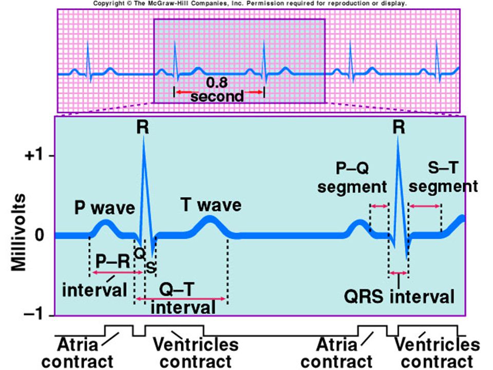
Components of the ECG correlate to cardiac events (Fig in the next slide) P wave— atrial depolarization when the electrical impulse spreads across the atria QRS complex— T wave— ventricular repolarization R T P P Q S
 P wave— atrial depolarization when the electrical impulse spreads across the atria. QRS complex— T wave— ventricular repolarization. R. T. P. P. Q. S.")
96
P T QRS

Similar presentations


 in thorax, in inferior mediastinum>")


