Download presentation
Presentation is loading. Please wait.

1
Bone Transport with TSF
S. Robert Rozbruch, MD Orthopaedic Trauma Service Director, Institute for Limb Lengthening & Reconstruction

5
Problems with classic Ilizarov frame
Complicated frame application Need to achieve alignment at surgery Arched olive wire technique Poor control of transport fragment Docking difficulty Mal-alignment Poor bony contact Require frame modification Deformity of lengthening regenerate

6
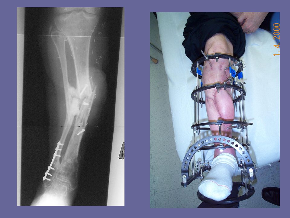
3 cm Infected rod, draining sinuses

7
equinus

8
Infected nonunion 3 cm LLD 5 cm defect Retained hardware flaps

9
Removal hardware Resect dead bone Square edges Insert antibiotic beads Rings first method

10
6 weeks later Apply proximal ring Establish mounting parameters Perform proximal tibia osteotomy Remove beads Begin transport

12
Establish Mounting parameters

13
Mounting parameters:intraop
stacking cubes

16
Virtual hinge “global positioning” Defined point Relative to the Reference ring

17
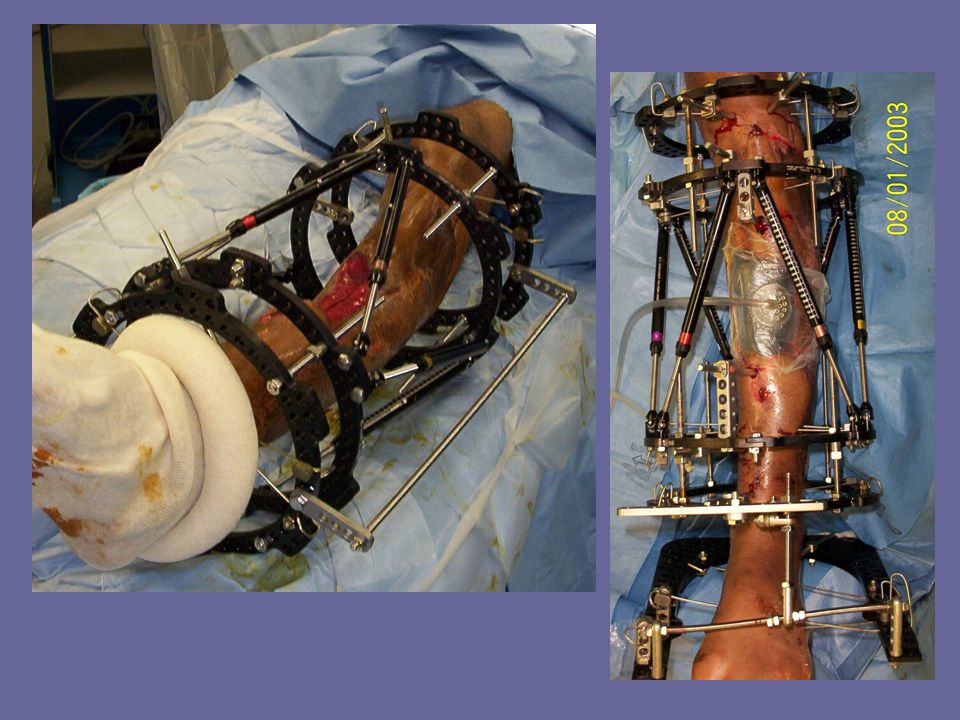
MCA Open grade 3B Bone loss

18
8 cm defect

19
Control each Segment separately

20
BG Docking site

21
Fibula plating Was already Done -prefer not To have them

23
Tibia-talar Arthrodesis and Simultaneous tibia lengthening
Bone transport for segmental tibia defect with osteomyelitis

24
Boat explosion Needs 7.5 cm lengthening and tibio-talar fusion Infected

25
Residual correction

26
Ilizarov rods

27
15 months 8 cm lengthening Tibia Diaphysis To talus fusion

28
Bumper injury Paramedic loading Patient Grade 3C Vascular surgery Flap coverage 1 year later with Draining sequestrum

29
8 cm

30
Not candidate for another flap

32
Trifocal Transport Of bone And Soft-tissue

35
Residual correction Of recurvatum and LONG

36
Apply spatial Struts When there Is room To achieve Excellent alignment

37
He had An equinus contracture

38
Simultaneous Treatment of Bone and Soft-tissue Defects With the Ilizarov Method
S. Robert Rozbruch, MD Adam M. Weitzman, BA J. Tracey Watson, MD Howard V. Katz, MD Paul Freudigman, MD Arkady Blyakher, MD

39
Can the Ilizarov method be used to treat bone and soft-tissue defects simultaneously without the use of flap coverage?

40
Monofocal method

41
Bifocal method

42
Method 25 patients from multiple University Centers
Not candidates for flap coverage Limb salvage undertaking in all cases Retrospective review

43
Defect size after debridement
Bone defect: 9.7 cm (range 2-25) Soft-tissue defect: 5.8 cm (range 2-14)
 Soft-tissue defect: 5.8 cm. (range 2-14)")
44
Infections Bone infections in 10 patients Soft-tissue infections in 16 patients

45
Flap Coverage 2 patients had previous flap coverage which then had partial necrosis leading to a soft-tissue defect

46
Time to closure Frame compression: 17 weeks (range 5-39)
Soft-tissue closure: 14.8 weeks (range 3-41)
")
47
Bone Healing Bony union: 24/25 patients
Bone grafting of docking site in 12 patients 3 patients needed a secondary IM nail after frame removal to achieve union. One patient has stiff nonunion, is satisfied, and does not want additional treatment

48
Time in frame 41.5 weeks average (range 10-82)
")
49
Infection Infections were cleared in bone and soft-tissue and there were no recurrences

50
Lengthening at another site
11 patients Proximal tibia, middle tibia, fibula, femur 5.5 cm lengthening (range 2-11 cm) Final LLD: 1.4 cm (range 0-5)
 Final LLD: 1.4 cm (range 0-5)")
51
8 cm Open femur fx 8 cm infected defect 6mos after injury

52
8 cm

54
LLRS ASAMI-NA AAOS specialty day Annual Meeting March 13, 2004
San Francisco, CA Annual Meeting July 23-25, 2004 Toronto, Canada

55
Thank You

56
Libby Rozbruch Jason Rozbruch

Similar presentations


Extension of traumatized wound to allow identification of zone of injury 2)Detection & removal of foreign material, especially.>")


:1799-1810 October 1, 2002 ©2002 by The Journal of Bone.>")


