Download presentation
Presentation is loading. Please wait.

1
Prepared by : Maha Hmeidan RN,MsN
Pyloric stenosis Prepared by : Maha Hmeidan RN,MsN

3
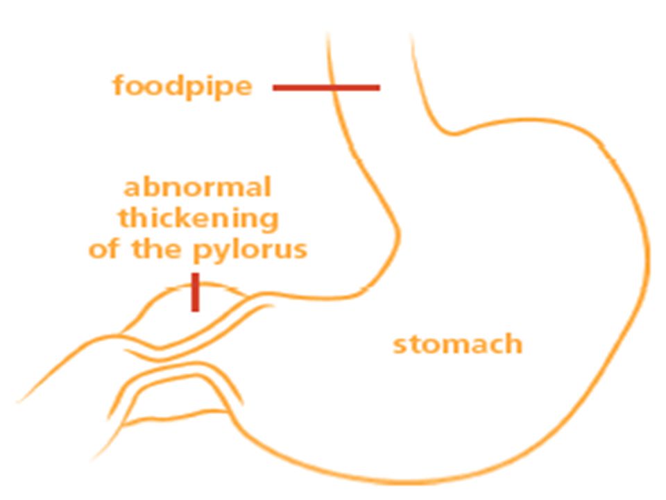
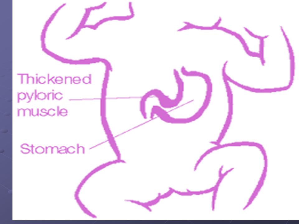
Pyloric Stenosis It is a condition that can affect babies in the first few weeks of life, (usually at around six weeks) It affects more boys than girls. It is a narrowing of the pylorus which obstructs the passage of milk or food into the intestine. In this case, the pyloric muscle around the pylorus becomes thicker soon after birth. The cause is not known but it can run in families.

5
Signs and symptoms of pyloric stenosis
A baby with pyloric stenosis will begin vomiting small amounts of milk. Over a few days this will become worse until the baby can no longer keep any milk down. This vomiting may become so forceful that the milk may be projected for several feet out of the baby's mouth. This is called projectile vomiting.

6
The milk can curdle and become yellow in color
The milk can curdle and become yellow in color. If the condition is not treated, the baby will become dehydrated and not gain weight.

7
Diagnostic measures The thickened pyloric muscle can be felt especially during feeding as a small, hard lump (OLIVE size) on the right side of the baby's stomach. The muscles around the stomach can sometimes be seen straining, moving from left to right as they try to push milk through the pylorus.
 on the right side of the baby s stomach. The muscles around the stomach can sometimes be seen straining, moving from left to right as they try to push milk through the pylorus.")
8
Diagnostic measures The baby is examined by the Doctor while the baby is feeding to see if the reversed movement of peristalsis happens and to observe any vomiting. Other investigations may be necessary such as ultrasound. For this test, the baby will be given a drink that can be seen on the screen.

10
Management and treatment
Surgery: this involves making a small cut in the stomach to reach the pylorus and cutting some of the surrounding muscle fibers. This widens the opening to the intestine and allows food to pass through. The cut will be closed with dissolvable stitches. You may sign a consent form for your baby.

11
Management and treatment
An anesthetist will also see the family to explain the child's anesthetic in more detail. If your child has any medical problems, such as allergies, please tell the doctors. As well as a drip, your child will need a naso-gastric tube through the nose and into the stomach Before the operation, intravenous drip to give fluid if the child becomes dehydrated through vomiting.

12
A naso-gastric tube is needed to empty the stomach of its contents
A naso-gastric tube is needed to empty the stomach of its contents. Before the baby wakes up from the operation, pain- relieving drugs will be given After surgery, antibiotic treatment may be needed to reduce the risk of infection

13
Post operative progress and care?
The child should be kept on nothing by mouth for about hours to allow the stomach and bowel to rest. Intravenous fluids is given during this time

14
Post operative progress and care
Then feeding is started gradually and may be increased according to child’s condition and the surgeon advice. Some times it is quite normal for your child to continue to vomit after the operation but it will not be the large amounts of projectile vomit as before.

15
Care of the wound; The wound will be about three centimeters long and will heal quickly, although the area around it may become a little red. Avoid using cream around the wound site as this may irritate it, and avoid long soaks in the bath until the wound has healed completely.

16
Care of the wound; The child can be able discharged in two to four days and will soon gain weight again. The child must be seen by the surgeon in several weeks after the operation.

17
Esophageal Atresia and Tracheoesophageal Fistula

18
Esophageal Atresia and Tracheoesophageal Fistula
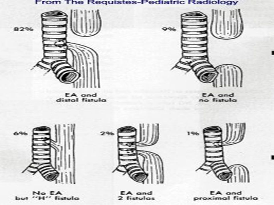
Atresia: absence of a normal opening Fistula: abnormal passage from a body organ to the body surface or between two internal body organs. Congenital esophageal atresia (EA) represents a failure of the esophagus to develop as a continuous passage. Instead, it ends as a blind pouch.
 represents a failure of the esophagus to develop as a continuous passage. Instead, it ends as a blind pouch.")
20
Types of EA,TEF

21
Tracheoesophageal fistula (TEF) represents an abnormal opening between the trachea and esophagus.
EA and TEF can occur separately or together. EA and TEF are diagnosed in the ICU at birth and treated immediately.

22
Diagnosis During pregnancy: maternal polyhydramnios and premature labor Newborn: Signs /symptoms: cough, chocking, cyanosis, excessive salivation, drooling, O Gastric tube: inability to pass down the esophagus into the stomach X-Ray: catheter curled up in the upper pouch of the esophagus air/gas bubbles in the stomach and intestine U/S: diagnosis of any associated cardiac, musculoskeletal, GI or GU abnormalities

23
How are Esophageal Atresia and Tracheoesophageal Fistula diagnosed?
♣ The presence of EA is suspected in an infant with excessive salivation (drooling) and in a newborn with drooling that is frequently accompanied by choking, coughing and sneezing. ♣ When fed, these infants swallow normally but begin to cough and struggle as the fluid returns through the nose and mouth. ♣
 and in a newborn with drooling that is frequently accompanied by choking, coughing and sneezing. ♣ When fed, these infants swallow normally but begin to cough and struggle as the fluid returns through the nose and mouth. ♣")
24
How are Esophageal Atresia and Tracheoesophageal Fistula diagnosed?
The infant may become cyanotic (turn bluish due to lack of oxygen) and may stop breathing as the overflow of fluid from the blind pouch is aspirated (sucked into) the trachea. ♣ The cyanosis is a result of laryngo-spasm (a protective mechanism that the body has to prevent aspiration into the trachea). Over time respiratory distress will develop.
 and may stop breathing as the overflow of fluid from the blind pouch is aspirated (sucked into) the trachea. ♣ The cyanosis is a result of laryngo-spasm (a protective mechanism that the body has to prevent aspiration into the trachea). Over time respiratory distress will develop.")
25
♣ If any of the above signs/symptoms are noticed, a catheter is gently passed into the esophagus to check for resistance. If resistance is noted, other studies will be done to confirm the diagnosis. ♣ A catheter can be inserted and will show up as white on a regular x-ray film to demonstrate the blind pouch ending. Sometimes a small amount of barium is placed through the mouth to diagnose the problems.

26
Treatment of EA and TEF If EA or TEF is suspected, all oral feedings should be stopped and intravenous fluids are started. The infant should be positioned to help drain secretions and decrease the likelihood of aspiration Babies with EA may sometimes have other problems. Therefore investigations should be continued to look for the heart and spine and sometimes studies are done to look at the kidneys.

27
Surgical repair to the defect: Care for each infant is individualized.
Surgery to fix EA is rarely an emergency. Once the baby is in condition for surgery, an incision is made on the side of the chest. The esophagus can usually be connected together. Following surgery, the baby may be hospitalized for a variable length of time.

28
Questions What are preoperative concerns in a patient with TEF?
How will you induce and maintain general anesthesia? During ligation of the fistula, the infant’s oxygen saturation decreases to 86%, what will you do? Will you extubate at the end of the surgery? Why or why not?

29
The End of EA and TEF

Similar presentations


 Department of Pediatrics>")
