Download presentation
Presentation is loading. Please wait.

1
Tom Eck – ecktw@umdnj.edu
HAD UNIT II CALM REVIEW

2
Major Points Cranial nerves are tested directly or indirectly on the majority of questions Know the course of each nerve, especially the foramen each passes through Know cutaneous distribution for sensory nerves and muscles innervated for motor nerves Memorize presentation of deficits associated with loss of each nerve

3
Major topics Cranial Nerves: Sensory Cranial Nerves: Motor
Cranial Nerves: Autonomic Vasculature Neck Lymphatics Embryology Connections

4
CRANIAL NERVES: SENSORY
CN I – Olfactory – Olfaction CN II – Optic – Vision CN V – Trigeminal – Facial Sensation CN VII – Facial – Taste CN VIII – Vestibulocochlear – Balance, Hearing CN IX – Glossopharyngeal – Pharyngeal Sensation CN X – Vagus – Laryngeal Sensation

5
Cranial Foramina , Ophthalmic artery Middle meningeal artery ,
Labyrinthine artery , Posterior meningeal artery

6
Nasal Ethmoid Vomer Sphenoid Lacrimal
1. which bone, when fractured, may be associated with inability to smell (Anosmia) as well as leakage of CSF? Nasal Ethmoid Vomer Sphenoid Lacrimal 10
 as well as leakage of CSF Nasal. Ethmoid. Vomer. Sphenoid. Lacrimal. 10.")
7
Ethmoidal fracture May result in damage to CN I fibers as they pass through the cribriform plate of the ethmoid Ethmoid is particularly vulnerable to trauma Also associated with CSF leakage (CSF rhinorrhea)
")
8
Don’t Do this! Nasogastric Tube

9
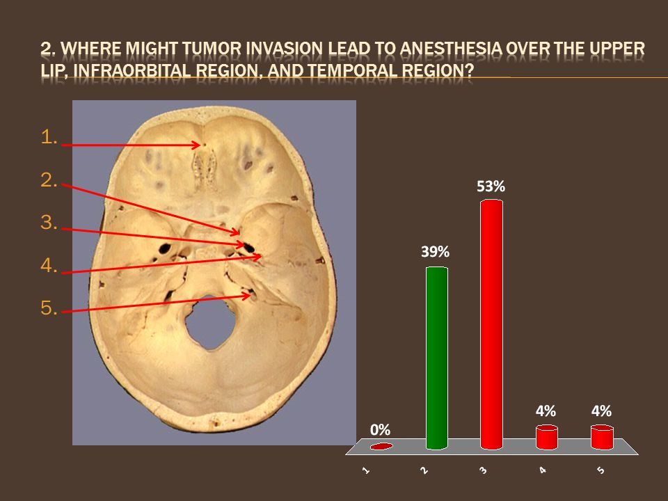
2. Where might tumor invasion lead to anesthesia over the upper lip, infraorbital region, and temporal region? 1 2 3 4 5 10
10
Maxillary Nerve Injury (V2)
Numbness in the upper lip, infraorbital region, and temporal region suggests the tumor has invaded the maxillary nerve (V2), which exits the skull via the foramen rotundum. It becomes the infraorbital nerve as it exits the skull via the infraorbital foramen You would also expect loss of sensation in the nasal mucosa and oral cavity
, which exits the skull via the foramen rotundum. It becomes the infraorbital nerve as it exits the skull via the infraorbital foramen. You would also expect loss of sensation in the nasal mucosa and oral cavity.")
11
Glossopharyngeal nerve (CN IX) Vagus nerve (CN X)
3. Which of the following Nerves does not contribute to the cutaneous innervation of the ear? Maxillary nerve (V2) Mandibular nerve (V3) Facial nerve (CN VII) Glossopharyngeal nerve (CN IX) Vagus nerve (CN X) 10
 Mandibular nerve (V3) Facial nerve (CN VII) Glossopharyngeal nerve (CN IX) Vagus nerve (CN X) 10.")
12
Cutaneous Innervation of Ear
Auriculotemporal nerve (CN V3) Root, superior helix, crus, tragus, ext auditory canal, tympanic membrane Auricular branch (CN VII) Concha, ext auditory canal Jacobsen’s nerve (CN IX) Arnold’s nerve (CN X) Concha, ext auditory canal, antihelix Lesser occipital nerve (C2) Superoposterior ear Great auricular (C2,3) Lateral helix, lobule, posteroinferior ear
 Root, superior helix, crus, tragus, ext auditory canal, tympanic membrane. Auricular branch (CN VII) Concha, ext auditory canal. Jacobsen’s nerve (CN IX) Arnold’s nerve (CN X) Concha, ext auditory canal, antihelix. Lesser occipital nerve (C2) Superoposterior ear. Great auricular (C2,3) Lateral helix, lobule, posteroinferior ear.")
13
Chorda tympani Lingual Hypoglossal Glossopharyngeal Vagus
4. What nerve has been compromised if a patient experiences numbness of the anterior tongue? Chorda tympani Lingual Hypoglossal Glossopharyngeal Vagus 10

14
Tongue Innervation Lingual nerve = sensation to anterior 2/3 of tongue
Chorda tympani = taste to anterior 2/3 of tongue Hypoglossal = motor to intrinsic and extrinsic tongue muscles (except palatoglossus) Glossopharyngeal = taste AND sensation to posterior 1/3 of tongue Vagus = taste for small patch near epiglottis
 Glossopharyngeal = taste AND sensation to posterior 1/3 of tongue. Vagus = taste for small patch near epiglottis.")
15
5. Which of the following nerves carries the afferent limb of the corneal reflex?
Ophthalmic Maxillary Facial Occulomotor Mandibular

16
Corneal Reflex Afferent Limb: Ophthalmic Nerve, V1 (Nasociliary Branch) Efferent Limb: Zygomatic Branch of Facial Nerve (CN VII) to palpebral portion of orbicularis oculi Remember: V1 includes the eyes and the tip of the nose
 to palpebral portion of orbicularis oculi. Remember: V1 includes the eyes and the tip of the nose.")
17
6. Which nerve supplies the skin overlying the vertex of the skull?
Lacrimal Supratrochlear Infratrochlear Infraorbital Supraorbital

18
Vertex

19
Supraorbital nerve Supplies much of the forehead and scalp
a branch of the ophthalmic nerve (V1 frontal nerve supraorbital) Exits the skull via the supraorbital foramen
 Exits the skull via the supraorbital foramen.")
20
Angle of jaw Lower lip Upper lip Buccal region Zygomatic region 10
7. Which area of the face would be expected to experience anesthesia following a fracture of the body of the mandible? Angle of jaw Lower lip Upper lip Buccal region Zygomatic region 10

21
Mental Nerve branches off the inferior alveolar nerve (V3), which courses through the mandible, supplying the skin of the chin (mental region) and lower lip exits the mandible via the mental foramen
, which courses through the mandible, supplying the skin of the chin (mental region) and lower lip. exits the mandible via the mental foramen.")
22
CRANIAL NERVES: MOTOR CN III – Oculomotor
CN IV – Trochlear extraocular muscles CN VI – Abducens CN V3 – Mandibular – muscles of mastication CN VII – Facial – muscles of expression CN IX – Glossopharyngeal – stylopharyngeus CN X – Vagus – muscles of pharynx and larynx CN XI – Spinal Accessory – trapezius, SCM CN XII – Hypoglossal – tongue muscles

23
swallowing phonation chewing shrugging taste 10
8. A posterior fossa tumor impinges on the jugular foramen. Which of the following will be entirely preserved? swallowing phonation chewing shrugging taste 10

24
Jugular foramen syndrome
Jugular foramen: glossopharyngeal (CN IX), vagus (X), spinal accessory (CN XI) Swallowing = vagus, glossopharyngeal, etc. Phonation = vagus (laryngeal muscles) Taste = vagus, glossopharyngeal, (and facial) Shrugging = spinal accessory, etc. Chewing = mandibular nerve (V3)
, vagus (X), spinal accessory (CN XI) Swallowing = vagus, glossopharyngeal, etc. Phonation = vagus (laryngeal muscles) Taste = vagus, glossopharyngeal, (and facial) Shrugging = spinal accessory, etc. Chewing = mandibular nerve (V3)")
25
Masseter Infrahyoid muscles Lateral pterygoid Medial pterygoid
9. Which of the following most directly opposes the action of the temporalis? Masseter Infrahyoid muscles Lateral pterygoid Medial pterygoid 10

26
Muscles of Mastication (V3)
temporalis = elevate and retract the mandible lateral pterygoid = depress and protrude mandible Elevation: Temporalis, Masseter, Medial Pterygoid Depression: Lateral Pterygoid, Suprahyoid/Infrahyoid Muscles Protrusion: Lateral Pterygoid, Masseter, Medial Pterygoid Retrusion: Temporalis, Masseter Lateral Movements: Temporalis of same side, Pterygoids of Opposite Side, Masseter Remember: Unilateral V3 lesion causes deviation to same side as lesion due to unopposed action of the contralateral medial and lateral pterygoid Temporalis Lateral Pterygoid

27
Levator veli palatini Palatopharyngeus Stylohyoid Stylopharyngeus
10. Patients with paralysis of the trigeminal nerve Lose function in which of the following muscles? Levator veli palatini Palatopharyngeus Stylohyoid Stylopharyngeus Tensor veli palatini 10

28
CN V3 - Mandibular nerve (motor)
Temporalis Masseter Lateral pterygoid Medial pterygoid Mylohyoid Anterior belly of digastric Tensor tympani Tensor veli palatini

29
11. Which of the following dominates the efferent Limb of the gag reflex?
CN V CN IX CN X CN XII 10

30
Gag reflex The nerve supply to the pharynx is derived from the pharyngeal plexus Glossopharyngeal = sensory supply (afferent limb) Vagus = motor supply (efferent limb) Sensory Exceptions: upper nasopharynx supplied by V2 (along with nasal mucosa); lower laryngopharynx supplied by internal laryngeal (CN X) Motor Exceptions: stylopharyngeus (CN IX), tensor veli palatini (CN V3) *These are also involved in the reflex
 Sensory Exceptions: upper nasopharynx supplied by V2 (along with nasal mucosa); lower laryngopharynx supplied by internal laryngeal (CN X) Motor Exceptions: stylopharyngeus (CN IX), tensor veli palatini (CN V3) *These are also involved in the reflex.")
31
CN V CN VII CN IX CN X CN XII
12. Which nerve is damaged if a person must constantly press their cheek in while eating? CN V CN VII CN IX CN X CN XII 10

32
Buccinator innervated by the Facial Nerve
keeps food out of the oral vestibule meets the superior pharyngeal constrictor (CN X) posteriorly at the pterygomandibular raphe
 posteriorly at the pterygomandibular raphe.")
33
Superior rectus Lateral rectus Superior Oblique Sphincter pupillae
13. Which of the following would remain functional following compression of the common tendinous ring? Superior rectus Lateral rectus Superior Oblique Sphincter pupillae Dilator pupillae 10

34
Common tendinous ring Through the common tendinous ring Outside:
OPTIC NERVE Ophthalmic artery Motor (Occulomotor n. , Abducens n.), except the Trochlear nerve Nasociliary nerve Outside: Opthalmic vein Sensory (Lacrimal n., Frontal n.), except the nasociliary nerve (which supplies the eyeball) Trochlear nerve
, except the Trochlear nerve. Nasociliary nerve. Outside: Opthalmic vein. Sensory (Lacrimal n., Frontal n.), except the nasociliary nerve (which supplies the eyeball) Trochlear nerve.")
35
Left glossopharyngeal Right glossopharyngeal Left hypoglossal
14. When a patient sticks out her tongue, it deviates to the right side. Which nerve has been damaged? Left glossopharyngeal Right glossopharyngeal Left hypoglossal Right hypoglossal 10

36
Hypoglossal nerve Unilateral lesion causes the tongue to deviate to the SAME side when protruded The intact genioglossus pulls the back of the tongue forward, deviating the tongue to the other side

37
CRANIAL NERVES: AUTONOMICS
COPS 3977 (Parasympathetic Ganglia) Ciliary = CN 3 (pupillary constriction and accomodation) Otic = CN 9 (salivation) Pterygopalatine = CN 7 (lacrimation) Submandibular = CN 7 (salivation) Sympathetic fibers carried by arteries from superior cervical ganglion
 Ciliary = CN 3 (pupillary constriction and accomodation) Otic = CN 9 (salivation) Pterygopalatine = CN 7 (lacrimation) Submandibular = CN 7 (salivation) Sympathetic fibers carried by arteries from superior cervical ganglion.")
38
Ophthalmic Oculomotor Long Ciliary Facial Maxillary 10
15. A patient complains of dry eyes following trauma. Which of the following nerves may have been damaged at its origin? Ophthalmic Oculomotor Long Ciliary Facial Maxillary 10

39
PTERYGOPALATINE GANGLION

40
Trigeminal (semilunar)
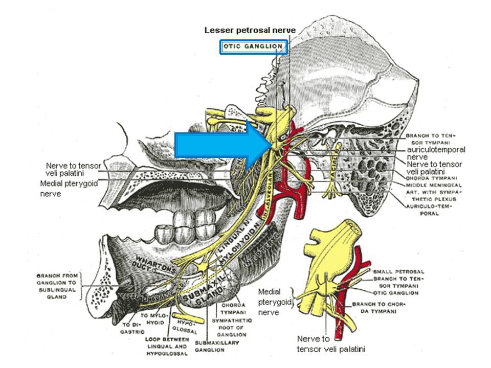
16. Which ganglion is located just below the foramen ovale and, when damaged, leads to Dry mouth (xerostomia)? Ciliary Pterygopalatine Otic Submandibular Geniculate Trigeminal (semilunar) 10
 Ciliary. Pterygopalatine. Otic. Submandibular. Geniculate. Trigeminal (semilunar) 10.")
42
OTIC GANGLION

43
Greater palatine Lesser petrosal Greater petrosal Chorda tympani
17. Which of the following nerves carries presynaptic parasympathetic fibers to the submandibular gland? Greater palatine Lesser petrosal Greater petrosal Chorda tympani Inferior alveolar 10

44
SUBMANDIBULAR GANGLION

45
CILIARY GANGLION Oculomotor Nerve (Pre) Ciliary Ganglion Short Ciliary Nerves (Post)
 Ciliary Ganglion Short Ciliary Nerves (Post)")
46
Vasculature External Carotid and its branches
Anterior: superior thyroid, lingual, facial Posterior: Occipital, Posterior Auricular Medial: Ascending Pharyngeal Terminal: Superfical Temporal, Maxillary Internal Carotid and Circle of Willis Dural Venous Sinuses Basic Venous Drainage Be familiar with major branches of maxillary, facial arteries

47
Sphenopalatine foramen
18. When significant trauma is inflicted at the pterion, an epidural hematoma often results. By what route does the involved artery enter the cranium? Foramen ovale Foramen rotundum Foramen spinosum Foramen lacerum Sphenopalatine foramen 10

48
Middle Meningeal artery
pterion = major weak point in skull; location where the frontal, sphenoid, temporal, and parietal bones meet fracture here associated with laceration of underlying middle meningeal artery (responsible for 70-80% of epidural hematomas) enters the skull via the foramen spinosum
 enters the skull via the foramen spinosum.")
49
Anterior cerebral artery Middle cerebral artery
19. Which Of the following does Not typically branch from the internal carotid artery? Anterior cerebral artery Middle cerebral artery Posterior cerebral artery Posterior communicating artery Ophthalmic artery 10

50
Circle of willis Represents a major site of anastomosis between the two vertebral arteries (via the basilar artery) and the two internal carotid arteries, which together supply the brain
 and the two internal carotid arteries, which together supply the brain.")
51
Superior sagittal sinus Occipital sinus Straight sinus Sigmoid sinus
20. Which of the following drains blood away from the confluence of the sinuses? Transverse sinus Superior sagittal sinus Occipital sinus Straight sinus Sigmoid sinus 10

52
The Confluence of the sinuses
Receives blood from the superior sagittal, straight, and occipital sinuses Blood drains into the (R/L) transverse sinuses, and from there to the (R/L) sigmoid sinuses
 transverse sinuses, and from there to the (R/L) sigmoid sinuses.")
53
Neck Fascial planes Major vessels
Strap muscles – innervated by ansa cervicalis (C1 to C5) * know segments contributed to each muscle Larynx – structure, muscles, innervation
 * know segments contributed to each muscle. Larynx – structure, muscles, innervation.")
54
Superficial (investing) fascia Prevertebral fascia Pretracheal fascia
21. Upon swallowing, what helps to elevate the thyroid along with the trachea and larynx? Superficial (investing) fascia Prevertebral fascia Pretracheal fascia Suspensory ligaments Pyramid lobe of the thyroid 10
 fascia. Prevertebral fascia. Pretracheal fascia. Suspensory ligaments. Pyramid lobe of the thyroid. 10.")
55
Fascial Layers of Neck Superficial investing fascia Pretracheal fascia
Suprahyoid muscles SCM Trapezius Pretracheal fascia Thyroid Trachea Move as a unit Prevertebral fascia Scalenes Paravertebral muscles

56
22. Between which structures would a pharyngeal infection be most likely to spread to the mediastinum? Between the trachea and the carotid sheath Between the trachea and the strap muscles Between the trachea and the esophagus Between the esophagus and the prevertebral muscles Between the trapezius and the prevertebral muscles 10

57
Retropharyngeal space
Situated between the buccopharyngeal fascia and the alar fascia Permits spread of infections into the mediastinum from the head and neck

58
posterior cricoarytenoid lateral cricoarytenoid vocalis
23. Which of the following muscles would be most active in forced respiration, as in when taking a deep breath? cricothyroid thyroarytenoid posterior cricoarytenoid lateral cricoarytenoid vocalis 10

59
The Larynx Know actions of muscles (use names to give you clues)
Know motor innervation (all inferior laryngeal, except cricothyroid = external laryngeal) Know sensory innervation Superior to vocal ligament = internal laryngeal Inferior to vocal ligament = inferior laryngeal (from recurrent laryngeal)
 Know sensory innervation. Superior to vocal ligament = internal laryngeal. Inferior to vocal ligament = inferior laryngeal (from recurrent laryngeal)")
60
Lymphatics Expect 2 or 3 lymph questions
Lymph drainage of the face and tongue are key Remember to keep an eye out for small details when studying

61
submental submandibular Inferior jugular buccal parotid
24. To which lymph nodes would you expect this squamous cell carcinoma to metastasize first? submental submandibular Inferior jugular buccal parotid 10

62
Major Drainage patterns
submental: central lower lip, chin, apex of tongue submandibular: upper lip, lateral lower lip, lateral part of anterior 2/3 of tongue Inferior deep cervical: medial part of anterior 2/3 Superior deep cervical: posterior 1/3 of tongue Parotid: lateral face and scalp, eyelids

63
Embryology Branchial Arches Eye Development Ear Development
Know the precursors

64
25. from which of the branchial arches is the affected nerve derived?
First Second Third Fourth Fifth

65
Second branchial pouch
26. The external auditory meatus develops from which of the following embryologic structures? First branchial arch Second branchial arch Third branchial arch First branchial cleft Second branchial pouch 10

66
Branchial arch derivatives
Nerve Skeletal Structures Muscles 1 CN V Mandible, malleus, incus, greater wing of sphenoid Muscles of mastication, mylohyoid, anterior belly of digastric, tensor tympani, tensor veli palatini 2 CN VII Stylohyoid process, stapes, upper body & lesser horn of hyoid Muscles of facial expression, stylohyoid, stapedius, postberior belly digastric 3 CN IX Lower body & greater horn of hyoid Stylopharyngeus 4 CN X Superior laryngeal Thyroid cartilage Cricothyroid, levator veli palatini, palatopharyngeus, palatoglossus, pharyngeal constrictors 5-6 Recurrent laryngeal Cricoid cartilage Intrinsic muscles of larynx (except cricothyroid & stylopharyngeus)
")
67
Branchial Clefts & Pouches

68
Utricular part; surface ectoderm Utricular part; neural ectoderm
27. The semicircular canals develop from the _________________ of the otic vesicle, which is itself derived from _______________. Utricular part; surface ectoderm Utricular part; neural ectoderm Saccular part; surface ectoderm Saccular part; neural ectoderm

69
Inner Ear Development otic placode otic pit otic vesicle
Dorsal Utricular Part utricle, semicircular canals (U looks like canals) Ventral Saccular Part saccule, cochlea (S for Spiral shape of cochlea)
 Ventral Saccular Part saccule, cochlea (S for Spiral shape of cochlea)")
70
Connections the cranium is a maze learn the major passageways
especially true for the face

71
28. Where do tears Enter the nasal cavity?
Inferior meatus Middle meatus Superior meatus Sphenopalatine foramen Pterygomaxillary fissure 10

72
Sinus Drainage Inferior meatus Middle meatus Superior meatus
Nasolacrimal duct Middle meatus Frontal sinus Anterior ethmoidal air cells Maxillary sinus Superior meatus Posterior ethmoidal air cells Sphenoethmoidal recess Sphenoidal sinus (associated with pituitary gland)
")
73
Superior orbital fissure Inferior orbital fissure Optic canal
29. A tumor from the infratemporal fossa gains entrance to the orbit. What is the most likely route? Superior orbital fissure Inferior orbital fissure Optic canal Sphenopalatine foramen Pterygoid canal 10

74
TO ORBIT A tumor could also invade the nasal cavity by passing through the pterygomaxillary fissure and sphenopalatine foramen.

75
Practical Be sure to spend some time with the models; there are a lot more on this exam Be able to identify structures with the head in various positions No mock practical this time, but use the structured lab review as a guide

76
How soon before the exam would you like the review?
Earlier A week is good Later 10

77
Good luck!

Similar presentations


>")


