Download presentation
Presentation is loading. Please wait.

1
Facial Resurfacing

2
Historically, people in many societies have searched for methods to restore youthful appearance and rejuvenate aging skin: 1. Stories about mythical substances 2.During the Dark Age 3.Modern society

3
The desire for cosmetic enhancement of facial skin with minimal risk and rapid recovery has inspired laser-mediated means of wrinkle and photodamage reduction.

4
More recently, the spectrum of skin issues for which these methods have been utilized has expanded past the treatment of photo-aging to include many skin problems more common in youth, such as pigmentary disorders and acne scarring.

5
Laser use on the skin has become one of the most popular methods for achieving a younger and smoother facial appearance.

6
Unfortunately, the increasingly widespread availability of cosmetic laser therapy coupled with attendant publicity has created extraordinary, often unrealistic, expectations. Many patients may believe that lasers are magic wands, capable of restoring their skin to the flawless perfection of infancy. Of course, the truth is that cosmetic lasers are not the answer for all dermatologic ills.

7
سکندر را نمی بخشند ابی به زور و زر میسر نیست این کار

9
While lighter skinned individuals are more likely to present with deep-seated wrinkles,
darker patients are usually seenwith finer wrinkles, mottled hyperpigmentation, and unique skin lesions such as dermatosis papulosa nigra and actinic lentigines .

10
patients with Fitzpatrick skin types IV-VI respond to UV injury by producing more melanin. Thus, they are at higher risk for the development of dyschromias after a laser resurfacing procedure.

11
Although laser resurfacing for skin types IV to VI is not contraindicated, resurfacing on these persons can be more difficult because of the almost certain occurrence of postinflammatory hyperpigmentation. This usually begins approximately 1 month postoperatively and lasts about 2 months on average, but can somerimes last as long as 9 months.

13
The Glogau classification system helps the physician determine the depth of damage, and thus offers some indication of what the depth of resurfacing should be. Patients with minimal photo-damage may require ablation of only the upper part of the epidermis. Those with moderate photo-damage may require more extensive resurfacing to the level of the papillary dermis, and so on.

15
The word LASER is an acronym, which stands for light amplification by the stimulated emission of radiation. Laser light has unique properties that allow it to be used therapeutically. Consequently, the popularity of laser procedures has skyrocketed in the past decade as the indications for use and types of lasers available continue to multiply. Laser light is monochromatic (single wavelength), coherent (in phase, both in time and space), and collimated (light waves are parallel). These properties make possible the generation and delivery of high fluence (energy per area), which can interact with the skin. Additionally, the monochromaticity of laser light is essential for selective targeting of structures in the skin that preferentially absorb light of that wavelength.
, coherent (in phase, both in time and space), and collimated (light waves are parallel). These properties make possible the generation and delivery of high fluence (energy per area), which can interact with the skin. Additionally, the monochromaticity of laser light is essential for selective targeting of structures in the skin that preferentially absorb light of that wavelength.")
18
hemoglobin, water, and collagen .
The endogenous chromophores of importance are: melanin, hemoglobin, water, and collagen .

19
and for dermis in the range of 280-1300.
In normally epidermis, absorption is usual in the range of nm and for dermis in the range of

20
Rejuvenation . ablate the epidermis, cause dermal wounding and provide a significant thermal effect (e.g. CO2 lasers) · ablate the epidermis, cause dermal wounding, and provide minimal thermal effects (e.g. short-pulsed Er:YAG lasers) · ablate the epidermis, cause dermal wounding and provide variable thermal effects (e.g. combined COrEr:YAG lasers, variable-pulsed Er:YAG lasers, and ablative radiofrequency devices) .do not ablate the epidermis, cause dermal wounding, and provide minimal thermal effects (e.g. nonablative lasers and light sources) .
 · ablate the epidermis, cause dermal wounding, and provide minimal thermal effects (e.g. short-pulsed Er:YAG lasers) · ablate the epidermis, cause dermal wounding and provide variable thermal effects (e.g. combined COrEr:YAG lasers, variable-pulsed Er:YAG lasers, and ablative radiofrequency devices) .do not ablate the epidermis, cause dermal wounding, and provide minimal thermal effects (e.g. nonablative lasers and light sources) .")
23
Ablative resurfacing was first introduced in the mid 1990s.
In an effort to decrease the risk/side effect profile, the use of erbium lasers was explored (Zachary 2000) .
 .")
24
Carbon dioxide lasers

26
water, hemoglobin, and collagen .
The endogenous chromophores of importance are: melanin, hemoglobin, water, and collagen . .Producing a wavelength of I 0,600 nm, the CO2 laser penetrates approximately nm into the skin by the absorption and vaporization of water-containing tissues. The epidermis is composed of 80% water. At the energies used for resurfacing, instant heating of water to 100°C occurs and vaporization of tissue ensues.

27
The thermal relaxation time(TRT) the amount of time required for a material to lose 63% (1/e) of its heat by thermal diffusion. If energy is delivered to a target in a period of time faster than the time it takes for the target to lose its heat by conduction to its surroundings, then no (or minimal) thermal damage will occur beyond the target. To achieve heating and destruction of target tissue in a period of time that is shorter than TRT the laser beam must be pulsed such that it is only on for a time t < TRT.

28
A chromophore is heated by laser light absorption in a time period shorter than its thermal relaxation time. The latter is the amount of time required for a material to lose 50% of its heat by conduction to its surroundings. Thus, when a chromophore is heated by selective photothermolysis, only the intended target is damaged and there is minimal diffusion of heat and no consequent injury to the surrounding structures. The mechanism of injury involves both thermal coagulation and/or photoacoustic injury in the form of supersonic high pressure shock waves. For both the CO2 and Er:YAGlasers, the predominant mechanism is photothermal.

29
.Energy densities of approximately 5J/cm2 must be applied in order to achieve tissue ablation.
.With each subsequent laser pass, vaporization of very thin ( nm) layers of skin occurs, leaving a small amount of residual thermal necrosis. With each subsequent laser pass, further tissue ablation occurs, but, because the area of residual thermal necrosis increases (effectively reducing the amount of tissue water and hence the targeted chromophore), the amount of ablation with each pass diminishes until a peak of approximately nm is reached. .Delivering more than three to four passes, or the use of excessive energy densities, significantly increases the risk of excessive thermal injury and subsequent scarring.
 layers of skin occurs, leaving a small amount of residual thermal necrosis. With each subsequent laser pass, further tissue ablation occurs, but, because the area of residual thermal necrosis increases (effectively reducing the amount of tissue water and hence the targeted chromophore), the amount of ablation with each pass diminishes until a peak of approximately 100 nm is reached. .Delivering more than three to four passes, or the use of excessive energy densities, significantly increases the risk of excessive thermal injury. and subsequent scarring.")
30
The pulse fluence necessary to achieve vaporization and thus ablation of skin tissue with the CO2 laser is 5J/cm2. This can be calculated based on two factors: The latent heat of vaporization of water is 2500J/cm3, and the energy (Ev)absorbed per unit volume of tissue is Ev= fluence x µa. The depth of penetration of the 10,600nm wavelength has been calculated to be 20µm, or l/µa, where µa is the absorption coefficient for water (equal to 500cm-1). Solving for fluence in the above equation, fluence = 2500/500 = 5J/cmz. The actual thickness of tissue ablation varies from 20 to 60nm.
absorbed per unit volume of tissue is Ev= fluence x µa. The depth of penetration of the 10,600nm wavelength has been calculated to be 20µm, or l/µa, where µa is the absorption coefficient for water (equal to 500cm-1). Solving for fluence in the above equation, fluence = 2500/500 = 5J/cmz. The actual thickness of tissue ablation varies from 20 to 60nm.")
31
Cutaneous CO2 laser resurfacing, as currently performed, has been shown to be highly effective in the treatment of photo-damaged skin. In addition to superficial ablation, there is a "tissue tightening" effect following use of these lasers. This effect is thought to be related to heat-induced collagen shrinkage, which occurs maximally at 63°C. Long-term collagen remodeling and neocollagenesis also occur after resurfacing, although the mechanisms behind this are not fully known. It is believed that these effects result from thermal desiccation associated with the concomitant collagen shrinkage. In addition, because there is increased expression of smooth muscle actin after laser treatment, the contracted area may serve as a scaffolding on which new collagen is formed and deposited during wound phase remodeling. These factors are most likely responsible for the long-term clinical improvement seen after resurfacing.

32
Significant collagen shrinkage and remodeling requires at least two passes.

34
Several CO2 laser systems are available and can be separated into two distinct groups: pulsed and scanned.

35
The high-energy pulsed CO2 lasers (e. g
The high-energy pulsed CO2 lasers (e.g. Ultrapulse, Lumenis) produce single, short (1 ms) pulses o fvery high energy (up to 7 J/cm2). A computerized pattern generator (CPG) attached to the laser delivery system can rapidly and precisely place S-mm spots in any of several patterns while maintaining appropriate ablation parameters. A large square pattern can be used to treat large areas rapidly. The density of the pattern can be low (nonoverlapping spots) or high (10-60%overlapping spot) BOLOGNIA: The first type is the high-powered pulsed CO2 laser system, which delivers energy in individual pulses of about 1 ms or less (e.g. UltraPulseM). This laser produces up to mJ of energy in each individual µS-l ms pulse. Vaporization can be performed either with a 3 mm spot size or by a computer pattern generator, which can deliver various patterns of up to 80 pulses, each pulse measuring mm in diameter.
 produce single, short (1 ms) pulses o fvery high energy (up to 7 J/cm2). A computerized pattern generator (CPG) attached to the laser delivery system can rapidly and precisely place 2.2S-mm spots in any of several patterns while maintaining appropriate ablation parameters. A large square pattern can be used to treat large areas rapidly. The density. of the pattern can be low (nonoverlapping spots) or high (10-60%overlapping spot) BOLOGNIA: The first type is the high-powered pulsed CO2 laser system, which delivers energy in individual pulses of about 1 ms or less (e.g. UltraPulseM). This laser produces up to 500 mJ of energy in each individual 600 µS-l ms pulse. Vaporization can be performed either with a 3 mm spot size or by a computer pattern generator, which can deliver various patterns of up to 80 pulses, each pulse measuring 2.25 mm in diameter.")
36
Scanned laser systems (e. g
Scanned laser systems (e.g. FeatherTouch, Lumenis; Silk- Touch, Lumenis) utilize a computerized scanning device to deliver lower power CO2 laser energy in the continuous mode rapidly over the skin, thus limiting the tissue dwell time in one area. This system achieves high peak powers by focusing the laser beam to small spot size, and rapidly scans the focused beam over a predetermined geometric pattern, exposing the individual tissue sites for less than 1ms. BOLOGNIA: The second type of CO2 resurfacing laser achieves well-controlled tissue ablation by rapidly scanning the focal spot of a focused continuous-wave CO2 laser over the skin (e.g. FeatherTouch and SilkTouch flash scanners). Computer-driven mechanical devices can scan a 0.2 mm spot in a spiral manner, ranging in diameter from 8 to 16 mm in several shapes at a constant velocity. No individual spot is irradiated more than once and the dwell time on any individual spot is less than 1 ms, while achieving fluences above the ablation threshold.
 utilize a computerized scanning device to deliver lower power CO2 laser energy in the continuous mode rapidly over the skin, thus limiting the tissue dwell time in one area. This system achieves high peak powers by focusing the laser beam to small spot size, and rapidly scans the focused beam over a predetermined geometric pattern, exposing the individual tissue sites for less than 1ms. BOLOGNIA: The second type of CO2 resurfacing laser achieves well-controlled tissue ablation by rapidly scanning the focal spot of a focused continuous-wave CO2 laser over the skin (e.g. FeatherTouch and SilkTouch flash scanners). Computer-driven mechanical devices can scan a 0.2 mm spot in a spiral manner, ranging in diameter from 8 to 16 mm in several shapes at a constant velocity. No individual spot is irradiated more than once and the dwell time on any individual spot is less than 1 ms, while achieving fluences above the ablation threshold.")
37
2 more popular CO2 lasers are UltraPulse laser (Coherent) and the SilkTouch scanner (Sharplan).
 and the SilkTouch scanner (Sharplan).")
38
WHAT IS DWELL TIME? Laser dwell time is the amount of time that the beam is on in one location. Low power densities require longer dwell times to achieve the same effect as high power densities. The longer the dwell time and the slower the heating, the more desiccation and charring of tissue that results. Further heating of charred tissue results in extremely high temperatures of °C. This is because carbonized and desiccated tissue acts as a heat sink for laser absorption. There is no buffer of water to absorb the heat and thus temperatures escalate rapidly. Continuous wave lasers must be rapidly scanned across the treated area in order to keep the dwell time less than the thermal relaxation time.

39
A study comparing four different CO2 lasers found that pulsed systems produced the least amount of thermal necrosis with the greatest subsequent collagen formation (compared with the scanned systems), but equivalent clinical outcomes between all four lasers were observed. BOLOGNIA: In general, two passes with the SilkTouch are approximately equal to three pulses with the UltraPulse and four passes with the FeatherTouch in terms of the amount of tissue removed and the depth of residual thermal damage.

41
(A) To generate identical fluence at skin surface, a chopped pulse (continuous wave) must be maintained for a longer period than with ultrapulsed or superpulsed lasers, which generate a higher peak power over a shorter time. (B) Comparison of ultrapulse, superpulse, and chopped-pulse CO2 laser fluence. At same average power, superpulsed laser must deliver 4 to 5 pulses for each ultra pulse. A chopped pulse is seven times longer than an ultra pulse for this same energy.
 Comparison of ultrapulse, superpulse, and chopped-pulse CO2 laser fluence. At same average power, superpulsed laser must deliver 4 to 5 pulses for each ultra pulse. A chopped pulse is seven times longer than an ultra pulse for this same energy..")
43
CW CO2 LASER:Because the peak powers were low (the same as the average power, or 10–30 W), the power densities were often subablative or marginally supra-ablative, and the only advantage was confinement of thermal injury to a level not observed with longer exposures (and higher fluences). The shortest possible exposures available with these devices were typically 50–100 ms. The next generation of CO 2 lasers used so-called “superpulsed (SP) technology”. Pulses were on the order of 50–200 mJ and were delivered with varying repetition rates (50–500 Hz). The term “UltraPulse,” used by a leading manufacturer, refers to a subset of superpulsed lasers with a rectangular pulse profile, capability of low repetition rates, and higher peak powers than older first-generation SP lasers.
 technology . Pulses were on the order of 50–200 mJ and were delivered with varying repetition rates (50–500 Hz). The term UltraPulse, used by a leading manufacturer, refers to a subset of superpulsed lasers with a rectangular pulse profile, capability of low repetition rates, and higher peak powers than older first-generation SP lasers.")
48
The short-pulsed Er:YAG laser was developed in the mid 1990s in an attempt to replicate the results of the CO2 laser while minimizing the side-effect profile. The emitted wavelength of nm has a higher affinity for water and is therefore absorbed 12 to 18 times more efficiently by superficial cutaneous tissues. Approximately µm of ablation occurs per pass, with very narrow zones of thermal necrosis. Clinically, this translates into a shorter postoperative healing time with a lower risk of post-treatment erythema and hyperpigmentation than with CO2 lasers.

49
Fleming has shown that to achieve equal depth of injury the Er:YAG must ablate deeper than the CO2 laser because there is minimal laser-induced thermal damage. Hence, multiple passes with the laser are necessary to ablate a similar depth as one pass of the CO2 laser. With the Er:YAG laser at 2940nm, the energy is so efficiently absorbed by water (12-18 times that of the l0,600nm wavelength) that its narrow depth of penetration (2-5 J/m) results in a very superficial layer of tissue ablation with minimal surrounding thermal injury (20- 50J/m compared to J/m for CO2). Because of this high absorbance in tissue, the Er:YAGlaser achieves ablation primarily through a photomechanical rather than a photothermal effect.
 that its narrow depth of penetration (2-5 J/m) results in a very superficial layer of tissue ablation with minimal surrounding thermal injury (20- 50J/m compared to J/m for CO2). Because of this high absorbance in tissue, the Er:YAGlaser achieves ablation primarily through a photomechanical rather than a photothermal effect.")
50
DISADVANTAGES large amount of plume produced.
1)In addition, because the Er:YAG effects are photomechanical rather than photothermal (like the CO2), intraoperative hemostasis is often difficult to achieve. The thermal damage zone created by this procedure is fixed and very small. In fact, this thermal damage is so shallow that it is insufficient to coagulate dermal capillaries. This explains why Er:YAG-lasered skin bleeds. 2)The lack of thermal damage may also be considered a disadvantage when treating patients with severe wrinkles. 3)Other limitations include the associated noise level and the large amount of plume produced.
In addition, because the Er:YAG effects are photomechanical rather than photothermal (like the CO2), intraoperative hemostasis is often difficult to achieve. The thermal damage zone created by this procedure is fixed and very small. In fact, this thermal damage is so shallow that it is insufficient to coagulate dermal capillaries. This explains why Er:YAG-lasered skin bleeds. 2)The lack of thermal damage may also be considered a disadvantage when treating patients with severe wrinkles. 3)Other limitations include the associated noise level and the. large amount of plume produced.")
51
ERB:YAG in comparison with CO2:
1)minimal surrounding thermal injury 2)poor hemostasis 3)Reduced collagen shrinkage
minimal surrounding thermal injury. 2)poor hemostasis. 3)Reduced collagen shrinkage.")
52
FOR OVERCMING THE POOR HEMOSTASIS
.The Derma-K is a hybrid that combines a conventional ablative Er:YAGwith(short pulse) a low-power CO2 laser for coagulation. .The dual mode Contour consists of two Er:YAGlaser heads. One delivers short-pulsed ablative energy and the other provides long-pulsed subablative coagulative energy. .The CO3 laser is a variable pulse system that delivers both short ablative and long coagulative pulses from the same Er:YAGlaser head.
 a low-power CO2 laser for coagulation. .The dual mode Contour consists of two Er:YAGlaser heads. One delivers short-pulsed ablative energy and the other provides long-pulsed subablative coagulative energy. .The CO3 laser is a variable pulse system that delivers both short ablative and long coagulative pulses from the same Er:YAGlaser head.")
53
METHOD Extensive prepping of the skin prior to laser treatment is unnecessary because the heat of the laser sterilizes the skin. The treated areas should be wrapped with wet towels to decrease the risk of fire.

54
Preoperatively, patients apply topical anesthetic cream EMLA (eutectic mixture of lidocaine and prilocaine) with occlusion 2.5 h prior to the procedure time. Forty-five minutes before the procedure, EMLA is reapplied with occlusion.

55
.The first CO2 ablative laser pass is performed mainly to remove the epidermis and feather peripherally to minimize any demarcation with surrounding nontreated skin. .The second laser pass, and, if used, a third pass is for heat deposition to promote tightening. .Finally, the erbium laser (in the ablation mode) can be used to remove superficial thermal necrosis for further sculpting of deeper rhytides and/or acne scars. Sculpting: to make sth a particular shape
 can be used to remove superficial thermal necrosis for further sculpting of deeper rhytides and/or acne scars. Sculpting: to make sth a particular shape.")
56
When the UltraPulse CO2 laser specifically is used, the first pass is usually performed at a density of 7 for the main treatment areas. The previously described preoperative topical anesthetic technique leads to increased skin hydration and, consequently, allows the use of a higher density setting to more efficiently remove the epidermis. If no hydration is used, the first pass is performed at a density of 6. Overlap:10% Hair-bearing areas must be avoided, including eyebrows and eyelashes.

57
Hydration is mandatory, however, for treatment of the neck.
Mandatory: comulsory , necessory by the law

58
When moving towards the jawline and hairline, the density is decreased to 6 and possibly 5 for higher risk patients. Thus: For initiating:-7 if hydration is performed(neck should be hydrated necessarily) -6 if hydration is not performed -6 in jawlines(5 if the pt is highrisk) -Progressing down the neck, density settings are decreased by one per row until the lowest setting of 1 is reached, allowing skip areas in the final row. NECK:INITIATING WITH 7 AND REACHES TO 1 IN THE LOWEST ROW
 -6 if hydration is not performed. -6 in jawlines(5 if the pt is highrisk) -Progressing down the neck, density settings are decreased by one per row until the lowest setting of 1 is reached, allowing skip areas in the final row. NECK:INITIATING WITH 7 AND REACHES TO 1 IN THE LOWEST ROW.")
59
The epidermis is then wiped free on the central face and other areas where a second pass is to be performed. The peripheral edges are usually left intact and the neck is never wiped. NECK:INITIATING WITH 7 AND REACHES TO 1 IN THE LOWEST ROW AND SHOULD NOT BE WIPED WIPE:to rub sth against a surface to remove dirt,liquid,etc.

60
The wet gauze also serves to rehydrate the desiccated tissue.
Dry gauze should be used to remove any remaining water and exudates prior to another pass because this surface water would absorb the energy and prevent further tissue ablation.

61
The second CO2 laser pass is performed at a density of 4–5 depending on the tightening needed and the risk for the area. .The upper eyelids and the central face are typically treated at densities of 5 ,whereas mid cheeks and some lower eyelids may be treated with densities of 4. NOTE: .Delivered energies are also decreased towards the periphery. .A second pass is rarely done on the lateral cheeks unless acne scarring is present. .A third pass may be done on acne scars and in perioral and glabellar regions to deliver additional heat to enhance tightening. .When using the EMLA topical anesthetic technique, the face is typically treated in sections. All passes in a given area are performed before moving on to the next section.

62
.Always 'feather' the peripheral areas by decreasing the density of pulse application as well as the pulse energy (decrease to m), or angle the beam at 45 degrees to spread the fluenee over a larger surface area. .Feather into the hairline a distance of 5-15 mm and well under the jawline (3-5 cm).
.")
64
Computer pattern generator (CPG) settings should not use densities greater than 6??? if cumulative thermal damage from excessive pulse overlap is to be avoided.

65
FITZPATRICK: in general, thinner skin (e. g
FITZPATRICK: in general, thinner skin (e.g., periorbital) requires fewer laser passes, and laser resurfacing of non-facial skin (e.g., neck, chest) should be avoided due to the relative paucity of pilosebaceous units in these areas
 requires fewer laser passes, and laser resurfacing of non-facial skin (e.g., neck, chest) should be avoided due to the relative paucity of pilosebaceous units in these areas.")
66
The endpoint of treatment is when one of the following conditions is seen:
1. The wrinkle or scar is removed. 2. A yellow-brown discoloration indicating thermal damage is seen. 3. No further skin tightening is observed

67
In cases where deep rhytides or acne scars persist, the erbium laser in the ablative (shorter-pulsed) mode is helpful to sculpt the edges or to remove the superficial coagulative necrotic layer, which can hinder healing. The utilized erbium laser energy, and spot size, depends on the area to be treated, with a 3.5- to 5.0-mm spot size set at 1–2 J/cm2 most commonly used. Bleeding can occur in these areas as the thermal effect is insufficient to provide hemostasis.

68
One should not continue ablation once the distinctive yellowish color of the desiccated reticular dermis has been reached.

69
When the erbium laser is the sole utilized system, the first pass is performed to most efficiently debride the epidermis. This is undertaken typically at 100 µm of ablation with no coagulation. The ablation depth is decreased at the periphery to minimize the final demarcation between treated and untreated areas. For the second pass, erbium laser coagulative pulses or, alternatively, ablation with concomitant coagulation is used to provide the heat needed for the tightening effect. Finally, the third pass utilizes the ablation mode to remove superficial necrosis but can also include additional coagulation to enhance the thermal effect. . To treat the neck, pure ablation is used with a graduated drop in setting to feather while proceeding lower and laterally on the neck. As with the pulsed CO2 laser, careful feathering to blend the treated and untreated areas is critical to ensure a natural and cosmetically pleasing result.

70
As with CO2 ablation, the area to be treated should be wrapped with wet towels. Eye protection with intraocular anti-reflective metal eyeshield(s) should be used if treating the eyelid .Wiping between laser passes is unnecessary because proteinaceous debris is ablated with each subsequent pass. Patient preparation and anesthesia are performed similarly to those for the CO2 laser. However, because the Er:YAG laser causes minimal pain, less anesthesia may be necessary for many patients, and topical and local anesthesia will suffice.

71
The fluence required for ablation using the Er:YAGlaser is 1 J for every 4µm of tissue vaporized. One adjusts the fluence to an appropriate setting for the amount of ablation desired. A tluence of 10J/cm2 will reliably vaporize 40 J/m of tissue, thus ablating epidermis in about two passes. The endpoint is visible effacement of the lesion(s) or until reticular dermis has been reached. Because of the relative lack of visual cues as to the depth of ablation, one must keep track of how many passes have been delivered once the papillary dermis has been reached.

72
With the Er:YAG, approximately 4 µmof tissue is vaporized per J/cm2 of energy applied. Therefore, a fluence of 5 J/cm2 will ablate the epidermis in four passes. Settings can be adjusted, depending on the depth of ablation desired. A repetition rate from I to 10 Hz and spot sizes from 3 to 7 mm can be selected with most Er:YAG lasers.

73
WHAT IS NECESSARY TOWARD BLEEDING?
With the traditional Er:YAG, bleeding may prevent deeper ablation. However, rapid pulse stacking can allow deeper ablation by ablating the accumulating blood along with additional tissue. Bleeding may be controlled with pressure or with soaks of 1% lidocaine and epinephrine. In this case, the surface must be dried prior to the next pass to prevent surface water absorption by the laser.

74
Various spot sizes can be used
Various spot sizes can be used. Repetition frequencies of I-10Hz with spot sizes of 3-7mm are typical. One should use 50% overlap of spots either freehand or with a computer pattern generator (CO2 LASER ovrlap spot:10 -60%)
")
75
long Pulse Er:YAG The Cynosure C03 utilizes a variable pulse Er:YAGlaser (0.5-l0ms) to effect ablation and coagulation. The long pulse duration may be below the tissue vaporization threshold resulting in thermal coagulation effects approaching those of short-pulsed CO2 lasers. The endpoint is similar to CO2 lasers with vaporization of epidermis and papillary dermis to leave behind coagulated collagen indicated visually by a visible yellow color. The color change is more subtle with the Er:YAGlaser, even given the long pulses delivered for coagulation. The amount of thermal damage is intermediate between that obtained using purely short pulsed Er:YAGlasers and resurfacing CO2 lasers. Also, wiping between passes rehydrates the tissue and can eliminate the yellow color. The Cynosure CO3 laser has been shown to result in less thermal necrosis than both the Sciton optically multiplexed Er:YAG laser and the CO2 laser but with approximately equivalent clinical efficacy. Settings used with long-pulsed Er:YAGlasers are generally similar to the short pulsed Er:YAGlaser. Ablation at a setting of 10J/cm2 fluence will vaporize 40J/m per pass. Freehand technique or use of a scanner may be performed. When performing freehand ablation, 50% overlap of spots and pulsing at 5 Hz or more efficiently removes tissue equivalent to two passes without overlap.
 to effect ablation and coagulation. The long pulse duration may be below the tissue vaporization threshold resulting in thermal coagulation effects approaching those of short-pulsed CO2 lasers. The endpoint is similar to CO2 lasers with vaporization of epidermis and papillary dermis to leave behind coagulated collagen indicated visually by a visible yellow color. The color change is more subtle with the Er:YAGlaser, even given the long pulses delivered for coagulation. The amount of thermal damage is intermediate between that obtained using purely short pulsed Er:YAGlasers and resurfacing CO2 lasers. Also, wiping between passes rehydrates the tissue and can eliminate the yellow color. The Cynosure CO3 laser has been shown to result in less thermal necrosis than both the Sciton optically multiplexed Er:YAG laser and the CO2 laser but with approximately equivalent clinical efficacy. Settings used with long-pulsed Er:YAGlasers are generally similar to the short pulsed Er:YAGlaser. Ablation at a setting of 10J/cm2 fluence will vaporize 40J/m per pass. Freehand technique or use of a scanner may be performed. When performing freehand ablation, 50% overlap of spots and pulsing at 5 Hz or more efficiently removes tissue equivalent to two passes without overlap.")
76
BOLOGNIA: Overall efficacy of the Er:YAG is rather similar to the CO2 laser, although the CO2 laser has been found to be superior in most compar- ative studies. The Er:YAG laser has been associated with less tissue tightening or contraction as compared to the CO2 laser, which may impact the long-term outcome in photoaged skin. The variable-pulsed Er:YAG laser (pulse durations of ms) demonstrates immediate tissue contraction and a healing rate that is intermediate between the short-pulsed Er:YAG (pulse durations of µs) and CO2 lasers. In comparative studies, the variable-pulsed Er:YAG laser was very effective in the removal of rhytides, although the CO2 laser was still found to be slightly more efficacious.
 demonstrates immediate tissue contraction and a healing rate that is intermediate between the short-pulsed Er:YAG (pulse durations of µs) and CO2 lasers. In comparative studies, the variable-pulsed Er:YAG laser was very. effective in the removal of rhytides, although the CO2 laser was still found to be slightly more efficacious.")
77
Ablative resurfacing with the CO2 and Er:YAG lasers results in
histologic evidence of neocollagenesis approximately weeks postoperatively. Early on, however, the inflammatory cell infiltrates differ, with a band-like infiltrate of neutrophils following CO2 treatment as opposed to a mild perivascular infiltrate of neutrophils and eosinophils following Er:YAG treatment.

81
POST LASER CARE .Studies indicate that closed wound care regimens utilizing occlusive dressings for h postoperatively may hasten reepithelialization and reduce crusting, discomfort, erythema, and swelling. .Pain management using this technique may require only acetaminophen. Dilute acetic acid soaks several times per day (a capful of white vinegar in a pint of warm water makes an approximately 0.25% acetic acid solution) to Move Exudate and debris followed immediately by application of moisturizing ointments such as Aquaphor Healing ointment, Theraplex, Crisco, or Catrix-I0.
 to. Move Exudate and debris followed immediately by application of moisturizing ointments such as Aquaphor Healing ointment, Theraplex, Crisco, or Catrix-I0.")
83
Contraindications: There are few true contraindications. A personal or family history of vitiligo should be considered a relative contraindication. Theoretically, a Koebner phenomenon could occur and bring out vitiligo in the laser-treated areas. Scleroderma patients should be counseled that ablative resurfacing could exacerbate their disease, although reports of successful treatment exist (T. Alster, personal communication). Darker-skinned patients need to understand the likelihood of hyperpigmentation, which is usually temporary but may be long-lasting. The use of hydroquinone preparations with vitamin A derivatives, glycolic acid and/or topical corti- costeroids, and good sunscreen minimized this problem. Patients with very fair and fine-pored skin appear to be at greatest risk for delayed hypopigmentation, which can be permanent. Unrealistic expectations and inability or unwill- ingness to perform wound care are contraindications for ablative skin resurfacing.
. Darker-skinned patients need to understand the likelihood of hyperpigmentation, which is usually temporary but may be long-lasting. The use of hydroquinone preparations with vitamin A derivatives, glycolic acid and/or topical corti- costeroids, and good sunscreen minimized this problem. Patients with very fair and fine-pored skin appear to be at greatest risk for delayed hypopigmentation, which can be permanent. Unrealistic expectations and inability or unwill- ingness to perform wound care are contraindications for ablative skin resurfacing.")
84
BOLOGNIA: Patients with a history of keloids, radiation therapy to the area or scleroderma are not candidates. Diseases that exhibit koebnerization, such as psoriasis and vitiligo, are relative contraindications Prior isotretinoin therapy has been associated with atypical scarring after dermabrasion or chemical peeling, even if the procedure were performed more than I year after isotretinoin treat- ment1. Therefore, it is generally recommended that patients wait for at least years before undergoing this procedure. Resurfacing performed at the same time or soon after facelifting or blepharoplasty increases the risk of skin necrosis and scarring due to the altered blood circulation of the undermined skin following these procedures. Hence, laser resurfacing of undermined skin should be deferred for at least 6 months after the original surgical procedure. Laser resurfacing is not used on the hands, neck and chest due to the unacceptably high risk of scarring.

89
By targeting water as a chromophore, the nm erbium fiber laser induces a dense array of microscopic, columnar, thermal zones of tissue injury that do not perforate or impair the function of the epidermis. In addition, for every MTZ (microthermal zone, or microscopic treatment zone) that the laser targets and treats intensively, it leaves the surrounding tissue unaffected amd intact. This "fractional" treatment allows the skin to heal much faster than if the entire area were treated at once, owing to the presence of residual viable epidermal and dermal cells. The stratum corneum remains intact during the process, thereby maintaining epidermal barrier function.

90
BOLOGNIA: This approach thermally ablates a fraction of the skin, leaving intervening regions of normal skin that serve to rapidly repopulate the ablated columns of tissue. It causes cylindrical areas of thermal damage to the epidermis and upper dennis, which are spaced at a density of approximately microscopic treatment zones of photothermolysis per cm2.

91
BOLOGNIA: As in ablative laser resurfacing, the areas of thermally ablated tissue are repopulated by fibroblast neocollagenesis and epidermal proliferation. In contrast to ablative resurfacing, fractional resurfacing provides faster recovery and fewer side effects, with resolution of erythema and edema within a few days in most patients. However, the improvement in rhytides and photodamage is not as impressive as with ablative resurfacing. Mild to moderate improvement requires multiple (5-61 treat- ment sessions at to 4-week intervals.

92
Pigmentary improvement is comparable to that
seen with pigment-specific Q-switched lasers and IPL, but acne scars and wrinkles appear to improve faster and to a greater extent than with the non-ablative devices.

93
The fractional laser contains an intelligent optical tracking system that utilizes OptiGuide Blue 1M tint, a water-soluble Federal Food, Drug, and Cosmetic Act (FD&C) dye. The optical mouse in the laser handpiece recognizes subtle differences in the density of blue dye on the skin's dermatogliphs. The mouse communicates with the laser to lay down an even MTZ spot pattern independent of handpiece velocity. This system allows for a more even placement of MTZs, which is important in fractional tissue treatment where the optimal spacing between lesions allows for rapid re-epithelialization and prevents negative sequelae associated with fully ablative treatment at depths of flm.
![]()
94
Using the mm handpiece on the nm erbium-doped fiber laser (Fraxel, Reliant Technologies), it is recommended that facial skin receive eight passes at a fluence of 8 mJ/cm2 and a density of 250 MTZ/cm2 to an endpoint of approximately MTZ/cm2, or approximately3 kJ. Treatment may be used consecutively every 3-4 weeks until desired results are achieved. Clinical improvement is greatest 3 months after a series of fractional photothermolysis treatments ,a finding that stands in contrast to results seen after purely non ablative, mid-infrared lasers where optimal efficiency was obtained 6 months after treatment.

96
Each column or 'microthermal zone' is approximately µm in width with a vertical thermal injury depth of µm into the dermis.

97
FITZPATRICK:Microscopic epidermal necrotic debris exfoliates over the next several days, producing a bronzed appearance to the skin.The wound healing response differs from the response after the use of ablative techniques because the epidermal tissue that is spared between thermal zones contains viable transient amplifying cells capable of rapid re epithelialization. Furthermore, because the stratum corneum has a low water content, it remains intact immediately after treatment. Therefore, the wound createdby fractional resurfacing is unique not simply that of an ablative laser used to make "holes" in the skin.

98
FRACTIONAL LASER

99
What ideas and information support the theory of fractional laser?
The biological response to the laser resurfacing wound raise the question of whether a treatment could be devised that would stimulate the remodeling response discussed above without removing or ablating tissue or creating an open wound with all the associated risks and inconveniences .Essentially, the goal of this nonablative remodeling (also called subsurface remodeling) treatment as it came to be called was to separate the two modalities of clinical improvement and rely solely on remodeling without any tissue removal .Several strategies were devised utilizing a laser pulse of energy directed to the mid-dermis with a synchronized cooling application to protect the epidermis and upper dermis from injury.
 treatment as it came to be called was to separate the two modalities of clinical improvement and rely solely on remodeling without any tissue removal .Several strategies were devised utilizing a laser pulse of energy directed to the mid-dermis with a synchronized cooling application to protect the epidermis and upper dermis from injury.")
100
Frustrations with the unpredictable outcomes associated with non-ablative remodeling treatment and the undesirable recovery associated with ablative laser resurfacing led to the development of fractional treatments as an extension of this approach. The skin surface is exposed to a succession of thousands of microscopic beams of light each of which is separated from the neighboring beams whether applied sequentially or simultaneously. The skin underlying each small beam of light was coagulated but not ablated. By limiting the diameter of the wounds created and the total percentage of skin exposed in a single treatment, the depth of treatment could be extended safely to less than that which was associated with scarring in classical wounds.

101
The limited nature of the tissue iniury and the large reservoir of unwounded tissues surrounding each area of exposed tissue allow rapid healing with a minimum of risk compared to all preceding photothermal laser therapies There has been considerable research performed as to how the MTZ heal. They appear to extrude the desiccated tissue from the surface leaving behind reiuvenated collagen.

103
The concept behind this approach is to thermally alter a fraction of the skin, leaving intervening areas of normal skin untouched, which rapidly repopulate the ablated columns of tissue. The 1550-nm erbium-doped mid-infrared fiber laser induces cylindrical areas of thermal damage to the epidermis and upper dermis spaced at 2000 microscopic treatment zones of photothermolysis per cm ( )
")
104
700 microns in depth into the dermis, referred to as
Each column is approximately 70 to 150 microns in width and induces vertical thermal injury of 400 to 700 microns in depth into the dermis, referred to as ‘‘micro thermal zones.’’ These zones comprise approximately 15% to 25% of the skin surface area per treatment session. Similar to ablative laser resurfacing, the areas of thermally ablated tissue are repopulated by fibroblast activity of neocollagenesis and epidermal stem cell reproduction. As compared to ablative resurfacing, fractional resurfacing results in faster recovery and fewer side effects.

105
.What is the the outcome of fractional laser in comparison with other lasers?
1-Although erythema and edema resolve within a few days in most patients, the improvement in rhytides and photodamage is not as impressive as with ablative resurfacing. 2-Pigmentary improvement is similar to that seen with pigment specific Q-switched lasers and IPL photorejuvenation, but acne scars and wrinkles appear to improve faster and to a greater extent than with the other nonablative techniques.

106
Thus: 1. Acne scar and wrinkles:
Ablatve resurfacing>Fractional>Nonablative 2.Pigmentory lesions: Fractional=Nonablative

107
Acne scar and wrinkles:
Ablatve resurfacing>Fractional>Nonablative requiring multiple treatment sessions, totaling 5 to 6 and spaced at 1- to 4-week intervals. (Sun-induced pigmentary alteration improves more quickly, while wrinkles require more treatments to see significant improvement.) The need for quicker results has led to a parallel development of ablative fractional resuffacing. Carbon dioxide and erbium wavelengths are available with others such as YSGG to come.
 The need for quicker results has led to a parallel development of ablative fractional resuffacing. Carbon dioxide and erbium wavelengths are available with others such as YSGG to come.")
108
Parameters? Typical fractionated devices have spot sizes in the range of pm How large the spot size can become while preserving the rapid healing of skin even with deep treatments is unclear but is likely to be 750 pm or less . Early work by the authors compared three spot sizes for fractional erbium lasers including round 250pm, round 750pm and square 430pm and failed to show a difference in healing times between these spot sizes. Results were similar but may be slightly better with the larger spot sizes. Second, spacing between laser exposures must be preserved to some degree. Clinical experience shows that depth of non-ablative fractional resurfacing needs to be deeper than with ablative devices to see clinical results Quantification of this is not known at this time . Increased density or closeness of ablation channels appears to improve results. Optimal density is not known and its relationship with spot size not known.

109
What are different kinds of fractional laser?
The original Fraxel SR (Reliant Technologies) was the aforementioned 1550-nm erbium fiber laser which was approved for use at 40 J/cm by the FDA in for soft tissue coagulation, periorbital rhytides and pigmented lesions in 2004, and skin resurfacing and melasma and acne and surgical scarring in 2006. The new addition is the Fraxel SR1500, which was approved by the FDA in January 2007 at 70 mJ/cm. This dose allows for greater penetration depth (up to 1.4 mm) as compared to the 300- to 800-micron depth attained previously, and is aimed at treating deeper rhytides. In addition, the Fraxel AFR, a fractionated CO 2 laser that provides a deeper penetration depth, is currently in development.
 was the aforementioned 1550-nm erbium fiber laser which was approved for use at 40 J/cm by the FDA in 2003 for soft tissue coagulation, periorbital rhytides and pigmented lesions in 2004, and skin resurfacing and melasma and acne and surgical scarring in The new addition is the Fraxel SR1500, which was approved by the FDA in January 2007 at 70 mJ/cm. This dose allows for greater penetration depth (up to 1.4 mm) as compared to the 300- to 800-micron depth attained previously, and is aimed at treating deeper rhytides. In addition, the Fraxel AFR, a fractionated CO 2 laser that provides a deeper penetration depth, is currently in development.")
110
Competitors in fractional resurfacing include the Lux 1540 Fractional (Palomar Medical Technologies, Burlington, Mass), a 1540-nm pulsed device which is approved by the FDA for soft tissue coagulation. This device contains a handpiece that divides pulsed light into microbeams which penetrate up to 1 mm. The advantages of the Palomar system include the practicality and versatility of a handpiece that attaches to its pulsed light and laser system and the fact that it is painless. Another version of fractional resurfacing by the same manufacturer is a noncoherent infrared light source, which generates pulses in the 825 to 1350 nm range of the spectrum (Lux IR Fractional infrared handpiece attachment for the StarLux pulsed light and laser system). This technology delivers an array of small beams that create a periodic lattice of isolated hyperthermic columns ranging from 1.5 to 3.0 mm in diameter to the reticular dermis. Finally, a Lux2940 fractional laser handpiece has been added, using delivery of erbium laser light to deliver very deep ablative columns
. This technology delivers an array of small beams that create a periodic lattice of isolated hyperthermic columns ranging from 1.5 to 3.0 mm in diameter to the reticular dermis. Finally, a Lux2940 fractional laser handpiece has been added, using delivery of erbium laser light to deliver very deep ablative columns.")
111
Another fractional resurfacing device is the Affirm laser (Cynosure Inc, Westford, Mass), which sequentially emits 1320-nm and 1440-nm wavelengths at fixed intervals. A microlens array is employed to diffuse the laser light into a lattice of microbeams, with targeting of superficial and deeper penetration depths through the two wavelengths. Finally, a fractional CO 2 with versatile settings is also under investigation (Mixto, DEKA).
.")
112
Nearly every type of laser or light-based treatment is being re-examined to determine if it would benefit from fractionated delivery The indications, efficacy and advantages of this approach are unproven, though promising, for these other application areas.

114
Indications of fractional laser?
Common indications for fractionated lasers in the near and mid-infrared range include treatment of acne scars, hlpertrophic scars, and traumatic scars .Many effects of photoaging can also be treated including particularly rhytids and solar lentigines and to a lesser extent skin laxity and vascular changes (e.g. telangiec- tasias) Specifically melasma but also other pigmentory disorders that manifest both superficial and deep placement of pigment are amenable to correction. Any other indication that would be improved by skin resurfacing could also potentially benefit from these treatments.
 Specifically melasma but also other pigmentory disorders that manifest both superficial and deep placement of pigment are amenable to correction. Any other indication that would be improved by skin resurfacing could also potentially benefit from these treatments.")
115
Patients with dyspigmentation and lentigines require 2 to 3 treatments, whereas those with significant rhytides require at least 5 or more treatment sessions. Patients with melasma require multiple treatments, and long-term follow-up is needed to properly assess how effective this treatment is for a recalcitrant condition that has a high recurrence rate with other modalities. Given the report of hypertrophic scarring following ablative resurfacing in a patient with a history of recent isotretinoin use, we currently recommend a 12-month waiting period following discontinuation of isotretinoin before commencing fractional resurfacing. All patients, regardless of whether they have a history of herpes labialis, receive prophylactic oral antivirals, such as acyclovir, famciclovir, or valacyclovir, starting 1 day before fractional resurfacing and continuing for 5 days postoperatively or until reepithelialization is complete. Oral antibiotics, such as dicloxacillin or azithromycin, may be prescribed to patients with a history of bacterial infections of the facial skin to reduce the chance of secondary bacterial infection.

116
Anesthesia Topical anesthesia is required, typically involving the application EMLA or LMX cream for 60 minutes before the procedure. During the procedure, cold air cooling (Zimmer MedizinSystems, Irvine, Calif) is required to minimize discomfort. Some of the newer fractional resurfacing devices are reportedly painless. Topical anesthetics are recommended prior to treatment with the 1450nm diode and the Fraxel restore laser.
 is required to minimize discomfort. Some of the newer fractional resurfacing devices are reportedly painless. Topical anesthetics are recommended prior to treatment with the 1450nm diode and the Fraxel restore laser.")
117
Technique Dry gauze is used to remove the anesthetic cream and a blue dye applied (original Fraxel protocol) in order to optimize contrast for the optical scanner, which is a component of the device. Newer versions of the Fraxel device (and newer fractional resurfacing devices) do not require blue dye application. A thick layer of gel is then reapplied and treatment begins. The standard treatment parameters employ mJ/Fraxel and a Fraxel density of 250/cm . The treatment time is approximately 20 to 30 minutes to treat the entire face. Approximately 8 passes are conducted over each treatment area, to generate a total Fraxel density of 2000/cm. The face is cleaned with soap and water and moisturizer applied.
 in order to optimize contrast for the optical scanner, which is a component of the device. Newer versions of the Fraxel device (and. newer fractional resurfacing devices) do not require blue dye application. A thick layer of gel is then reapplied and treatment begins. The standard treatment parameters employ 8-10 mJ/Fraxel and a Fraxel density of 250/cm . The treatment time is approximately 20 to 30 minutes to treat the entire face. Approximately 8 passes are conducted over each treatment area, to generate a total Fraxel density of 2000/cm. The face is cleaned with soap and water and moisturizer applied.")
118
By 24 h, the lower epidermis and basal cell layer are restored, and microscopic epidermal necrotic debris (MEND) has formed. MEND represents damaged keratinocytes and melanin that migrate upward through viable keratinocytes at the margin of the MTZ, and is extruded by day 7. MEND formation is associated with a mild bronze color clinically, which can persist for several weeks.

119
The current Fraxel re:store1M laser protocol involves a topical anesthetic, followed by application of a thin layer of LipoThene gel to the treatment area. Reflections from the lubricant are used to sense handpiece motion The use of anesthetic or too much gel can impede treatment The handpiece is held perpendicular to the skin, and rolled evenly along the skin Adjacent tracks should not be overlapping, and multiple passes are used to achieve the desired coverage. Superficial lesions such as lentigos are treated with lower energy levels, and deeper lesions such as acne scars with higher energy levels. Less auxillary cooling may be used to optimize the treatment of superficial targets. If more aggressive treatment is desired, treatment levels (corresponding to percentage coverage) are increased After treatment, gel, smudges, and debris are removed from the handpiece window with a cotton swab.
 are increased . After treatment, gel, smudges, and debris are removed from the handpiece window with a cotton swab.")
120
Postoperative management
Some advocate the administration of oral prednisone 30 mg the morning of the procedure and an additional dose the subsequent two mornings following the procedure if edema persists.

121
TIPS &TRACKS Areas that heal more slowly than the face can be safely treated using fractionated treatments due to the faster healing and high risk cases can often also be treated safely. As treatments are performed 2-4 weeks apart and collagen may take many months to rejuvenate the final results may not be seen for several months after stafting treatment.

123
Nonablative skin laser resurfacing

124
In cosmetic dermatology, there are three main area of clinical aging which all have to be accessed when planning a holistic facial rejuvenation: (1) lines and wrinkles (i.e., dynamic wrinkles, static wrinkles, and wrinkle folds), (2) volume loss and loss of facial contour (e.g., loss of subcutaneous fat in mid-face, with subsequent gravitational folds), and (3) skin surface and textural changes(pigmentation changes,impaired skin firmness and elasticity,atrophic crinkling,crepe-like textural changes,etc) Only if all three key areas are addressed, a successful and natural appearing rejuvenation can be achieved.

127
Historically, ablative lasers were the optimal treatment for photodamaged skin.
However, ablative skin resurfacing has become increasingly unpopular with both patients and physicians due to the significant risks of prolonged recovery time, possible permanent hypopigmentation,and/or scarring. Nonablative skin resurfacing has become the treatment of choice for photorejuvenation. It offers an elegant, effective, noninvasive treatment for problems related to photodamage and aging (mild to moderate photodamage).
.")
128
Nonablative lasers attempt similarly to heat and stimulate the wound healing process in the dermis, but without removing epidermis. This is often referred to as dermal remodeling, subsurface resurfacing, or laser toning. In theory, dermal heating should be aimed at tissue µm below the skin surface.

129
Rejuvenation . ablate the epidermis, cause dermal wounding and provide a significant thermal effect (e.g. CO2 lasers) · ablate the epidermis, cause dermal wounding, and provide minimal thermal effects (e.g. short-pulsed Er:YAG lasers) · ablate the epidermis, cause dermal wounding and provide variable thermal effects (e.g. combined COrEr:YAG lasers, variable-pulsed Er:YAG lasers, and ablative radiofrequency devices) .do not ablate the epidermis, cause dermal wounding, and provide minimal thermal effects (e.g. nonablative lasers and light sources) .
 · ablate the epidermis, cause dermal wounding, and provide minimal thermal effects (e.g. short-pulsed Er:YAG lasers) · ablate the epidermis, cause dermal wounding and provide variable thermal effects (e.g. combined COrEr:YAG lasers, variable-pulsed Er:YAG lasers, and ablative radiofrequency devices) .do not ablate the epidermis, cause dermal wounding, and provide minimal thermal effects (e.g. nonablative lasers and light sources) .")
131
The role of UV light Ultraviolet-induced photodamage accelerates and magnifies the physiologic changes of the normal aging process. Ultraviolet exposure produces a myriad of changes in the skin, including free radical formation, apoptosis, angiogenesis, melanogenests, DNA mutations, oncogenesis, immunosuppression, matrix metalloproteinase induction, and degradation of connective tissue. The histologic manifestations of photodamaged skin include loss of collagen and abnormal clumping of elastic fibers in the superficial dermis. In addltion, ultrastructural analysis shows a thin epidermis, flattened rete, increased vasculature,chronic Inflammation,elastotic changes including the accumulation of large amounts of elastic material, wide spaces between the collagen bundles, and random deposition of collagen fibers.

132
Photo-damaged skin contains increased levels of metalloproteinases, which degrade and disorganize collagen fibrils. Ultraviolet (UV) radiation induces free radicals, which further damage collagen. Damaged collagen is replaced by increased glycosaminoglycans and thickened elastic fibers (solar elastosis). Most of these changes occur between and 500 µm below the skin surface.
 radiation induces free radicals, which further damage collagen. Damaged collagen is replaced by increased glycosaminoglycans and thickened elastic fibers (solar elastosis). Most of these changes occur between 100 and 500 µm below the skin surface..")
133
Clinical photodamage is classified into three types
Clinical photodamage is classified into three types. Type I photodamage includes telangiectasias, solar lentigines, increased skin coarseness, and symptoms of rosacea. Type II photodamage includes rhytides, dermatochalasis, comedones, and skin laxity. Type III photodamage includes actinic keratoses, nonmelanoma skin cancers, and melanoma. Standard nonablative skin resurfacing is successful in patients with types I and II photodamage. Generally, photorejuvenation treatments are undertaken on the sun-exposed areas of the face, neck, upper chest, and hands.

135
Each type of laser is associated with a different depth of penetration , which is reciprocal to the absorption coefficient of water (µ water).
.")
137
Different kinds of nonablative resurfacing
Nonablative skin resurfacing technology can be categorized into four different general modalities; vascular lasers, mid-infrared lasers, intense pulsed light systems, and radiofrequency devices. Recently developed, light emitting diode (LED), devices may also play a role in improving photodamaged skin.
, devices may also play a role in improving photodamaged skin.")
138
Vascular nonablative laser
Hemoglobin absorbs light between and 595 nm; the PDL or a low-fluence potassium- titanyl-phosphate (KTP) laser can be used to heat dermal blood vessels and adjacent perivascular collagen. Damage to the endothelium may lead to cytokine-mediated induction of collagen remodeling.
 laser can be used to heat. dermal blood vessels and adjacent perivascular collagen. Damage to the endothelium may lead to cytokine-mediated induction of collagen remodeling.")
139
Melanin is concentrated in the basal layer, located µm below the skin surface. Heating melanin may result in subjacent dermal collagen heating and contribute to desired histologic changes.

144
Some physicians recommend that patients stop topical retinoids, a-hydroxy acids, and vitamin C derivatives 48 h before and after each treatment. Relative contraindications are a previous history of skin cancer, Kaposi's sarcoma, lupus erythematosus, or other photosensitivity. Patients should wait months after completing isotretinoin therapy before undergoing laser treatment. Individuals with a suntan or history of keloid scarring should not be treated. Although there is no scientific evidence regarding this, patients who are pregnant should not undergo laser treatment.

150
Patient selection Patient selection for nonablative skin resurfacing is based on an evaluation of the individual's degree of photodamage and aging. The ideal patient is 35-55 years old with moderate signs of photodamage and aging. Conversely, patients with deep rhytides and severe laxity may show minimal to no response. Such patients may be better candidates for ablative resurfacing or other more invasive cosmetic techniques.

151
PDL The exact mechanism of action of pulsed dye laser-induced collagen formation is unclear. Theoretically, laser- induced damage to vascular endothelium produces cytokines that lead to dermal remodeling of collagen and improvement in the appearance of rhytides. Despite approval by the US Food and Drug Administration (FDA) for treating photodamage with the LP PDL(595nm), only modest results have been observed with these short wave-lengths, presumably because of predominantly vascular targeting and superficial penetration to the papillary dermis.
 for treating photodamage with the LP PDL(595nm), only modest results have been observed with these short wave-lengths, presumably because of predominantly vascular targeting and superficial penetration to the papillary dermis.")
152
Recently, the application of the precursor photosensitizer aminolevulinic acid (ALA) in combination with the LP PDL has enhanced the ability of this laser to treat photoaging. Photodynamic therapy mediated by LP PDL is effective in the removal of actinic keratoses (AK), actinic cheilitis (AC), lentigines, fine rhytides, and textural changes caused by photoaging. The mechanism of this effect appears to be the activation by the LP PDL at 595 nm of the photosensitizer protoporphyrin IX which preferentially accumulates in photodamaged cells, resulting in their destruction either by apoptosis or an immune-mediated response. Thus, the effects of PDL on photodamaged skin have been significantly augmented by ALA application.
, actinic cheilitis (AC), lentigines, fine rhytides, and textural changes caused by photoaging. The mechanism of this effect appears to be the activation by the LP PDL at 595 nm of the photosensitizer protoporphyrin IX which preferentially accumulates in photodamaged cells, resulting in their destruction either by apoptosis or an immune-mediated response. Thus, the effects of PDL on photodamaged skin have been significantly augmented by ALA application.")
153
Tips &tracks in PDL rejuvenation
Normally, no topical anesthesia is required. However, such anesthesia may be applied for 1 hour prior to treatment especially if hlgher laser fluences are to be used. Full face treatment without overlapping pulses is recommended. If purpura is noted, the utilized fluence is generally too high. The periorbital area is particularly prone to developing purpura whereas the paranasal area requires higher fluences as compared with the cheeks.

155
Nd:YAG Nd:YAG lasers are currently available in 1320-nm long-pulsed, nm long-pulsed, nm short-pulsed, and nm Q-switched versions. This category of laser has been used to benefit photo-damage , mild rhytides, and acne scarring, and is historically safe in darker skin types.

156
At 1064 nm, there is weak melanin, hemoglobin, and water absorption
At 1064 nm, there is weak melanin, hemoglobin, and water absorption. The absorption by these discrete chromophores is low compared to vascular-specific and pigment-specific lasers, and the water absortion is much lower at 1064 nm compared to the mid-infrared wavelengths of 1.3–1.5 mm . Nd:YAG laser irradiation produces deeply penetrating photons into the dermis due to decreased scattering of light at this wavelength . The lack of very strong absorbing chromophores coupled with good dermal penetration results in deep tissue heating. Skin irradiation at 1064 nm produces volumetric heating of a cylinder of tissue below the laser pulse, extending millimeters into the dermis . A 5 and 10 mm spot produce depth of penetrations of 5 and 10 mm, respectively, in skin.

158
The theory of selective photothermolysis developed by Anderson and Parris describes the necessary requirements for selective destruction of site-specific targets in tissue using electromagnetic radiation. Selective targeting of tissue targets requires (a) the use of a wavelength preferentially absorbed by the chromophore, (b) a pulse duration less than or equal than the thermal relaxation time (TRT), or cooling time, of the targeted structure, and (c) sufficient fluence to produce irreversible damage. In 2001, Drs. Altshules, Anderson, and colleagues proposed the extended theory of selective photothermolysis , which describes the pulse duration requirements for nonuniformly pigmented structures in tissue, such as blood vessels and hair follicles. When treating a tattoo or pigmented lesion, heating of the structure will destroy the lesion, and the heat does not flow out of the target until it is fully damaged. When targeting a nonuniform structure, such as a blood vessel or a hair follicle, there are portions of the structure that exhibit much greater absorption than others. The weakly absorbing portions of the structure are then damaged by heat diffusion from the highly absorbing areas of the structure. In the case of a leg vein, the blood is the “absorber,” but closure of the vein requires coagulation of the vein wall which must be heated by diffusion from the blood. Similarly, the hair shaft and matrix cells are the “absorbers” for hair follicles, but the other follicular tissues including the stem cells do not contain chromophores absorbing in the near-infrared. Consequently, the treatment pulse duration for nonuniformly pigmented targets is significantly longer than the thermal relaxation time, and has been termed the thermal damage time (TDT).
.")
159
The main indications of Nd:YAG laser:

160
Water absorption at 1064 nm is weak compared to the mid-infrared wavelengths used for rejuvenation. The deep scattering at this wavelength and relatively weak absorption by the skin’s major chromphores results in volumetric heating of the dermis. When epidermal cooling methods are properly employed, the dermis is heated without the creation of an epidermal wound. This results in fibroblast activation and new collagen production. Electron microscopic analysis of skin following irradiation with a 1064 nm laser, pulse duration of 300 ms, spot size of 5 mm, and fluence of 13 J/cm, showed a decrease in the collagen fiber diameter in the papillary dermis 1 and 3 months after treatment . This finding is consistent with the deposition of new collagen.

163
The Nd:YAG laser has two major emission wavelengths in the near-infrared range, one at 1064 nm, and another at 1300 nm with selectivity determined by different optical resonators. NOTE:Passing the nm beam through potassium titanyl phosphate (KTP) crystal in the laser cavity halves the wavelength (i.e., doubles the frequency) to 532 nm, which is in the green visible light range.
 crystal in the laser cavity halves the wavelength (i.e., doubles the frequency) to 532 nm, which is in the green visible light range.")
164
The currently available model of the 1320nm Nd:YAG laser is accompanied by a unique hand-piece with three portals. One portal contains the cryogen spray that cools the epidermis prior to, during and after treatment, one portal emits the l320nm Nd:YAG laser irradiation, and one portal contains a thermal sensor. Emitted I320 nm Nd:YAG laser fluences lead to peak measured epidermal temperatures of 42-48C. An epidermal surface temperature of 40-48'C correlates with a dermal temperature of 70'C. This is the required dermal temperature for collagen denaturation and the subsequent wound healing response. The handpiece thermal sensor captures the surface T max after the initial test spot allowing the clinician to adjust the fluence accordingly. For example, T max after an initial test spot at the setting of 14J/cmz may be 37'C. For optimal results, the clinician should increase the fluence by 1 J /cmz increments until the surface Tmax is between a n d 4 8 C .

166
DIOD LASER The 1450nm diode laser is quite similar in its effect to the l320nm Nd:YAG laser. This mid-infrared wavelength laser also vaporizes water in the dermis, creates an imperceptible wound, and subsequent neocollagenesis for the treatment of rhytides and atrophic acne scars. The 1450nm diode and l320nm Nd:YAG laser systems are often used interchangeably with similar efficacy. However, it remains to be seen whether more specific treatment parameters will show one to be superior to the other. One study did suggest the 1450nm diode to be superior in the recontouring of atrophic scars when used at fluences ranging from 9 to 14J/cm2. Another study, by Friedman et al, found the 1450nm diode laser damages sebaceous glands selectively and is effective for the treatment of inflammatory acne on the back . Finally, a study compared the effect of the cryogen alone to the 1450 nm laser with cryogen cooling and found the laser effect led to significantly more collagen in the papillary dermis

167
1.Photorejuvenation 2.Active acne lesions
The 1450nm diode laser utilizes an integrated cooling device that delivers cryogen before, during, and after irradiation in a manner similar to that seen with the 1320nm Nd:YAG laser. This laser has a slightly longer emitted pulse duration of 250ms compared to the 200 ms pulse duration seen with 1320 nm Nd:YAG laser. There is no thermal sensor in the l450nm diode laser handpiece but generally treatment fluences range between 9 and l4J/cmz. Theoretically, there should be no epidermal absorp- tion by melanin when this laser is used in darker skin types. However, there is still a risk of post-treatment hypopigmentation when this laser is used with skin types V or VI This may be secondary to cryoinjury and/ or nonspecific energy absorption. Clinical improvement was correlated with optical profilometry findings but not with the number of treatments. Perioral sites were least improved. All in all 2 indications of 1450 diod laserare: 1.Photorejuvenation 2.Active acne lesions

168
1540nm erbium:glass laser The nm erbium-doped phosphate glass laser is another mid-infrared range laser that targets intracellular water. This wavelength has the least amount of melanin absorption compared with the and nm laser systems - an advantage when approaching darker skin types.

169
The nm erbium:glass laser is widely used in Europe for the treatment of mild to moderate rhytides. As with all mid-infrared lasers, selective vaporization of water-containing tissue dermis leads to subsequent collagen remodeling and reduction of rhytides. This laser penetrates up to a depth of 2 mm. Theoretically, this depth correlates with the depth of maximum solar elastosis. This system differs from the l320nm and l450nm lasers in several ways: Instead of a three-phase cryogen cooling system, the 1450 nm erbium:glass handpiece delivers continuous contact cooling with a sapphire lens cooled to 5'C. The efficacy of the nm laser has been demonstrated by photography, profilometry and ultrasound imaging showing a 40o/o reduction in wrinkles and a 170lo increase in epidermal thickness at 6 weeks after the fourth treatment . In another study, histologic evidence of significant dermal remodeling, clinical satisfaction, and few side effects were noted after treatment with the l540nm laser.

170
IPL IPL treatment begins with a consultation to define the patient's goals. The IPL is used for a combination of essential telangiectasias, solar lentigines and, less commonly, early rhytides. Parameters are selected based on skin type and target tissue. For example, for facial telangiectasias in a patient with types l-lll skin the initial setting might be nm filtea l5-20J /cmz,withsingle or double varying pulse duration delivered pulses. Due to the extensive list of available devices, see each manufacturer's literature for suggested parameters.

171
Darker skin types may preclude the use of certain types of nonablative skin resurfacing. In such patients light sources and lasers that target pigment must be used with caution and at settings to minimize thermal damage. Side effects such as blisters, scars, focal atrophy, textural change, and hyper- or hypopigmentation are all more likely to be seen in darker complected individuals. Mid-infrared lasers with emitted wavelengths varying between 1320 and nm target water in the dermis and theoretically can be used safely in darker skin types. However, when irradiated at high fluences non- specific laser energy absorption by melanin can lead to thermal damage and side effects even in darker skin types. The most common albeit rare side effect experienced by patients with darker skin color after nonablative skin resurfacing is transient hyperpigmentation. This is usually seen with those nonablative devices that utilize cryogen epidermal cooling. The hyperpigmentation may be due to cryoinjury and can be avoided by reducing the amount of cryogen delivered with each pulse.

173
WHY COOLING? Melanin is concentrated in the basal layer, located µm below the skin surface. Heat- ing melanin may result in subjacent dermal collagen heating and contribute to desired histologic changes. Although protective, the presence of melanin makes it more difficult to treat photo-aging. Melanin absorption of laser energy can result in epidermal damage and decrease the amount of energy that reaches the intended dermal chromophores. Because the absorption coefficient of melanin decreases as wavelength increases , nearinfrared and infrared wavelengths can best provide non ablative rejuvenation for darker skin types. Epidermal cooling is the most important part of treating ethnic skin; however, too much cooling can result in postinflammatory hyperpigmentation .

175
Technique The most popular PDL system (Candela) is equipped with a cryogen cooling device The handpiece is held perpendicular to the skin and stamped over the target areas. Acne lesions may be treated individually, or the entire face from forehead to jaw line may be treated The Q-switched Nd: Y AG handpiece is held approximately 2 cm away from the skin, and moved in a "painting“ fashion to cover the entire skin. Because the longer wavelength penetrates deeper, the Nd:YAG laser is always kept outside the periorbital rim to avoid eye damage, even if internal eye shields are worn The diode systems use a "stamping" technique The entire face, or affected areas, can be treated with nonoverlapping pulses.
 is equipped with a cryogen cooling device. The handpiece is held perpendicular to the skin and stamped over the target areas. Acne lesions may be treated individually, or the entire face from forehead to jaw line may be treated. The Q-switched Nd: Y AG handpiece is held approximately 2 cm away from the skin, and moved in a painting fashion to cover the entire skin. Because the longer wavelength penetrates deeper, the Nd:YAG. laser is always kept outside the periorbital rim to avoid eye damage, even if internal eye shields are worn. The diode systems use a stamping technique. The entire face, or affected areas, can be treated with nonoverlapping pulses.")
178
When using long wavelengths, such as the diode or Nd: YAG systems, gauze should be placed inside the mouth to protect teeth and fillings.

179
A test spot in a nonauspicious area, such as the preauricular cheek, should be performed, and parameters adjusted to achieve mild erythema. Suggested starting parameters for fair-skinned individuals are listed in Table 8-2. The handpiece should be held exactly perpendicular to the skin. If there is a contact cooling device, the cooling window should be in complete contact with the skin, and should remain in contact even after light delivery. The cooling window is cleaned to remove debris that may heat the window and attenuate the beam. If there is a cryogen spray cooling system, the handpiece must be perpendicular to ensure alignment of the cryogen spray and laser pulse . Second passes are often used to provide subsequent and gentle heating. However, after the first pass, inflammation ensues. This results in increased perfusion and temperature, so a second pass over this mileu may result in higher temperatures and more damage.

181
The neck, chest, and hands are more hazardous
The neck, chest, and hands are more hazardous. These areas have decreased sebaceous glands and follicular units, so fewer stem cells are available for re-epithelialization and wound healing.

183
LED The science of light-emitting diodes (LEDs), which emit low-intensity noncoherent light of multiple wavelengths, is new. The light excites atoms and molecules, resulting in chemical reactionsm that are postulated to influence fibroblast proliferation. Gentlewaves (Light BioScience, Virginia Beach, VA) is the first LED to be approved by the FDA for the treatment of periorbital rhytides. This technology is being explored for prevention of radiation dermatitis and as a light source for photodynamic therapy. Light emitting diodes (LED) are emerging as another method of skin photomodulation The LED mechanism of action is thought to be through both new collagen formation and the inhibition of matrix metalloproteinase activity, the enzyme responsible for collagen breakdown. LED treatment is provided in a series of biweekly treatments over four weeks. It has shown virtually no side effects. Ideally, LED treatment is used in conjunction with other non-ablative modalities to improve signs of photodamage and to bolster dermal collagen.
, which emit low-intensity noncoherent light of multiple wavelengths, is new. The light excites atoms and molecules, resulting in chemical reactionsm that are postulated to influence fibroblast proliferation. Gentlewaves (Light BioScience, Virginia Beach, VA) is the first LED to be approved by the FDA for the treatment of periorbital rhytides. This technology is being explored for prevention of radiation dermatitis and as a light source for photodynamic therapy. Light emitting diodes (LED) are emerging as another method of skin photomodulation The LED mechanism of action is thought to be through both. new collagen formation and the inhibition of matrix metalloproteinase activity, the enzyme responsible for collagen breakdown. LED treatment is provided in a series of biweekly treatments over four weeks. It has shown virtually no side effects. Ideally, LED treatment is used in conjunction with other non-ablative modalities to improve signs of photodamage. and to bolster dermal collagen.")
Similar presentations
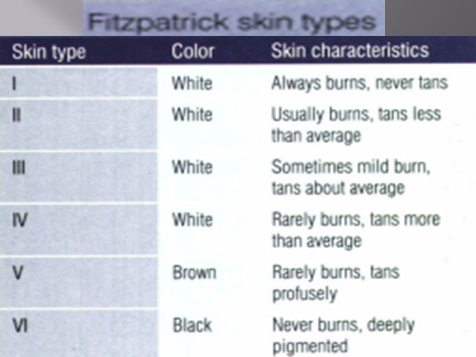
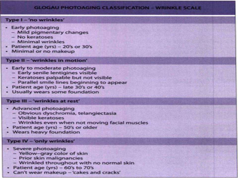
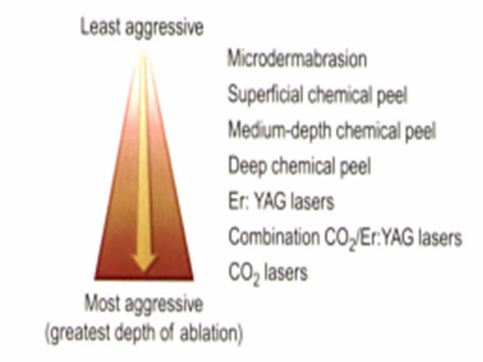

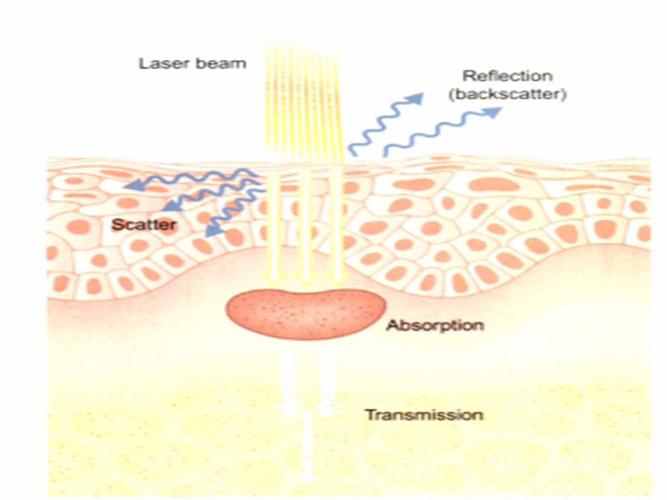


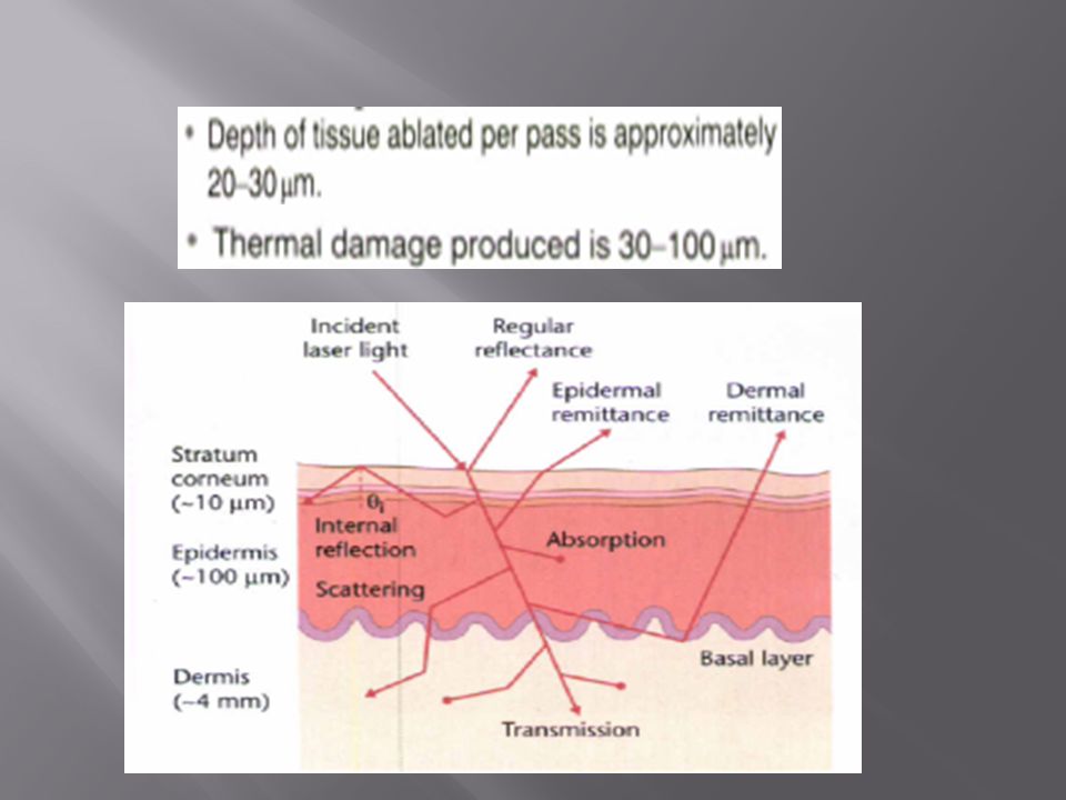
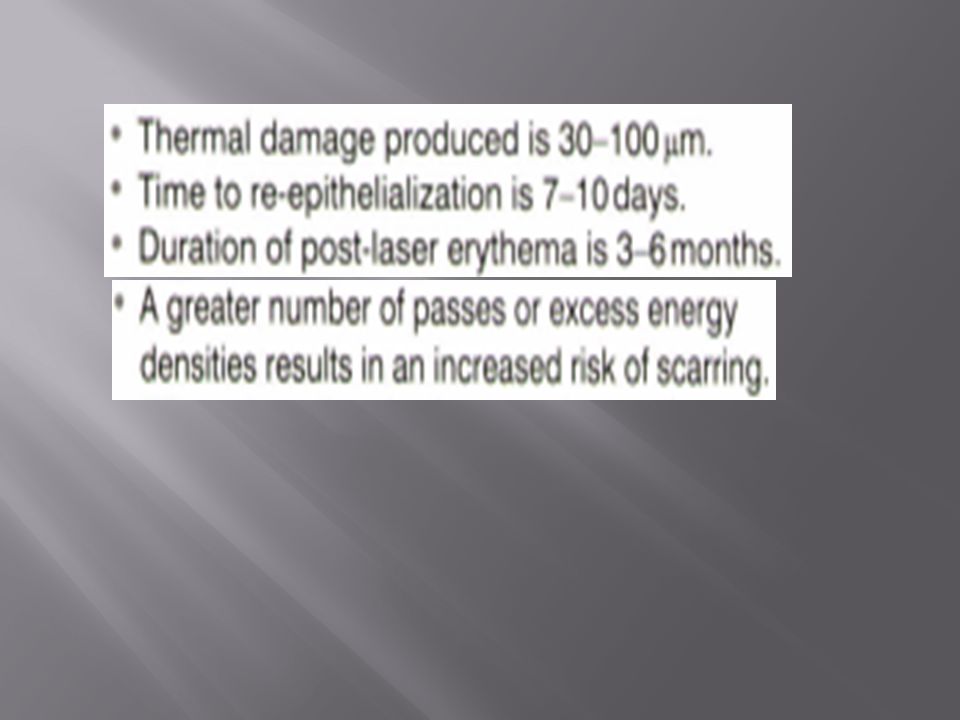

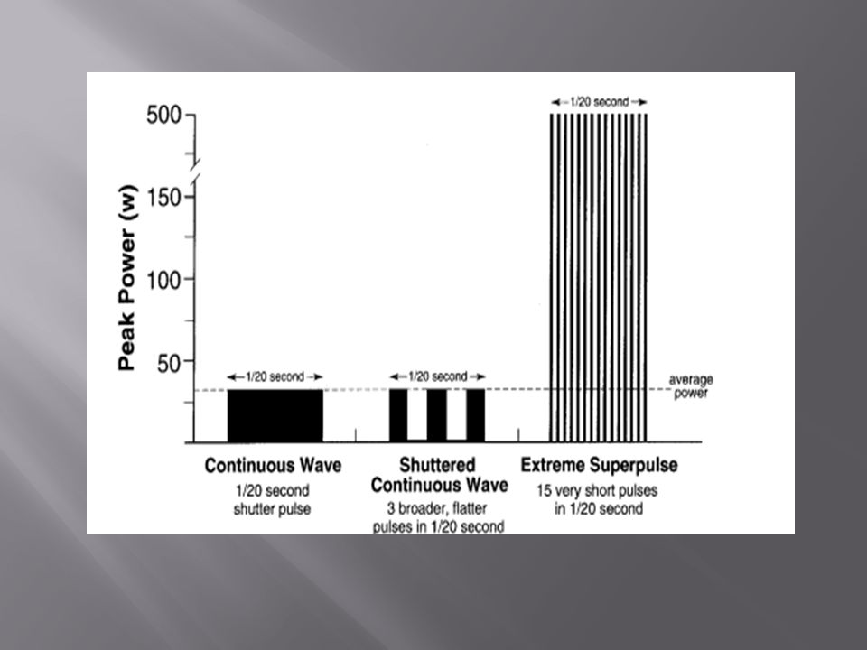

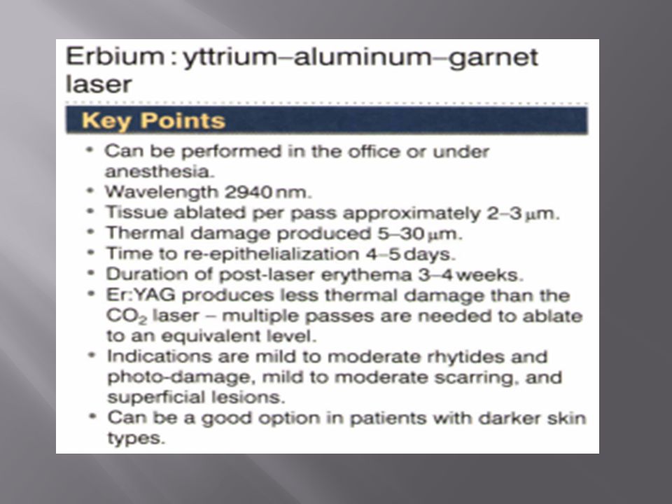


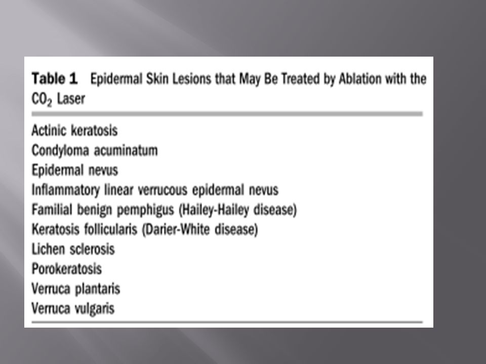


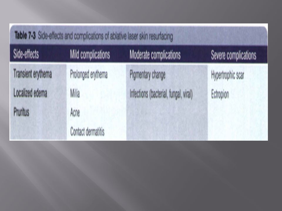


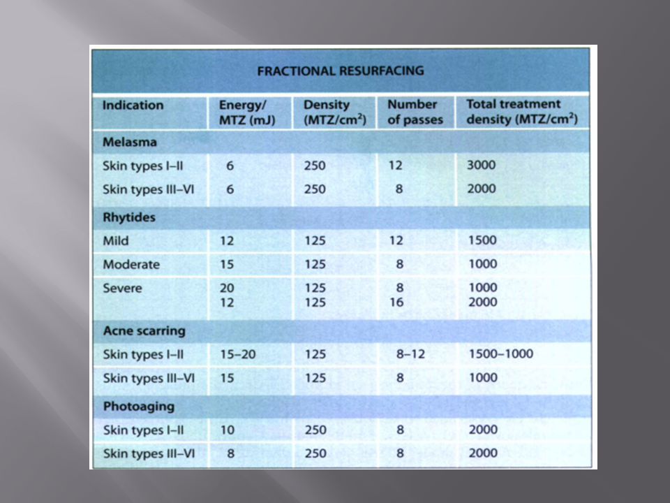
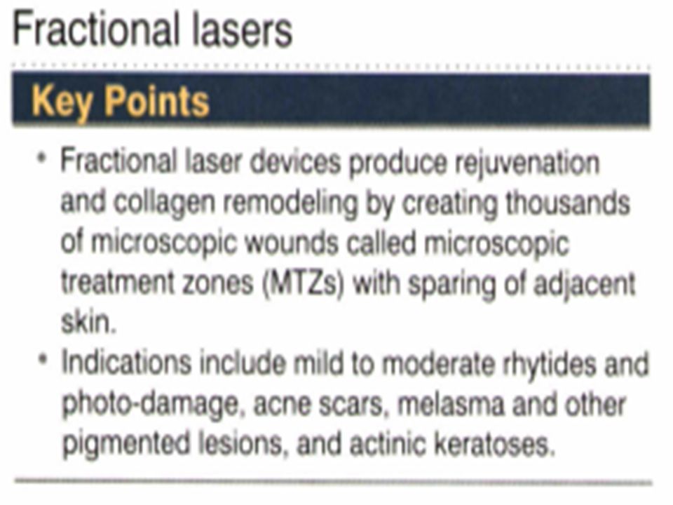


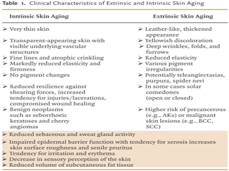


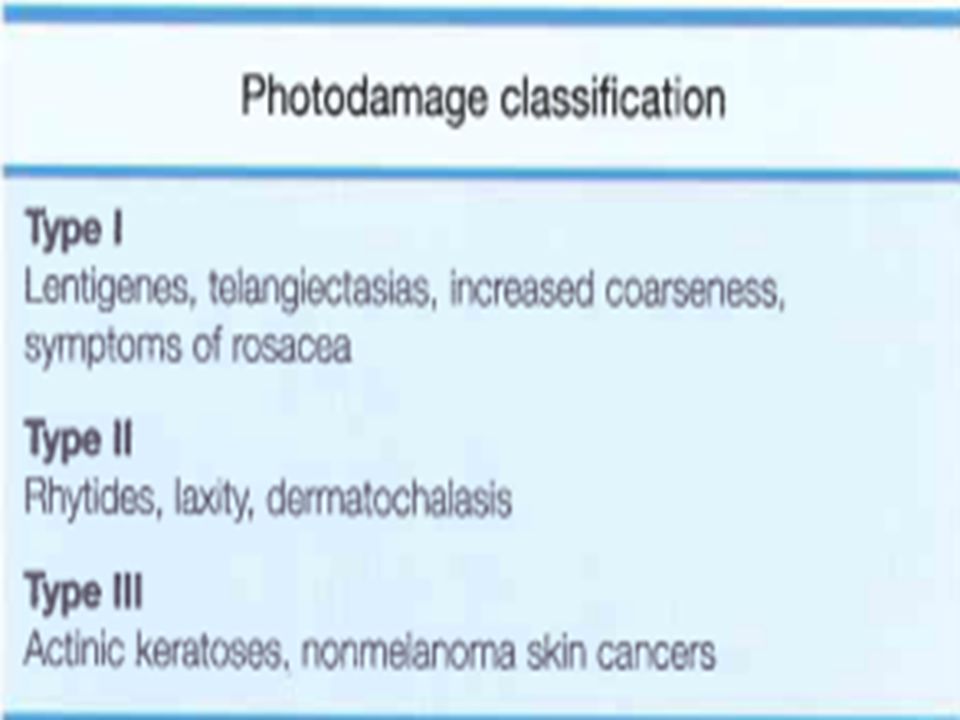


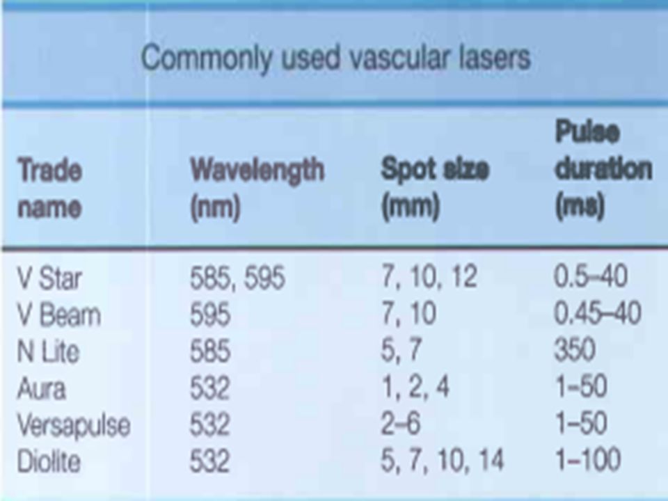


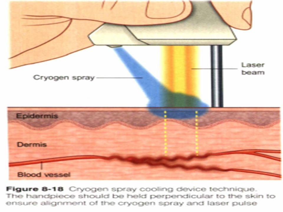

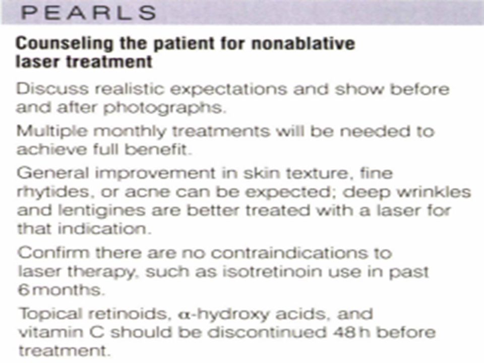
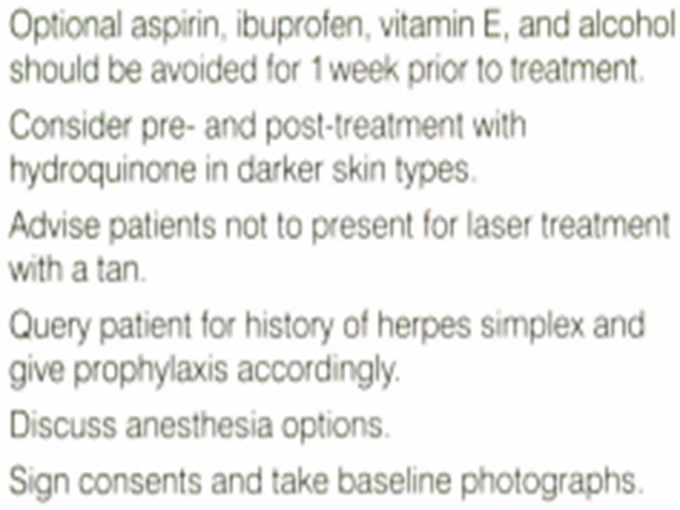
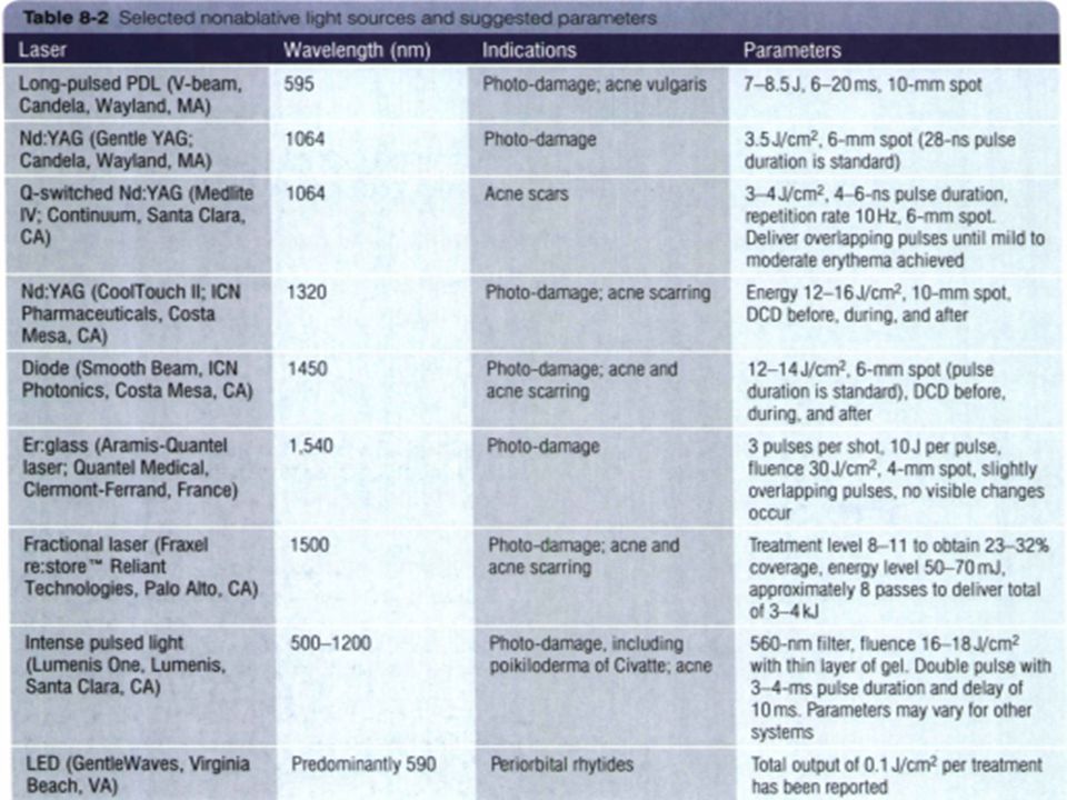
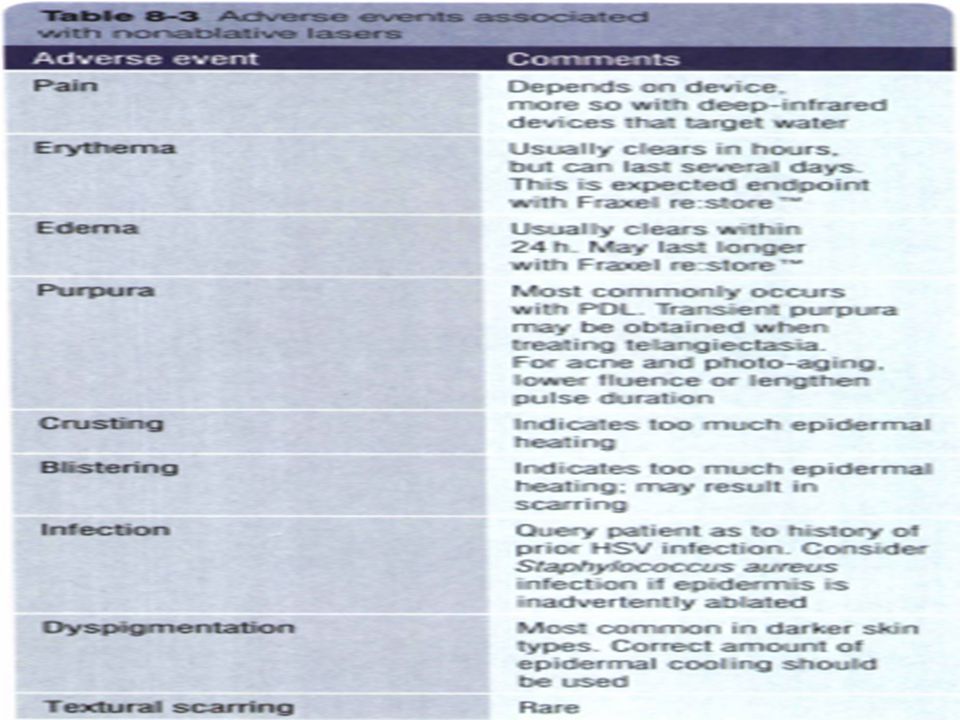


 In The.>")
 Immediate threat: –Dehydration and electrolyte.>")


